Review
. 2014 Jan-Feb;4(1):6-15.
doi: 10.4161/bioa.28014. Epub 2014 Jan 31.

Salient features of the ciliated organ of asymmetry

Affiliations
Review

Salient features of the ciliated organ of asymmetry

Jeffrey D Amack. Bioarchitecture. 2014 Jan-Feb.

Abstract

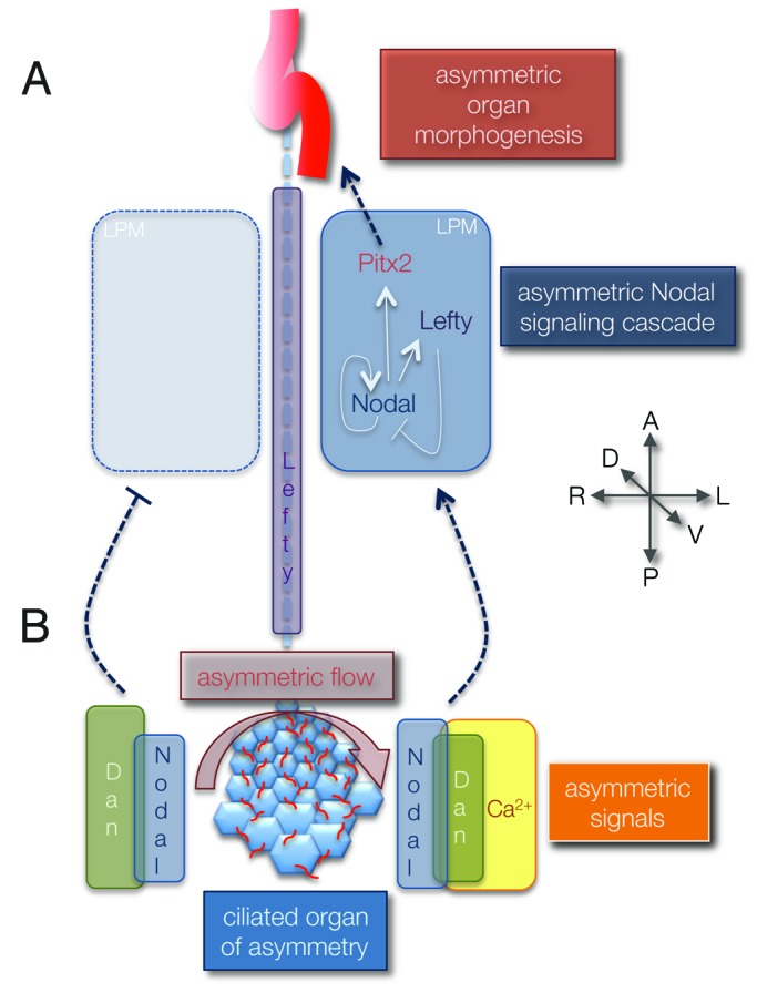
Many internal organs develop distinct left and right sides that are essential for their functions. In several vertebrate embryos, motile cilia generate an asymmetric fluid flow that plays an important role in establishing left-right (LR) signaling cascades. These 'LR cilia' are found in the ventral node and posterior notochordal plate in mammals, the gastrocoel roof plate in amphibians and Kupffer's vesicle in teleost fish. I consider these transient ciliated structures as the 'organ of asymmetry' that directs LR patterning of the developing embryo. Variations in size and morphology of the organ of asymmetry in different vertebrate species have raised questions regarding the fundamental features that are required for LR determination. Here, I review current models for how LR asymmetry is established in vertebrates, discuss the cellular architecture of the ciliated organ of asymmetry and then propose key features of this organ that are critical for orienting the LR body axis.

Keywords: Kupffer’s vesicle; Left-right asymmetry; calcium ion flux; cilia; congenital heart defects; gastrocoel roof plate; posterior notochordal plate.


Figures

None
Figure 1. Working model for how LR asymmetry is established in vertebrate embryos. (A) Asymmetric Nodal signaling guides asymmetric organ development. The TGFβ signaling molecule Nodal initiates its own expression in left lateral plate mesoderm (LPM) to expand asymmetric patterning along the left side of the embryo and induces expression of Lefty and Pitx2. Lefty molecules function as Nodal antagonists in the embryonic midline (dashed line) and LPM to place boundaries on Nodal signaling. Pitx2 is a transcription factor thought to control genes involved in asymmetric morphogenesis of visceral organs. (B) Asymmetric fluid flow in the ciliated organ of asymmetry is translated into asymmetric Nodal signaling. Motile LR cilia (red) generate a leftward flow that results in increased Ca2+ signals on the left side of the organ of asymmetry and elevated expression of Cerebrus/Dan family Nodal antagonists (Dan proteins) on the right side. These flow-dependent asymmetric signals are integrated with bilateral Nodal expression to trigger Nodal signaling exclusively in the left LPM. Embryonic axes: A, anterior; P, posterior; D, dorsal; V, ventral; L, left; R, right.
None
Figure 2. Salient features of the ciliated organ of asymmetry. (1–2) Close-up view of ciliated epithelial cells in the organ of asymmetry. (1) Motile monocilia (red) use a vortical motion to generate fluid flow. (2) Motile cilia must be tilted posteriorly to create leftward flow. Positioning of the basal body (yellow) at the posterior end of the cell is involved in cilia tilting. (3–4) Overview of the ciliated organ of asymmetry. (3) Asymmetric fluid flow (arrow) induces a transient left-sided calcium ion (Ca2+) flux that is essential for establishing asymmetric Nodal signaling. (4) The arrangement of ciliated cells is important for generating and sensing fluid flow. Shown as an example is a gradient of cell morphologies along the anteroposterior axis in the zebrafish Kupffer’s vesicle, where cells with small apical surfaces are tightly packed into the anterior pole and fewer, larger cells are placed in the posterior region. Embryonic axes: A, anterior; P, posterior; D, dorsal; V, ventral; L, left; R, right.

References

    1. Peeters H, Devriendt K. Human laterality disorders. Eur J Med Genet. 2006;49:349–62. doi: 10.1016/j.ejmg.2005.12.003. - DOI - PubMed
    1. Ramsdell AF. Left-right asymmetry and congenital cardiac defects: getting to the heart of the matter in vertebrate left-right axis determination. Dev Biol. 2005;288:1–20. doi: 10.1016/j.ydbio.2005.07.038. - DOI - PubMed
    1. Sutherland MJ, Ware SM. Disorders of left-right asymmetry: heterotaxy and situs inversus. Am J Med Genet C Semin Med Genet. 2009;151C:307–17. doi: 10.1002/ajmg.c.30228. - DOI - PubMed
    1. Brueckner M. Heterotaxia, congenital heart disease, and primary ciliary dyskinesia. Circulation. 2007;115:2793–5. doi: 10.1161/CIRCULATIONAHA.107.699256. - DOI - PubMed
    1. Bisgrove BW, Morelli SH, Yost HJ. Genetics of human laterality disorders: insights from vertebrate model systems. Annu Rev Genomics Hum Genet. 2003;4:1–32. doi: 10.1146/annurev.genom.4.070802.110428. - DOI - PubMed

Publication types

LinkOut - more resources