. 2014 Jun;35(6):1195-201.
doi: 10.3174/ajnr.A3829. Epub 2014 Jan 30.

Surgical and clinical confirmation of temporal bone CT findings in patients with otosclerosis with failed stapes surgery

Affiliations

Surgical and clinical confirmation of temporal bone CT findings in patients with otosclerosis with failed stapes surgery

J Whetstone et al. AJNR Am J Neuroradiol. 2014 Jun.

Abstract

Background and purpose: Prior descriptions of imaging after failed stapes procedures for otosclerosis predated currently available CT technology and/or failed to assess commonly used metallic implants. The purpose of this study was to correlate temporal bone CT findings with clinically and intraoperatively determined causes of surgical failure.

Materials and methods: All patients with otosclerosis undergoing stapedectomy between December 1999 and December 2010 were identified from a search of neurotology clinical records. Patients presenting because of failed stapes surgery and having temporal bone CT scans at the time of revision surgery or clinical evaluation were included. Imaging and clinical records were retrospectively evaluated by a medical student, radiology resident, and senior neuroradiologist. Stapes prosthesis complications and relevant anatomic CT findings were correlated to clinical and intraoperative findings.

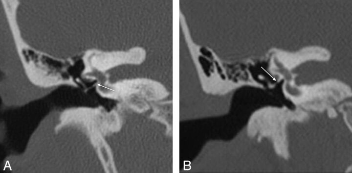
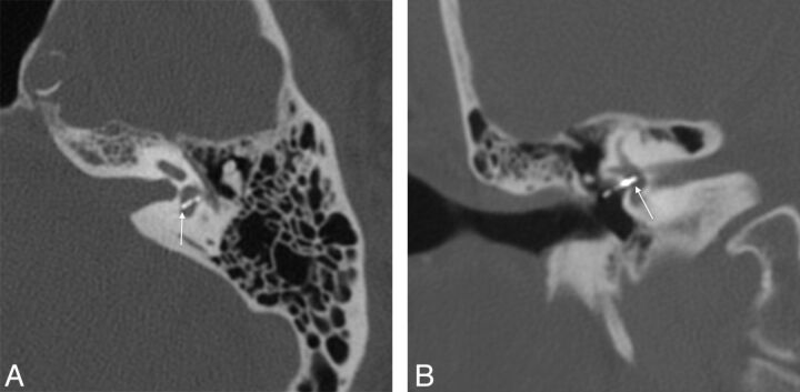
Results: Twenty-two of 340 patients met inclusion criteria. Temporal bone CT findings were correlated to intraoperative findings in 17 of 22 patients and to clinical findings in 5 of 22 patients. Surgically confirmed abnormalities included 7 of 7 incus erosions, 3 of 6 piston re-sizings, 3 of 5 granulation tissues, 3 of 5 prosthesis disconnections, 3 of 4 obliterative otosclerosis, 2 of 2 oval window dislocations, and 1 labyrinthine ossificans. Clinically confirmed abnormalities included 2 cases each of superior semicircular canal dehiscence, and wrong piston size, and 1 each of piston disconnection, labyrinthine ossificans, and intravestibular footplate.

Conclusions: CT evaluation in the setting of failed stapes surgery is challenging. Many postoperative complications such as piston migration, incus necrosis, and overt vestibular penetration are well recognized on temporal bone CT. Of particular note, superior semicircular canal dehiscence is an important contraindication to stapes surgery.


Figures

Fig 1.
Fig 1.
A, Axial CT in a 15-year-old girl with persistent conductive hearing loss poststapedectomy shows a small lucency in the left fissula antefenestrum (thin arrow), consistent with fenestral otosclerosis. Her piston prosthesis appeared short (thick arrow), without intravestibular penetration (surgically confirmed). B, Axial CT in a 56-year-old man with sensorineural hearing loss after stapedectomy shows extensive lucency surrounding the cochlea (arrows), consistent with cochlear otosclerosis.
Fig 2.
Fig 2.
A, Coronal CT in a 40-year-old woman with recurrent right conductive hearing loss after stapedectomy shows inferior dislocation of the prosthesis (arrow) with respect to the oval window. B, Coronal CT in a 15-year-old girl with CHL shows superior dislocation of the stapes prosthesis from the OW (arrow).
Fig 3.
Fig 3.
A, Axial CT in a 31-year-old woman with right conductive hearing loss after stapedectomy shows a gap (arrow) consistent with disconnected prosthesis. B, Coronal CT (same patient as in A) shows slender incus erosion and disconnection (arrow). This patient had intraoperatively confirmed disconnection and incus necrosis.
Fig 4.
Fig 4.
A, Axial CT in a 68-year-old man with recurrent mixed hearing loss shows findings suggesting the lateralized piston syndrome: prosthesis displacement inferior to the OW (arrow). Piston tip is encased in new otosclerotic bone. B, Coronal oblique multiplanar reformation (same patient as in A) shows incus erosion (arrow). Intraoperative findings confirmed scar tissue surrounding the stapes piston, causing prosthesis extrusion.
Fig 5.
Fig 5.
A, Stenver multiplanar reformation CT in a 55-year-old woman with mixed hearing loss shows findings of lateralized piston syndrome. The piston is in the oval window, but no vestibular penetration (thin arrow) is noted. Note piston lateralization to the tympanic membrane (thick arrow). B, Poschl MPR (same patient as in A) shows the piston traversing the expected location of the incus long process, which is eroded (arrow).
Fig 6.
Fig 6.
A, Axial CT in a 44-year-old woman with conductive hearing loss demonstrates no vestibular penetration (arrow) by the piston prosthesis. B, Coronal CT multiplanar reformation in the same patient as in A also shows no vestibular penetration (arrow), suggesting short piston. Intraoperative findings confirmed inadequate piston depth.
Fig 7.
Fig 7.
A, Axial CT in an 80-year-old man with sensorineural hearing loss shows deep intrusion into the vestibule (arrow). B, Coronal CT multiplanar reformation in a 68-year-old man with vestibular symptoms suggests deep intravestibular position of the stapes prosthesis (arrow). Long prosthesis and small labyrinthine fistula were confirmed intraoperatively.
Fig 8.
Fig 8.
A, Axial CT in a 56-year-old man with persistent mixed hearing loss after stapedectomy demonstrates heaped-up lucent bone formation at the right round window consistent with obliterative otosclerosis (OtoO) (arrow). This patient also had probable superior semicircular canal dehiscence (not shown). B, Axial CT in the contralateral ear in the same patient as in B also shows OtoO. Note tip of prior stapes piston embedded within otosclerotic new bone (arrow).
Fig 9.
Fig 9.
A, Axial CT in a 41-year-old man with vertigo and complete sensorineural hearing loss in the left ear after prior stapedectomy show focal hyperattenuation in the vestibule (arrow), consistent with intravestibular footplate dislocation. B, Coronal multiplanar reformation in the same patient as in A shows intravestibular footplate dislocation (arrow).
Fig 10.
Fig 10.
A, Poschl multiplanar reformation CT in a 67-year-old man with mixed hearing loss in the right ear after prior stapedectomy shows ipsilateral superior semicircular canal dehiscence (arrow) that probably explains surgical failure. B, Axial CT demonstrates ipsilateral labyrinthine ossificans (arrow) in the same patient as in A that might also have contributed to his surgical failure.

References

    1. Cummings CW, Fredrickson JM, Harker LA, et al. , eds. Otolaryngology-Head and Neck Surgery. 2nd ed. St Louis: Mosby; 1993
    1. Declau F, van Spaendonck M, Timmermans JP, et al. . Prevalence of histologic otosclerosis: an unbiased temporal bone study in Caucasians. Adv Otorhinolaryngol 2007;65:6–16 - PubMed
    1. Sakihara Y, Parving A. Clinical otosclerosis, prevalence estimates and spontaneous progress. Clin Otosc Prev Acta Otol 1999;199:468–72 - PubMed
    1. Vincent R, Sperling N, Oates J, et al. . Surgical findings and long-term hearing results in 3,050 stapedotomies for primary otosclerosis: a prospective study with the Otology-Neurotology Database. Otol Neurotol 2006;27(Suppl 2):S25–47 - PubMed
    1. Ozuer MZ, Olgun L, Gultekin G. Revision stapes surgery. Otolaryngol Head Neck Surg 2012;146:109–13 - PubMed