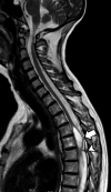
Familial adhesive arachnoiditis associated with syringomyelia
- PMID: 24481329
- PMCID: PMC7965138
- DOI: 10.3174/ajnr.A3858
Familial adhesive arachnoiditis associated with syringomyelia
Abstract
Adhesive arachnoiditis is a rare condition, often complicated by syringomyelia. This pathologic entity is usually associated with prior spinal surgery, spinal inflammation or infection, and hemorrhage. The usual symptoms of arachnoiditis are pain, paresthesia, and weakness of the low extremities due to the nerve entrapment. A few cases have had no obvious etiology. Previous studies have reported one family with multiple cases of adhesive arachnoiditis. We report a second family of Belgian origin with multiple cases of arachnoiditis and secondary syringomyelia in the affected individuals.
© 2014 by American Journal of Neuroradiology.
Figures


References
-
- Duke RJ, Hashimoto SA. Familial spinal arachnoiditis: a new entity. Arch Neurol 1974;30:300–03 - PubMed
-
- Nagai M, Sakuma R, Aoki M, et al. . Familial spinal arachnoiditis with secondary syringomyelia: clinical studies and MRI findings. J Neurol Sci 2000;177:60–64 - PubMed
-
- Benner B, Ehni G. Spinal arachnoiditis: the postoperative variety in particular. Spine (Phila Pa 1976) 1978;3:40–44 - PubMed
-
- Brammah TB, Jayson MI. Syringomyelia as a complication of spinal arachnoiditis. Spine (Phila Pa 1976) 1994;19:2603–05 - PubMed
Publication types
MeSH terms
Supplementary concepts
LinkOut - more resources
Full Text Sources
Other Literature Sources
Medical
