Review
. 2014 Mar;14(3):166-80.
doi: 10.1038/nri3607. Epub 2014 Jan 31.

Apoptotic cell clearance: basic biology and therapeutic potential

Affiliations
Review

Apoptotic cell clearance: basic biology and therapeutic potential

Ivan K H Poon et al. Nat Rev Immunol. 2014 Mar.

Abstract

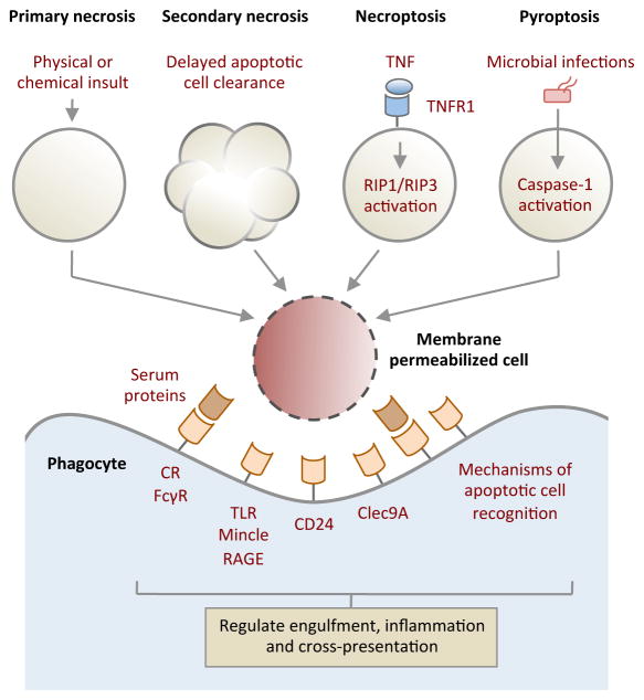
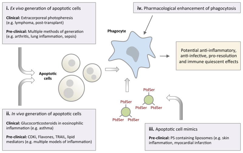
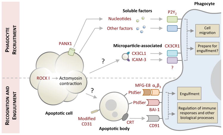
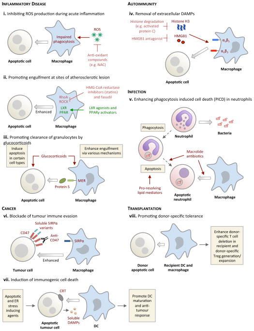
The prompt removal of apoptotic cells by phagocytes is important for maintaining tissue homeostasis. The molecular and cellular events that underpin apoptotic cell recognition and uptake, and the subsequent biological responses, are increasingly better defined. The detection and disposal of apoptotic cells generally promote an anti-inflammatory response at the tissue level, as well as immunological tolerance. Consequently, defects in apoptotic cell clearance have been linked with various inflammatory diseases and autoimmunity. Conversely, under certain conditions, such as the killing of tumour cells by specific cell-death inducers, the recognition of apoptotic tumour cells can promote an immunogenic response and antitumour immunity. Here, we review the current understanding of the complex process of apoptotic cell clearance in physiology and pathology, and discuss how this knowledge could be harnessed for new therapeutic strategies.


Figures

Box 1
Box 1
Handling membrane permeabilizwd (necrotic) cells
Box 2
Box 2
Apoptotic cells as a potential therapeutic intervention
Figure 1
Figure 1. Phases of apoptotic cell clearance
Cells undergoing apoptosis often exhibit morphological changes (for example membrane blebbing and cellular shrinkage) to facilitate cell detachment and organelle fragmentation. Prior to or during the onset of apoptotic morphology, apoptotic cells also release ‘find-me’ signals in the form of soluble factors (for example nucleotides) or microparticle-associated molecules (including CX3C chemokine ligand 1 (CX3CL1) and intercellular adhesion molecule 3 (ICAM3)) to recruit phagocytes for cell clearance. Nucleotides are released from apoptotic cells via caspase-activated pannexin 1 (PANX1) membrane channels. Whether detection of ‘find-me’ signals by phagocytes can prepare molecular machinery necessary for engulfment in addition to cell migration warrants further investigation. Exposure of ‘eat-me’ signals (such as phosphatidylserine (PtdSer) and calreticulin (CRT)) accompanied by modification of ‘don’t eat-me’ signals (CD31) on apoptotic cells or fragments of apoptotic cell (also referred to as apoptotic bodies) mediate their recognition by phagocytes. Phagocytes can engage ‘eat-me’ signals directly via cell surface receptors (such as brain-specific angiogenesis inhibitor (BAI-1) and CD91) or indirectly through bridging molecules (such as milk fat globule-EGF factor 8 (MFG-E8)) that are in turn detected by membrane receptors (αVβ3). Subsequent downstream signalling initiates engulfment and engulfment-associated responses from phagocytes. The mechanism underpinning the formation of apoptotic bodies and microparticles is not fully defined. P2Y2, purinergic receptor P2Y2; ROCK I, Rho-associated coiled-coil containing protein kinase I.
Figure 2
Figure 2. Potential approaches for targeting the apoptotic cell clearance process for therapeutic benefits
a. Bacterial infection. Following phagocytosis of invading bacteria, neutrophils frequently undergo a form of ‘phagocytosis induced cell death’ (PICD) with subsequent engulfment by surrounding phagocytes, providing a second round of pathogen destruction. The engulfing phagocytes also increase production of pro-resolving lipid mediator release (for example RvD1) with enhanced host-directed bacterial killing. b. Acute inflammation. Reactive oxygen species (ROS) that are produced at sites of acute inflammation impair phagocytosis through the activation of RhoA within phagocytes. Scavenging ROS (for example by using N-acetylcystein; NAC) enhances apoptotic cell clearance. c. Glucocorticoids can potentially augment eosinophil clearance by promoting both eosinophil apoptosis and cell clearance via a protein S–Mer-dependent pathway. d. Atherosclerosis. Impaired engulfment in atherosclerosis is, in part, mediated by increased activity of RhoA and its downstream mediator ROCK, both of which are negative regulators of apoptotic cell engulfment. RhoA inhibition by HMG-CoA reductase inhibitors (statins) or ROCK inhibition seems to have a beneficial effect in atherosclerosis, possibly by regulating engulfment. e. Rheumatoid arthritis. At sites of inflammation, extracellular damage-associated molecular patterns (DAMPs) such as histones and HMGB1 negatively regulate apoptotic cell engulfment by binding to integrins on the surface of phagocytes. Strategies to degrade DAMPs (for example through the degradation of histone H3 by antigen presenting cells (APCs)) can improve apoptotic cell clearance. f. Recognition and uptake of donor apoptotic cells by recipient DCs and macrophages can promote donor-specific tolerance in the recipients and limit allograft rejection. g. Induction of tumour cell death accompanied with CRT and DAMPs exposure can promote DC-mediated engulfment and DC maturation to initiate an anti-tumour immune response. h. Targeting tumour cells with anti-CD47 blocking antibodies or soluble SIRPα variants inhibits CD47-SIRPα interaction and facilitates tumour cell removal by macrophages.

Similar articles

Cited by

References

    1. Ravichandran KS. Find-me and eat-me signals in apoptotic cell clearance: progress and conundrums. J Exp Med. 2010;207:1807–17. - PMC - PubMed
    1. Wood W, et al. Mesenchymal cells engulf and clear apoptotic footplate cells in macrophageless PU.1 null mouse embryos. Development. 2000;127:5245–52. - PubMed
    1. Parnaik R, Raff MC, Scholes J. Differences between the clearance of apoptotic cells by professional and non-professional phagocytes. Curr Biol. 2000;10:857–60. - PubMed
    1. Monks J, Smith-Steinhart C, Kruk ER, Fadok VA, Henson PM. Epithelial cells remove apoptotic epithelial cells during post-lactation involution of the mouse mammary gland. Biol Reprod. 2008;78:586–94. - PubMed
    1. Juncadella IJ, et al. Apoptotic cell clearance by bronchial epithelial cells critically influences airway inflammation. Nature. 2013;493:547–51. - PMC - PubMed

Publication types