

The use of a neutral peptide aptamer scaffold to anchor BH3 peptides constitutes a viable approach to studying their function
- PMID: 24481451
- PMCID: PMC4040713
- DOI: 10.1038/cddis.2013.564
The use of a neutral peptide aptamer scaffold to anchor BH3 peptides constitutes a viable approach to studying their function
Abstract
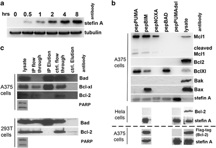
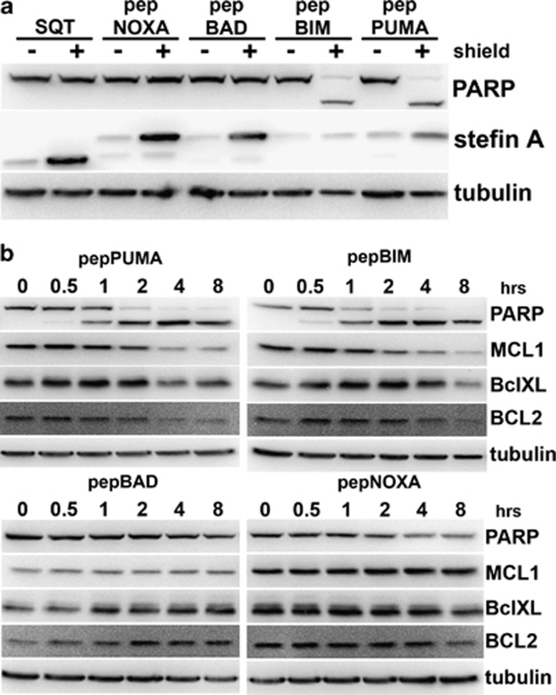
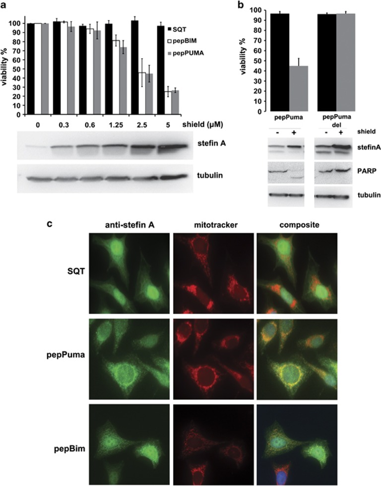
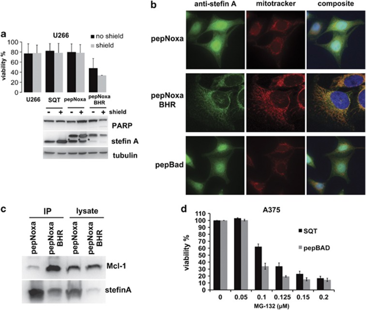
The B-cell CLL/lymphoma-2 (Bcl-2) family of proteins are important regulators of the intrinsic pathway of apoptosis, and their interactions, driven by Bcl-2 homology (BH) domains, are of great interest in cancer research. Particularly, the BH3 domain is of clinical relevance, as it promotes apoptosis through activation of Bcl-2-associated x protein (Bax) and Bcl-2 antagonist killer (Bak), as well as by antagonising the anti-apoptotic Bcl-2 family members. Although investigated extensively in vitro, the study of the BH3 domain alone inside cells is more problematic because of diminished secondary structure of the unconstrained peptide and a lack of stability. In this study, we report the successful use of a novel peptide aptamer scaffold - Stefin A quadruple mutant - to anchor and present the BH3 domains from Bcl-2-interacting mediator of cell death (Bim), p53 upregulated modulator of apoptosis (Puma), Bcl-2-associated death promoter (Bad) and Noxa, and demonstrate its usefulness in the study of the BH3 domains in vivo. When expressed intracellularly, anchored BH3 peptides exhibit much the same binding specificities previously established in vitro, however, we find that, at endogenous expression levels, Bcl-2 does not bind to any of the anchored BH3 domains tested. Nonetheless, when expressed inside cells the anchored PUMA and Bim BH3 α-helices powerfully induce cell death in the absence of efficient targeting to the mitochondrial membrane, whereas the Noxa helix requires a membrane insertion domain in order to kill Mcl-1-dependent myeloma cells. Finally, the binding of the Bim BH3 peptide to Bax was the only interaction with a pro-apoptotic effector protein observed in this study.
Figures

Similar articles
-
The BH3 alpha-helical mimic BH3-M6 disrupts Bcl-X(L), Bcl-2, and MCL-1 protein-protein interactions with Bax, Bak, Bad, or Bim and induces apoptosis in a Bax- and Bim-dependent manner.J Biol Chem. 2011 Mar 18;286(11):9382-92. doi: 10.1074/jbc.M110.203638. Epub 2010 Dec 9. J Biol Chem. 2011. PMID: 21148306 Free PMC article.
-
BH3 domains other than Bim and Bid can directly activate Bax/Bak.J Biol Chem. 2011 Jan 7;286(1):491-501. doi: 10.1074/jbc.M110.167148. Epub 2010 Nov 1. J Biol Chem. 2011. PMID: 21041309 Free PMC article.
-
Bid chimeras indicate that most BH3-only proteins can directly activate Bak and Bax, and show no preference for Bak versus Bax.Cell Death Dis. 2015 Apr 23;6(4):e1735. doi: 10.1038/cddis.2015.105. Cell Death Dis. 2015. PMID: 25906158 Free PMC article.
-
Structural biology of the Bcl-2 family of proteins.Biochim Biophys Acta. 2004 Mar 1;1644(2-3):83-94. doi: 10.1016/j.bbamcr.2003.08.012. Biochim Biophys Acta. 2004. PMID: 14996493 Review.
-
How to unleash mitochondrial apoptotic blockades to kill cancers?Acta Pharm Sin B. 2017 Jan;7(1):18-26. doi: 10.1016/j.apsb.2016.08.005. Epub 2016 Sep 21. Acta Pharm Sin B. 2017. PMID: 28119805 Free PMC article. Review.
Cited by
-
Mining prognostic markers of Asian hepatocellular carcinoma patients based on the apoptosis-related genes.BMC Cancer. 2021 Feb 18;21(1):175. doi: 10.1186/s12885-021-07886-6. BMC Cancer. 2021. PMID: 33602168 Free PMC article.
-
Affimer proteins are versatile and renewable affinity reagents.Elife. 2017 Jun 27;6:e24903. doi: 10.7554/eLife.24903. Elife. 2017. PMID: 28654419 Free PMC article.
-
Studies of the oligomerisation mechanism of a cystatin-based engineered protein scaffold.Sci Rep. 2019 Jun 21;9(1):9067. doi: 10.1038/s41598-019-45565-6. Sci Rep. 2019. PMID: 31227800 Free PMC article.
-
MicroRNAs Enable mRNA Therapeutics to Selectively Program Cancer Cells to Self-Destruct.Nucleic Acid Ther. 2018 Oct;28(5):285-296. doi: 10.1089/nat.2018.0734. Epub 2018 Aug 8. Nucleic Acid Ther. 2018. PMID: 30088967 Free PMC article.
-
Cell-penetrating Alphabody protein scaffolds for intracellular drug targeting.Sci Adv. 2021 Mar 26;7(13):eabe1682. doi: 10.1126/sciadv.abe1682. Print 2021 Mar. Sci Adv. 2021. PMID: 33771865 Free PMC article.
References
MeSH terms
Substances
Grants and funding
LinkOut - more resources
Full Text Sources
Other Literature Sources
Research Materials
Miscellaneous
