Review
. 2014 Apr;11(2):251-68.
doi: 10.1007/s13311-013-0251-0.

Epilepsy related to developmental tumors and malformations of cortical development

Affiliations
Review

Epilepsy related to developmental tumors and malformations of cortical development

Eleonora Aronica et al. Neurotherapeutics. 2014 Apr.

Abstract

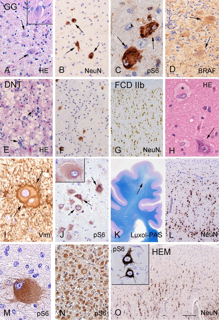
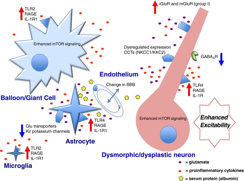
Structural abnormalities of the brain are increasingly recognized in patients with neurodevelopmental delay and intractable focal epilepsies. The access to clinically well-characterized neurosurgical material has provided a unique opportunity to better define the neuropathological, neurochemical, and molecular features of epilepsy-associated focal developmental lesions. These studies help to further understand the epileptogenic mechanisms of these lesions. Neuropathological evaluation of surgical specimens from patients with epilepsy-associated developmental lesions reveals two major pathologies: focal cortical dysplasia and low-grade developmental tumors (glioneuronal tumors). In the last few years there have been major advances in the recognition of a wide spectrum of developmental lesions associated with a intractable epilepsy, including cortical tubers in patients with tuberous sclerosis complex and hemimegalencephaly. As an increasing number of entities are identified, the development of a unified and comprehensive classification represents a great challenge and requires continuous updates. The present article reviews current knowledge of molecular pathogenesis and the pathophysiological mechanisms of epileptogenesis in this group of developmental disorders. Both emerging neuropathological and basic science evidence will be analyzed, highlighting the involvement of different, but often converging, pathogenetic and epileptogenic mechanisms, which may create the basis for new therapeutic strategies in these disorders.


Figures

Fig. 1
Fig. 1
Histopathological features of glioneuronal tumors, focal cortical dysplasia (FCD), tuberous sclerosis complex (TSC), and hemimegalencephaly (HME). (ad) Ganglioglioma (GG). (a) Hematoxylin/eosin (HE) staining of GG showing large dysplastic neurons (arrows and insert) and glial cells. (b) NeuN staining detects the neuronal component (nuclear staining) of GG. (c) Phosphorylated ribosomal S6 protein (pS6) expression in dysplastic neurons (arrows). (d) BRAF V600E immunostaining showing prominent expression in large dysplastic cells (arrows). (e, f) Dysembryoplastic neuroepithelial tumor (DNT). (e) HE staining of DNT showing a typical heterogeneous cellular composition, with floating neurons (arrow) surrounded by a prominent population of oligodendroglia-like cells. (f) NeuN staining detects the neuronal component of DNT. (gj) FCD. (g) FCD NeuN staining showing cortical dyslamination. (h) HE staining showing a dysmorphic neuron with enlarged nucleus and aggregates of Nissl substance (arrow) and a balloon cell (*). (i) Vimentin (Vim) staining of balloon cells. (j) pS6 expression in dysmorphic neurons (arrows) and a balloon cell (insert). (kn) TSC, cortical tubers. (k) Luxol–Periodic acid–Schiff (PAS) staining showing a cortical tuber (arrow) with decreased density of myelinated fibers. (l) NeuN staining showing cortical dyslamination within the tuber. (m, n) pS6 expression in a giant cell (m; cortical tuber from adult TSC patient) and within a focal lesion in a fetus at 23 gestational weeks. (o) HME. NeuN staining showing cortical dyslamination within the enlarged hemisphere, inset shows pS6 expression in dysmorphic neurons. Scale bar: A, B, D–F, J, N = 80 μm; C, I, M = 30 μm; H = 40 μm; G, L, O = 500 μm; K = 1.2 cm
Fig. 2
Fig. 2
Schematic of epileptogenic developmental brain lesions as mammalian target of rapamycin (mTOR)opathies. Overactivation of the mTOR signaling in glioneuronal tumors (GNT), focal cortical dysplasia (FCD) IIb, and hemimegalencephaly (HME), as well as tubers in tuberous sclerosis complex (TSC), suggests a pathogenic link between these malformations and reflects a spectrum of disorders of mTOR signaling (mTORopathies; see text). P = phosphorylation; IGF-1 = insulin-like growth factor-1; PI3K = PI3kinase; PDK1 = phosphoinositide-dependent kinase-1; LKB1 = tumor suppressor liver kinase B1; AMPK = AMP-activated protein kinase; HPV = human papilloma virus; S6K1 = p70S6kinase; 4E-BP1 = elongation binding protein 1; pS6 = phosphorylated ribosomal S6 protein; Erk = extracellular signal regulated kinase; IRS = insulin receptor substrate; PTEN = Phosphatase and tensin homologue; Akt = protein kinase B; Rheb = ras homolog enriched in brain; mTORC = mammalian target of rapamycin complex; REDD1 = regulated in DNA damage and development 1; DEPTOR = DEP domain containing MTOR-interacting protein
Fig. 3
Fig. 3
Schematic depicting mechanisms contributing to epileptogenesis in developmental tumors (glioneuronal tumors) and malformations of cortical development. A better understanding of these mechanisms may create the basis to develop effective therapeutic strategies to control seizures. TLR2 = Toll-like receptor 2; RAGE = receptor for advanced glycation end products; IL-1R1 = interleukin 1 receptor; mTOR = mammalian target of rapamycin; Glu = glutamate; Kir = inward-rectifying potassium channel; BBB = blood–brain barrier; iGluR = ionotropic glutamate receptor; mGluR = metabotropic glutamate receptor; CCT = cation–chloride (NKCC1 and KCC2) cotransporter; GABAAR = gamma-aminobutyric acid a receptor

References

    1. Blumcke I, Vinters HV, Armstrong D, Aronica E, Thom M, Spreafico R. Malformations of cortical development and epilepsies: neuropathological findings with emphasis on focal cortical dysplasia. Epileptic Disord. 2009;11:181–193. - PubMed
    1. Sisodiya SM, Fauser S, Cross JH, Thom M. Focal cortical dysplasia type II: biological features and clinical perspectives. Lancet Neurol. 2009;8:830–843. - PubMed
    1. Aronica E, Becker AJ, Spreafico R. Malformations of cortical development. Brain Pathol. 2012;22:380–401. - PMC - PubMed
    1. Thom M, Blumcke I, Aronica E. Long-term epilepsy-associated tumors. Brain Pathol. 2012;22:350–379. - PMC - PubMed
    1. Barkovich AJ, Guerrini R, Kuzniecky RI, Jackson GD, Dobyns WB. A developmental and genetic classification for malformations of cortical development: update 2012. Brain. 2012;135:1348–1369. - PMC - PubMed

Publication types