. 2014 Apr 1;127(Pt 7):1394-405.
doi: 10.1242/jcs.134692. Epub 2014 Jan 30.

N-WASP-directed actin polymerization activates Cas phosphorylation and lamellipodium spreading

Affiliations

N-WASP-directed actin polymerization activates Cas phosphorylation and lamellipodium spreading

Xian Zhang et al. J Cell Sci. .

Abstract

Tyrosine phosphorylation of the substrate domain of Cas (CasSD) correlates with increased cell migration in healthy and diseased cells. Here, we address the mechanism leading to the phosphorylation of CasSD in the context of fibronectin-induced early spreading of fibroblasts. We have previously demonstrated that mechanical stretching of CasSD exposes phosphorylation sites for Src family kinases (SFKs). Surprisingly, phosphorylation of CasSD was independent of myosin contractile activity but dependent on actin polymerization. Furthermore, we found that CasSD phosphorylation in the early stages of cell spreading required: (1) integrin anchorage and integrin-mediated activation of SFKs, (2) association of Cas with focal adhesion kinase (FAK), and (3) N-WASP-driven actin-assembly activity. These findings, and analyses of the interactions of the Cas domains, indicate that the N-terminus of Cas associates with the FAK-N-WASP complex at the protrusive edge of the cell and that the C-terminus of Cas associates with the immobilized integrin-SFK cluster. Thus, extension of the leading edge mediated by actin polymerization could stretch Cas during early cell spreading, priming it for phosphorylation.

Keywords: Actin dynamics; BCAR1; Cas; Crk-associatied substrate; FAK; Focal adhesion kinase; SFK; Src family kinase; p130Cas.


Figures

Fig. 1.
Fig. 1.
Inhibition of Cas phosphorylation in fibroblasts causes reduced spreading and loss of lamellipodial protrusions. (A–C) Time-lapse sequential images showing the spreading on fibronectin of cells that have been transfected with GFP plus control shRNA (A, supplementary material Movie 1) or GFP plus Cas shRNA (B,C; supplementary material Movies 2, 3). Mouse embryonic fibroblasts were transfected with the GFP-expressing plasmid and expression plasmids of nontargeting shRNA or Cas shRNAs. After 72 h of transfection, cells were harvested and plated on fibronectin-coated cover glass for live imaging for 30 min. (D) In the same sample as A–C, reduction of Cas was monitored by western blot (IB) for Cas. Reduction of CasSD phosphorylation was detected using antibodies against the Y165 and Y410 residues. sus, suspension. (E) Summary of the spreading behavior of control-shRNA- and Cas-shRNA-transfected cells in the first 30 min on fibronectin. w/, with; w/o, without. (F) Staining of the actin cytoskeleton showed loss of active edges that correlated with loss of CasSD phosphorylation in early spreading of cells transfected with GFP plus Cas shRNA. Cells were fixed 10 min after plating. The difference in the actin structure can be seen between cells with GFP co-transfection marker (inset 1) or left untransfected (inset 2). Scale bars: 10 µm. (G) Fibroblasts were fixed as in F and stained with the 9EG7 antibody against activated β1 integrin. The Cas-knockdown cell (inset 1) lacked the mature adhesion structure that is observed in untransfected cells (inset 2). Scale bars: 10 µm.
Fig. 2.
Fig. 2.
SFKs downstream of integrins phosphorylate CasSD in an anchorage-dependent manner. (A–D) Inhibition of SFK by use of PP2 diminished fibronectin-induced CasSD phosphorylation at residues Y165 and Y410 but caused hyperphosphorylation of SFK at residue Y416, as shown by western blot (IB) (A). sus, suspension. Cells were pre-incubated in 20 µM PP2 or its control chemical PP3 for 30 min before being plated on fibronectin-coated dishes (FN). The ratios of (B) pY165 CasSD∶total Cas and (C) pY410 CasSD∶total Cas in different experiments were normalized against control samples at the 30-min time-point and (D) pY416 Src∶total Src ratios against control samples at the 10-min time-point; mean±s.d., four repeats. P-values were calculated by two-tailed paired Student's t-test. A.U., arbitrary units. (E,F) Immunostaining of (E) PP3-treated or (F) PP2-treated early spreading cells. Cells treated with PP2 showed a delay in spreading and defective formation of active protrusions. Sites of limited Cas phosphorylation signal correlated with local actin polymerization (F). Cells were fixed 10 min after spreading initiation. Scale bar: 10 µm. (G) Interaction of β3 integrins with Src was detected in cells in suspension and adherent cells. Fibroblasts were transfected with a β3-integrin–GFP plasmid and harvested 24 h later, kept in suspension for 45 min and then directly lysed with RIPA buffer or plated onto a fibronectin-coated dish for 15 min before lysis. 2 µl of polyclonal antibody was used to pull down endogenous and overexpressed β3 integrins. The mock sample was prepared from the lysate of cells in suspension and no antibody was applied. Src associated with the immunoprecipitated (IP) β3 integrins was detected using an antibody against Src (shown in H). The level of β3 integrin pulled down was monitored using an antibody against GFP. (H–J) The SFK activity of suspension cells increased in response to RGD soluble peptide. Application of RGD did not affect the SFK activity in early spreading cells although CasSD phosphorylation showed a significant decrease. *P<0.05. Fibroblasts kept in suspension were collected by centrifugation and re-suspended in medium containing 1 mM RGD peptide for 5 min before being collected by centrifugation and lysed for western blot (H). The control (con) sample was similarly prepared, except that the RGD peptide was excluded from the re-suspension medium. For adherent samples, fibroblasts were spread on fibronectin for 5 min before the medium was removed and RGD containing medium (or plain medium for control) was applied for another 5 min. Cells were washed once with PBS and lysed directly. Both (I) pY416 Src∶total Src and (J) pY165 Cas∶total Cas ratios in different experiments were normalized against the adhesion sample treated with control medium; mean±s.d., four repeats. P-values were calculated by two-tailed paired Student's t-test.
Fig. 3.
Fig. 3.
Formation of FAK-Cas complex is crucial for CasSD phosphorylation. (A,B) FAK and Cas associated with each other in suspension and adherent cells. Fibroblasts were harvested, kept in suspension (sus) for 45 min and directly lysed with RIPA buffer or plated on fibronectin-coated dishes (FN) for 15 min before lysis. 5 µg of combined polyclonal and monoclonal antibodies against Cas (A) or against FAK (B) were used to pull down Cas and FAK from the lysates. The mock sample was prepared from the lysate of cells in suspension (sus) and lacked antibody. FAK and Cas associated with the immunoprecipitated (IP) complex were detected with monoclonal antibodies against FAK or Cas in the western blot assays (IB). Loading of pulldown samples was adjusted so that lysate content of Cas and FAK levels were comparable between adherent cells and cells in suspension . (C–E) Knockdown of FAK affected CasSD phosphorylation in fibroblasts. Plasmids encoding control shRNA, FAK shRNA, or FAK shRNA and an shRNA-resistant mouse FAK (mFAK) were transfected into fibroblasts by electroporation. After 72 h of transfection, cells were harvested and left in suspension for 45 min before being plated onto fibronectin-coated dishes for 10 min. Cells were directly lysed for western blot assay. (D) The ratios of pY165 Cas to total Cas, and (E) FAK to GAPDH were normalized against the control shRNA sample. Significant decrease of CasSD pY165 signal was seen with efficient FAK depletion, *P<0.05. A.U., arbitrary units. (F–H) Expression of FRNK interfered with CasSD phosphorylation. Fibroblasts were transfected with a GFP or GFP–FRNK construct. 16 h after transfection, cells were harvested and kept in suspension for 45 min. Cells were then plated on fibronectin and subjected to direct western blot (F). (G) pY165 Cas∶total Cas ratios in different experiments were normalized against a GFP sample at the 30-min time-point. (H) pY397 FAK:total FAK ratios in different experiments were normalized against a GFP sample at the 30-min time-point. (I–K) Inhibition of FAK kinase activity did not affect CasSD phosphorylation. Cells were pre-incubated in DMSO or 5 µM of FAK inhibitor PF-228 for 30 min before being plated on fibronectin and being subjected to western blot assay (I). (J) pY165 Cas to total Cas ratios in different experiments were normalized against a DMSO sample at the 30-min time-point. (K) pY397 FAK:total FAK ratios were normalized against the DMSO-treated sample at the 30-min time-point. (L,M) FRNK associated with Cas and disrupted the FAK–Cas interaction. Cells transfected with GFP or GFP–FRNK were plated on fibronectin for 15 min before being lysed in RIPA buffer. Cas was immunoprecipitated as in A, and associated FAK was detected using a monoclonal antibody against FAK, and FRNK was detected by using an antibody against GFP in western blot assays (L). The mock sample was prepared from the untransfected cells and no antibody was applied for immunoprecipitation. FAK∶Cas ratios in different pulldown experiments were normalized against the values of GFP samples (M). Significant decrease of full-length FAK:Cas ratio was seen, *P<0.05. (N,O) Immunofluorescence showed that phosphorylation of FAK at residue Y397 and Cas at residue Y165 was disrupted in GFP–FRNK-transfected cells. Cells were transfected as in F and fixed 12 min after plating. Untransfected cells in the same pool served as control for staining. Scale bar: 10 µm. Mean±s.d. are shown from three repeats. P-values were calculated by two-tailed paired Student's t-test.
Fig. 4.
Fig. 4.
Neither actomyosin contractility nor talin-dependent focal adhesion is required for CasSD phosphorylation. (A–D) CasSD phosphorylation during spreading on fibronectin did not decrease with the treatment of blebbistatin (Bb). However, FAK phosphorylation at residue Y397 was significantly impaired (*P<0.05.). Cells were pre-incubated in medium containing DMSO or 50 µM blebbistatin for 30 min and plated on fibronectin-coated dishes for the indicated time before being lysed for western blot assay (IB). sus, suspension. A.U., arbitrary units. (E–H) F-actin staining confirmed that stress fibers were missing in the cells treated with blebbistatin (F,H). The pY165 signal of Cas was comparable in control and treated cells (E,F). FAK phosphorylation at residue Y397 was greatly reduced with loss of focal adhesion structure in blebbistatin-treated cells (G,H). All samples were fixed 20 min after plating. (I–K) Western blots showed comparable CasSD phosphorylation at residue Y165 in talin-deficient cells 10 min after plating on fibronectin although levels of pY165 decreased at later time-points (I). The profile of phosphorylation at residue Y410 was similar to that for residue Y165, except that a smeared signal at a lower molecular mass was observed at later time-points. (J) pY165 Cas∶total Cas and (K) pY410 Cas∶total Cas ratios in different experiments were normalized against control samples (con) at the 60-min time-point. Depletion of talin2 in talin1−/− fibroblasts was examined by total talin antibody 8d4. tl2, talin2. Means±s.d., three repeats. Two-tailed paired Student's t-test was performed. (L) Immunostaining with talin antibody and an antibody against Cas phosphorylated at residue Y165 confirmed that reduction of total talin did not affect CasSD phosphorylation when the cell was still spread. Cells were fixed 15 min after plating. Scale bars: 10 µm.
Fig. 5.
Fig. 5.
CasSD phosphorylation is dependent on actin polymerization activity and Cas is associated with the FAK–N-WASP complex. (A–D) Fibroblasts in suspension (sus) were pre-incubated in medium containing DMSO, 200 nM cytochalasin D (CD) or 150 nM latrunculin A (LA) for 30 min before being plated on fibronectin (FN). At the indicated time, cells were lysed with sample buffer for western blot assay (IB) (A). CasSD phosphorylation and SFK activity were monitored using antibodies against pY165 Cas, pY410 Cas and pY416 Src. pY165 Cas∶total Cas and pY410 Cas∶total Cas ratios of each experiment were normalized against the DMSO sample at the 30-min time-point. The pY416 Src∶total Src ratios were normalized against the DMSO sample at the 10-min time-point. A.U., arbitrary units. (E–G) Fixation and immunostaining of spreading cells (12 min after plating) confirmed that the CasSD phosphorylation decreased during cell spreading, when actin polymerization was inhibited. The treated cells spread to their normal area at the 30-min time-point; however, the cytoskeleton structure was altered (not shown). The remaining phosphorylated Cas signal in cytochalasin-D-treated cells, as well as certain patterns observed in control cells, were correlated with sites of actin process formation (E,F, inset). The areas indicated by the white boxes are shown in the inset images. (H,I) N-WASP was associated with Cas and FAK in suspension and adherent cells. Suspension and adherent cell lysates were prepared as in Fig. 3A,B. 2 µg of polyclonal antibody against N-WASP (H) and 5 µg of combined antibodies against Cas (I) were used in the immunoprecipitation (IP). FAK and Cas were detected using monoclonal antibodies and N-WASP was detected using the same antibody that had been used for immunoprecipitation. The mock sample was prepared as in Fig. 3A,B. (J–L) Expression of FRNK disrupted Cas–N-WASP association but not FAK–N-WASP interaction. Cell lysates were prepared as in Fig. 3L and the antibody against N-WASP was applied in the pulldown assay. The amount of Cas pulled down with N-WASP IP showed a clear decrease when FRNK was expressed, *P<0.05. Mean±s.d., three repeats. Two-tailed paired Student's t-test was performed. (M–O) N-WASP localized at the distal edge of actin protrusions. Cells were transfected with a GFP–N-WASP plasmid for 24 h and treated and plated as in E–G. The areas indicated by the white boxes are shown in the inset images. Colocalization (inset, shown in white) was detected by ImageJ colocalization assays. Scale bars: 10 µm.
Fig. 6.
Fig. 6.
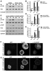
N-WASP is involved in the mechanism leading to CasSD phosphorylation. (A,B) Inhibition of N-WASP by wiskostatin disrupted CasSD phosphorylation. Cells were pre-incubated in DMSO or 10 µM wiskostatin for 20 min before being plated on fibronectin and lysed for western blot (IB). The ratio of pY165 Cas∶total Cas of control cells after 20 min of spreading was set as 1 and the ratio of other time-points were normalized accordingly. Mean±s.d., two repeats, two-tailed paired Student's t-test. A.U., arbitrary units; sus, suspension. (C–E) Knockdown of N-WASP decreased CasSD phosphorylation upon cell spreading on fibronectin. FAK phosphorylation at the Y397 site also decreased. Cells expressing scrambled or N-WASP-specific shRNA were harvested 2 days after transfection, re-suspended in serum-free medium and plated on fibronectin for the indicated time before being lysed for western blot assay (C). The ratios of pY165 Cas∶total Cas and pY397 FAK∶total FAK were normalized as in B. Mean±s.d., four repeats, two-tailed paired Student's t-test. (F,G) Cells co-transfected with GFP and N-WASP shRNA showed delayed spreading at the early time-point. Cells were plated on fibronectin for 10 min before being fixed and stained for N-WASP and pY165 Cas. Scale bars: 10 µm.

Similar articles

Cited by

References

    1. Albiges-Rizo C., Destaing O., Fourcade B., Planus E., Block M. R. (2009). Actin machinery and mechanosensitivity in invadopodia, podosomes and focal adhesions. J. Cell Sci. 122, 3037–3049 10.1242/jcs.052704 - DOI - PMC - PubMed
    1. Alexander N. R., Branch K. M., Parekh A., Clark E. S., Iwueke I. C., Guelcher S. A., Weaver A. M. (2008). Extracellular matrix rigidity promotes invadopodia activity. Curr. Biol. 18, 1295–1299 10.1016/j.cub.2008.07.090 - DOI - PMC - PubMed
    1. Arias-Salgado E. G., Lizano S., Sarkar S., Brugge J. S., Ginsberg M. H., Shattil S. J. (2003). Src kinase activation by direct interaction with the integrin beta cytoplasmic domain. Proc. Natl. Acad. Sci. USA 100, 13298–13302 10.1073/pnas.2336149100 - DOI - PMC - PubMed
    1. Auvinen M., Paasinen-Sohns A., Hirai H., Andersson L. C., Hölttä E. (1995). Ornithine decarboxylase- and ras-induced cell transformations: reversal by protein tyrosine kinase inhibitors and role of pp130CAS. Mol. Cell. Biol. 15, 6513–6525 - PMC - PubMed
    1. Boettner B., Van Aelst L. (2009). Control of cell adhesion dynamics by Rap1 signaling. Curr. Opin. Cell Biol. 21, 684–693 10.1016/j.ceb.2009.06.004 - DOI - PMC - PubMed

Publication types

Substances

LinkOut - more resources