. 2014 Apr 15;127(Pt 8):1684-98.
doi: 10.1242/jcs.133892. Epub 2014 Jan 30.

Regulation of Src trafficking and activation by the endocytic regulatory proteins MICAL-L1 and EHD1

Affiliations

Regulation of Src trafficking and activation by the endocytic regulatory proteins MICAL-L1 and EHD1

James B Reinecke et al. J Cell Sci. .

Abstract

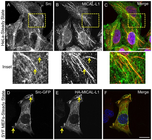
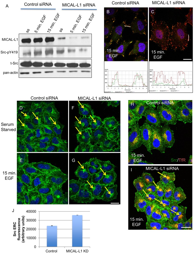
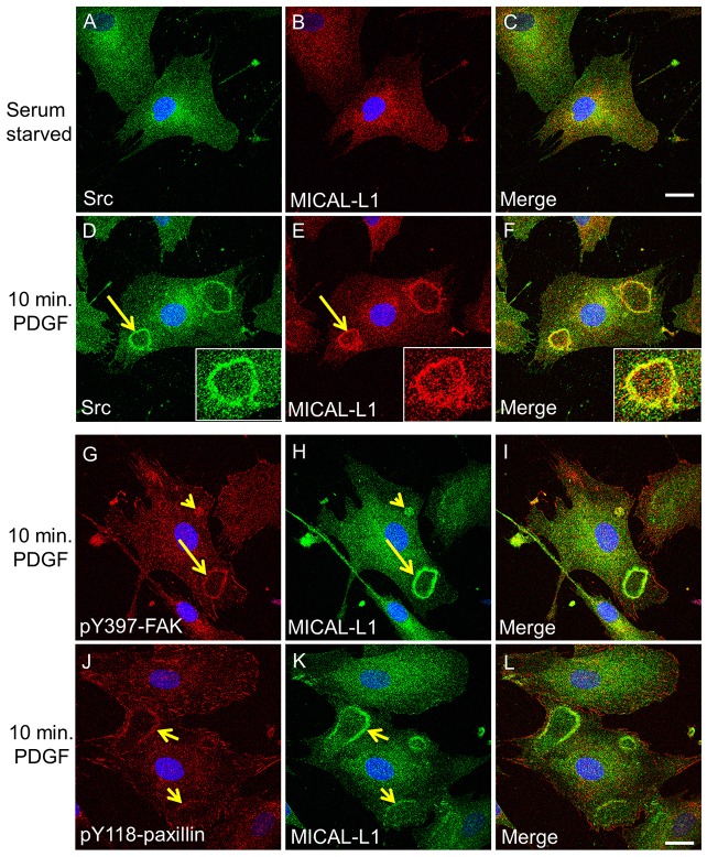
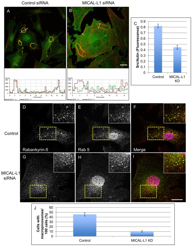
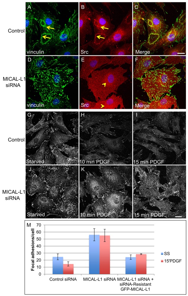
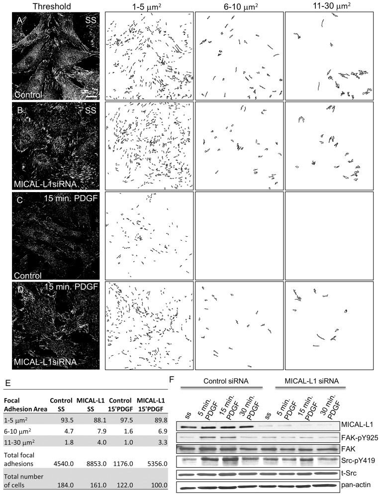
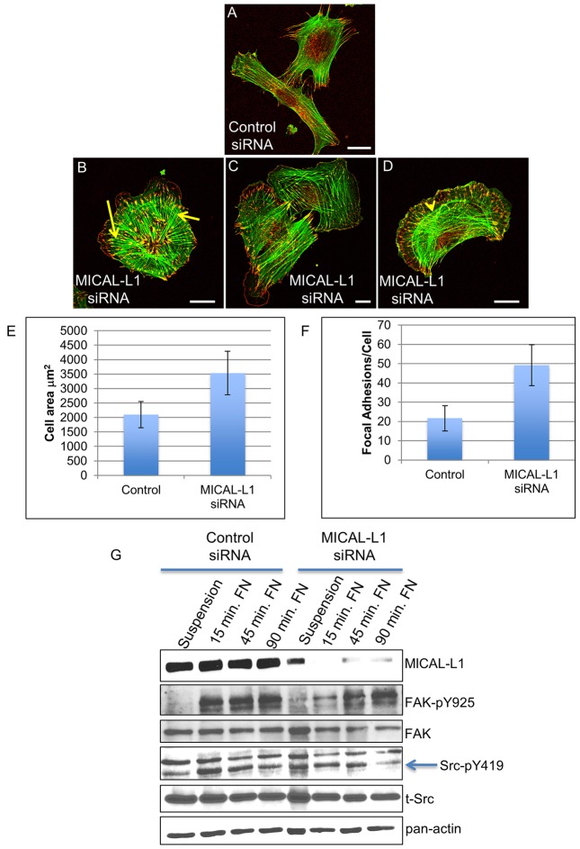
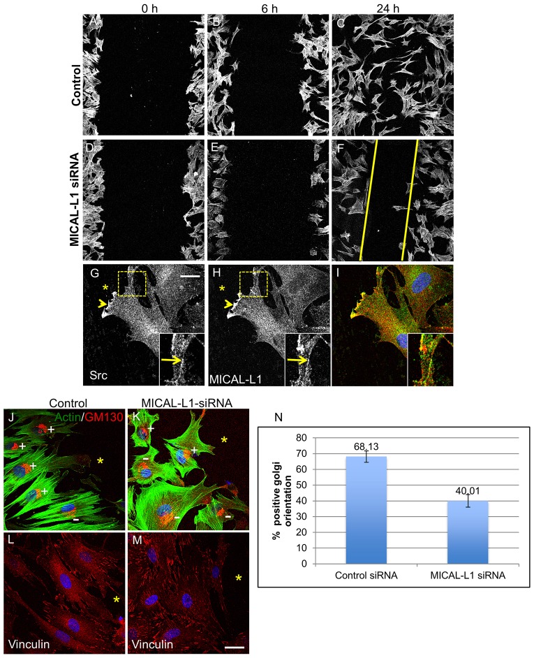
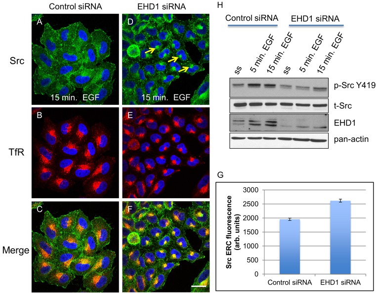
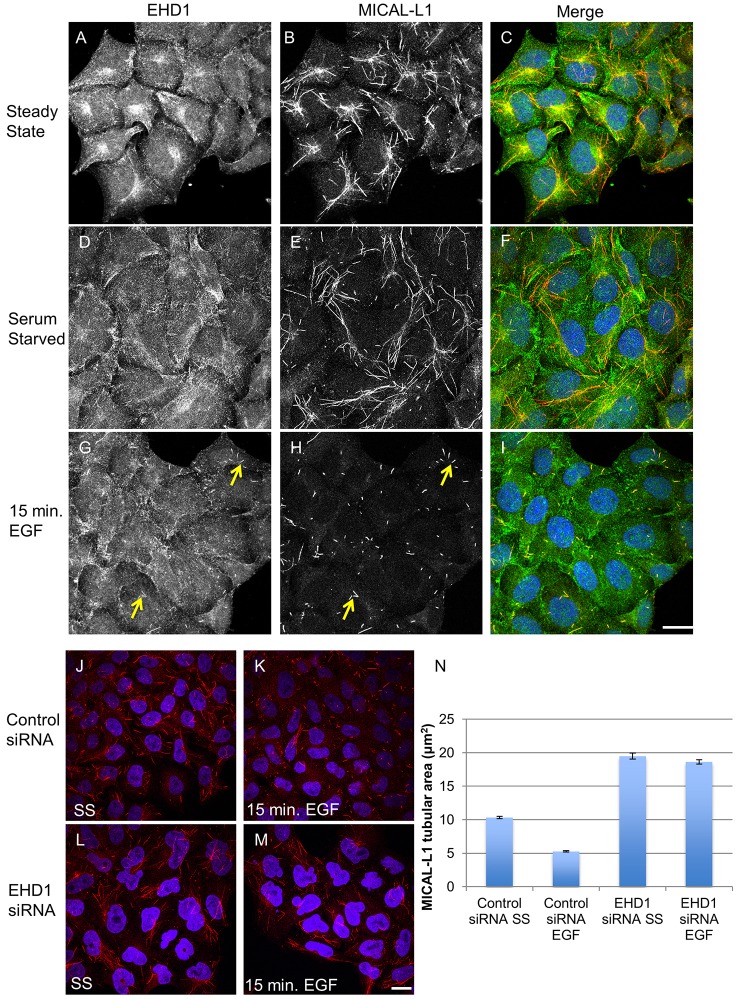
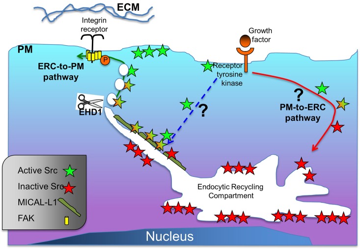
Localization of the non-receptor tyrosine kinase Src to the cell periphery is required for its activation and to mediate focal adhesion turnover, cell spreading and migration. Inactive Src localizes to a perinuclear compartment and the movement of Src to the plasma membrane is mediated by endocytic transport. However, the precise pathways and regulatory proteins that are responsible for SRC transport are incompletely understood. Here, we demonstrate that Src partially colocalizes with the endocytic regulatory protein MICAL-L1 (molecule interacting with CasL-like protein 1) in mammalian cells. Furthermore, MICAL-L1 is required for growth-factor- and integrin-induced Src activation and transport to the cell periphery in HeLa cells and human fibroblasts. Accordingly, MICAL-L1 depletion impairs focal adhesion turnover, cell spreading and cell migration. Interestingly, we find that the MICAL-L1 interaction partner EHD1 (EH domain-containing protein 1) is also required for Src activation and transport. Moreover, the MICAL-L1-mediated recruitment of EHD1 to Src-containing recycling endosomes is required for the release of Src from the perinuclear endocytic recycling compartment in response to growth factor stimulation. Our study sheds new light on the mechanism by which Src is transported to the plasma membrane and activated, and provides a new function for MICAL-L1 and EHD1 in the regulation of intracellular non-receptor tyrosine kinases.

Keywords: Circular dorsal ruffle; EHD1; Endocytic recycling; Focal adhesion; MICAL-L1; Migration; Src.


Figures

Fig. 1.
Fig. 1.
Partial colocalization between MICAL-L1 and Src in mammalian cells. (A–C) Immunofluorescence demonstrating partial colocalization of endogenous Src (A) along endogenous MICAL-L1-decorated tubular endosomes (B) in HeLa cells as marked by arrows in inset. (D–F) Overexpressed Src–GFP (D) partially colocalizes with HA–MICAL-L1 (E) in SYF fibroblasts (arrows). Blue, DNA stained with DAPI. Scale bars: 10 µm.
Fig. 2.
Fig. 2.
MICAL-L1 depletion in HeLa cells impairs EGF-induced Src activation and translocation out of the ERC. (A) An immunoblot of control (lanes 1–3) and MICAL-L1-depleted HeLa cells (lanes 4–6) demonstrating reduced EGF-induced Src activation as measured by Src autophosphorylation (Src-pY419) upon MICAL-L1 depletion. SS, serum starved. (B,C) Control (B) and MICAL-L1-KD (C) cells were stimulated with EGF for 15 min, fixed and labeled with antibodies against Src-pY419 (green) and paxillin (red). Profile analysis of two individual focal adhesions demonstrates the recruitment of active Src to paxillin-containing focal adhesions in control (B) but not MICAL-L1-KD cells (C). (D–G) Under serum-starved (SS) conditions, total Src (green) localizes to the perinuclear region (arrows) in control (D) and MICAL-L1-siRNA-treated cells (F). In response to EGF, Src translocates from the ERC to the plasma membrane in control cells (E) but is largely retained in the ERC in MICAL-L1-KD cells (G, arrows). (H–J) Cells were stimulated with EGF, and stained for Src (green) and transferrin receptor (TfR, red). Note the increased overlap of Src and TfR at the ERC in MICAL-L1-KD cells (I, yellow arrows) compared to control (H). (J) ImageJ was used to quantify Src fluorescence in the TfR-positive ERC after EGF stimulation, and one-way ANOVA showed that there was significantly more Src in MICAL-L1 KD compared to control (P<0.01). Error bars show s.e.m. Blue, DNA stained with DAPI. Scale bars: 10 µm.
Fig. 3.
Fig. 3.
MICAL-L1 colocalizes with Src and focal adhesion proteins along CDRs in human foreskin fibroblasts (BJ). (A–F) BJ cells were serum starved (A–C) or stimulated with 15 ng/ml PDGF (D–F) for 10 min. Immunofluorescence of Src (green) and MICAL-L1 (red) demonstrates their colocalization along CDRs (arrows, see inset). (G–L) BJ cells were stimulated with PDGF and stained for MICAL-L1 (green) and FAK-pY397 (red, G) or paxillin-pY118 (red, J). Arrows depict colocalization along CDRs. Arrowheads mark colocalization of MICAL-L1 and FAK-pY397 on macropinocytic-like vesicles. Blue, DNA stained with DAPI. Scale bars: 10 µm.
Fig. 4.
Fig. 4.
MICAL-L1 regulates Src recruitment to CDRs. (A–C) Control (A) and MICAL-L1-KD cells (B) were stimulated with PDGF for 10 min, fixed and stained with Src (green) and phalloidin to show F-actin (red). Src recruitment to CDRs was then quantified using profile analysis (C). (D–J) Control (D–F) and MICAL-L1-KD cells (G–I) were stimulated with PDGF for 20 min and stained with antibodies against Rabankyrin-5 (green) and Rab5 (red) to mark macropinosomes (yellow arrows). Cells containing macropinosomes were manually counted in 100 cells per experiment (J). Error bars show s.e.m. Blue, DNA stained with DAPI. Scale bars: 10 µm.
Fig. 5.
Fig. 5.
MICAL-L1 regulates PDGF-induced focal adhesion turnover. (A–F) Representative images of control and MICAL-L1-depleted fibroblasts stimulated with PDGF for 10 min and stained for vinculin (green) and Src (red). Arrows denote the colocalization of Src and vinculin at CDRs in control cells, whereas arrowheads mark the ERC-localized Src in MICAL-L1-depleted cells. (G–L) Control and MICAL-L1-depleted fibroblasts were serum-starved (SS) (G,J) or stimulated with PDGF for 10 min (H,K) or 15 min (I,L). Focal adhesions are labeled with vinculin. (M) The number of focal adhesions in SS or PDGF-stimulated cells was quantified using ImageJ. Error bars show s.e.m. from three independent experiments. The total number of cells and focal adhesions are shown in Fig. 6E. For rescue, focal adhesions were quantified from 45 cells from three independent experiments (15 per experiment). The Tukey test was used to calculate the statistical significance between treatments. The number of focal adhesions was significantly different (P<0.01) between control serum-starved and MICAL-L1-KD serum-starved cells but not between control and MICAL-L1-rescue SS cells. There was also a significant difference between control cells treated with PDGF and MICAL-L1-depleted cells treated with PDGF. MICAL-L1-rescue cells treated with PDGF had significantly fewer focal adhesions than MICAL-L1-KD cells under similar conditions, but the number was also significantly different from control cells treated with PDGF (P<0.05). Blue, DNA stained with DAPI. Scale bars: 10 µm.
Fig. 6.
Fig. 6.
MICAL-L1 depletion impedes focal adhesion turnover and leads to increased focal adhesion size. (A–D) Images from Fig. 5 were used to demonstrate the size-distribution quantification of focal adhesions in control and MICAL-L1-depleted BJ cells. Images were imported into ImageJ and fluorescence levels were set to a threshold to optimally depict focal adhesions. Focal adhesion area was calculated using the ‘measure particles’ function, and size distributions were set to group focal adhesions into three size categories: small (1–5 µm2), medium (6–10 µm2) and large (11–30 µm2). (E) A summary of focal adhesion number and size distribution in serum starved (SS) and PDGF-stimulated cells. Data are presented as the percentage of focal adhesions within each area category for three independent experiments. The Tukey test was used to calculate statistical significance. For small focal adhesions, there were significantly fewer focal adhesions in PDGF-treated MICAL-L1-KD cells compared to control. By contrast, MICAL-L1-KD cells had significantly more medium and large focal adhesions compared to control cells (P<0.01). (F) A representative immunoblot of BJ cells stimulated with PDGF in the presence (lanes 1–4) or absence of MICAL-L1 (lanes 5–8). FAK, total Src (t-Src) and actin were used as loading controls. Scale bar: 10 µm.
Fig. 7.
Fig. 7.
MICAL-L1 is required for normal cell spreading on fibronectin and optimal integrin-induced Src activation. (A–D) Control (A) and MICAL-L1-depleted cells (B–D) were serum starved (SS) in suspension for 1 h and then plated onto 10 µg/ml fibronectin-coated coverslips for 90 min and stained with phalloidin-488 (green) and anti-vinculin (red). During spreading of MICAL-L1-depleted cells, prominent short actin cables (B, arrows), polygonal cells with disorganized actin stress fibers (C) and dorsal actin arcs (D, arrowhead) were observed. Scale bars: 10 µm. (E) The cell area (mean±s.e.m.) was quantified from three independent experiments (control n = 65, MICAL-L1 siRNA n = 66) using Pascal LSM Image Examiner. One-way ANOVA demonstrated a significant increase in the area of MICAL-L1-KD cells compared to control cells (P<0.01). (F) The mean number of focal adhesions per cell was quantified from three independent experiments (control, n = 40; MICAL-L1 siRNA, n = 53). There were significantly more focal adhesions per cell in MICAL-L1 KD cells (P<0.01). (G) A representative immunoblot of BJ cells held in suspension or plated onto fibronectin-coated plates in the presence (lanes 1–4) or absence of MICAL-L1 (lanes 5–8). The arrow denotes the Src band. FAK, total Src (t-Src) and actin were used as loading controls.
Fig. 8.
Fig. 8.
MICAL-L1 is required for cell migration. (A–F) BJ cells were grown to confluence on fibronectin-coated coverslips and a scratch wound was created. Monolayers were washed and incubated in low-serum medium for the times indicated, and then were fixed and stained with phalloidin. Images were taken with a 10× objective and are representative of three independent experiments. The yellow line denotes the final degree of wound closure. (G–I) Cells were wounded, allowed to migrate for 6 h and then stained for Src (G) and MICAL-L1 (H). The arrowhead marks colocalization along the leading edge. Arrows in the inset show colocalization along tubulo-vesicular structures. (J–N) Loss of MICAL-L1 impairs wound polarization. Cell monolayers were wounded and allowed to migrate for 6 h prior to fixation and were then stained with phalloidin and anti-GM130 (J,K) or anti-vinculin (L,M). + and − denote proper polarization of the Golgi apparatus towards the wound, or lack of it, respectively. Representative images of three independent experiments are shown (J–M) and quantified (N, statistically significant, P<0.05, error bars show s.e.m.). The asterisk shows the location of the wound. Blue, DNA stained with DAPI. Scale bars: 10 µm.
Fig. 9.
Fig. 9.
EHD1 is required for EGF-induced Src translocation and activation in HeLa cells. Control cells (A–C) and EHD1-siRNA-treated cells (D–F) were stimulated with EGF and stained with Src (green) and TfR (red). Treatment with EHD1 siRNA results in the retention of Src in the ERC (overlap between Src and TfR, compare C to F). (G) The level of Src fluorescence in the ERC was quantified from three independent experiments and found to be significantly higher in EHD1-KD cells (one-way ANOVA, P<0.01). (H) An immunoblot demonstrating impaired Src activation in EHD1-depleted cells (lanes 4–6) compared to control cells (lanes 1–3). Actin and total Src (t-Src) were used as loading controls. SS, serum starved. Error bars show s.e.m. Blue, DNA stained with DAPI. Scale bar: 10 µm.
Fig. 10.
Fig. 10.
EHD1 is required for EGF-induced MICAL-L1-positive tubule vesiculation. (A–I) Compared to cells in the steady state (A–C) and serum-starved cells (SS) (D–F), EGF treatment (G–I) induces increased recruitment of EHD1 to MICAL-L1-positive tubules (arrows) resulting in the cleavage and vesiculation of MICAL-L1-decorated recycling tubules. (J–N) Treatment with EHD1 siRNA impairs the vesiculation of MICAL-L1-decorated tubular endosomes in response to EGF. In control cells, EGF stimulation leads to a reduction in the total area of MICAL-L1-containing tubular endosomes (compare K to J, quantified in N). Treatment with EHD1 siRNA results in a modest increase in the area of MICAL-L1-containing tubular endosomes in SS cells (L). In the absence of EHD1, EGF stimulation does not induce vesiculation of MICAL-L1-decorated tubular endosomes (M, quantified in N). Tubule area was quantified from three independent experiments (at least 30 cells per experiment). A Tukey test demonstrated that there were significant increases in the area of MICAL-L1-positive tubules in SS and EGF-treated EHD1-KD cells compared to control (P<0.01). No significant difference was observed between SS EHD1-KD and EGF-treated EHD1-KD cells. Error bars show s.e.m. Blue, DNA stained with DAPI. Scale bars: 10 µm.
Fig. 11.
Fig. 11.
Schematic model depicting the proposed roles of MICAL-L1 and EHD1 in mediating Src translocation from the ERC to the plasma membrane in response to growth factor or integrin stimulation. Inactivated Src is maintained at the ERC. Upon growth factor receptor stimulation (i.e. EGF), Src is sorted into MICAL-L1-decorated tubular recycling endosomes and becomes partially activated by an unknown mechanism. EHD1 is recruited to the tubular recycling endosomes by MICAL-L1, where it vesiculates recycling endosomes that contain Src and are transported to the plasma membrane where Src becomes fully activated.

Similar articles

Cited by

References

    1. Arias-Salgado E. G., Lizano S., Sarkar S., Brugge J. S., Ginsberg M. H., Shattil S. J. (2003). Src kinase activation by direct interaction with the integrin beta cytoplasmic domain. Proc. Natl. Acad. Sci. USA 100, 13298–13302 10.1073/pnas.2336149100 - DOI - PMC - PubMed
    1. Azimifar S. B., Böttcher R. T., Zanivan S., Grashoff C., Krüger M., Legate K. R., Mann M., Fässler R. (2012). Induction of membrane circular dorsal ruffles requires co-signalling of integrin-ILK-complex and EGF receptor. J. Cell Sci. 125, 435–448 10.1242/jcs.091652 - DOI - PubMed
    1. Cai B., Giridharan S. S., Zhang J., Saxena S., Bahl K., Schmidt J. A., Sorgen P. L., Guo W., Naslavsky N., Caplan S. (2013). Differential roles of C-terminal Eps15 homology domain proteins as vesiculators and tubulators of recycling endosomes. J. Biol. Chem. 288, 30172–30180 10.1074/jbc.M113.488627 - DOI - PMC - PubMed
    1. Caplan S., Naslavsky N., Hartnell L. M., Lodge R., Polishchuk R. S., Donaldson J. G., Bonifacino J. S. (2002). A tubular EHD1-containing compartment involved in the recycling of major histocompatibility complex class I molecules to the plasma membrane. EMBO J. 21, 2557–2567 10.1093/emboj/21.11.2557 - DOI - PMC - PubMed
    1. Chang J. H., Gill S., Settleman J., Parsons S. J. (1995). c-Src regulates the simultaneous rearrangement of actin cytoskeleton, p190RhoGAP, and p120RasGAP following epidermal growth factor stimulation. J. Cell Biol. 130, 355–368 10.1083/jcb.130.2.355 - DOI - PMC - PubMed

Publication types

MeSH terms