

doi: 10.1007/978-1-62703-989-5_9.
Whole-mount skeletal staining
Affiliations
- PMID: 24482169
- PMCID: PMC5384832
- DOI: 10.1007/978-1-62703-989-5_9
Item in Clipboard
Whole-mount skeletal staining
Methods Mol Biol.
2014.
Abstract
The first step in almost every investigation of skeletal phenotypes is analysis of whole-mount skeletal preparations. Whole-mount skeletal staining permits evaluation of the shapes and sizes of skeletal elements in their appropriate locations. The technique is thus the major method for detecting changes in skeletal patterning. Because cartilage and bone can be distinguished by differential staining, this technique is also a powerful means to assess the pace of skeletal maturation. This protocol covers staining of the pre- and postnatal mouse skeleton using Alcian blue and Alizarin red to identify cartilage and bone, respectively.
Figures
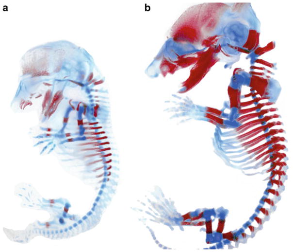
Alcian blue and Alizarin red staining of (a) E14.5 and (b) E16.5 embryos. Images were taken using bright field optics. Following adjustment of color levels using Photoshop, the embryos were pasted onto a white background
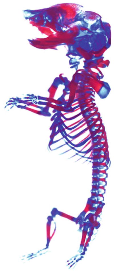
Alcian blue and Alizarin red staining of a P0 pup. Image was processed as in Fig. 1
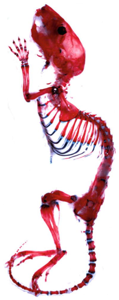
Alcian blue and Alizarin red staining of a 4-week-old postnatal mouse. The image was processed as in Fig. 1
Similar articles
-
Alizarin Red and Alcian Blue Preparations to Visualize the Skeleton.Methods Mol Biol. 2020;2043:207-212. doi: 10.1007/978-1-4939-9698-8_17. Methods Mol Biol. 2020. PMID: 31463914
-
Simultaneous differential staining of cartilage and bone in rodent fetuses: an alcian blue and alizarin red S procedure without glacial acetic acid.Biotech Histochem. 1994 Jul;69(4):181-5. doi: 10.3109/10520299409106284. Biotech Histochem. 1994. PMID: 7522588
-
A protocol for differential staining of cartilages and ossified bones in fetal and adult mouse skeletons using alcian blue and alizarin red S.J Histotechnol. 2020 Dec;43(4):204-209. doi: 10.1080/01478885.2020.1756081. Epub 2020 Sep 10. J Histotechnol. 2020. PMID: 32909916
-
Analysis of skeletal ontogenesis through differential staining of bone and cartilage.Methods Mol Biol. 2008;461:37-45. doi: 10.1007/978-1-60327-483-8_5. Methods Mol Biol. 2008. PMID: 19030790 Review. No abstract available.
-
Treatment of mice with retinoids in vivo and in vitro. Skeletal staining.Methods Mol Biol. 1999;97:33-9. doi: 10.1385/1-59259-270-8:33. Methods Mol Biol. 1999. PMID: 10443358 Review. No abstract available.
Cited by
-
BRD4 binds to active cranial neural crest enhancers to regulate RUNX2 activity during osteoblast differentiation.Development. 2024 Jan 15;151(2):dev202110. doi: 10.1242/dev.202110. Epub 2024 Jan 24. Development. 2024. PMID: 38063851 Free PMC article.
-
Proteus mirabilis fimbriae- and urease-dependent clusters assemble in an extracellular niche to initiate bladder stone formation.Proc Natl Acad Sci U S A. 2016 Apr 19;113(16):4494-9. doi: 10.1073/pnas.1601720113. Epub 2016 Apr 4. Proc Natl Acad Sci U S A. 2016. PMID: 27044107 Free PMC article.
-
Integration of 3D genome topology and local chromatin features uncovers enhancers underlying craniofacial-specific cartilage defects.Sci Adv. 2022 Nov 25;8(47):eabo3648. doi: 10.1126/sciadv.abo3648. Epub 2022 Nov 23. Sci Adv. 2022. PMID: 36417512 Free PMC article.
-
Elevated secretion of pro-collagen I-alpha and vascular endothelial growth factor as biomarkers of acetabular labrum degeneration and calcification in hip osteoarthritis: An explant study.J Orthop Translat. 2023 Dec 26;44:19-25. doi: 10.1016/j.jot.2023.08.007. eCollection 2024 Jan. J Orthop Translat. 2023. PMID: 38179125 Free PMC article.
-
The developing bird pelvis passes through ancestral dinosaurian conditions.Nature. 2022 Aug;608(7922):346-352. doi: 10.1038/s41586-022-04982-w. Epub 2022 Jul 27. Nature. 2022. PMID: 35896745
References
-
- Horobin RW. How do dyes impart color to different components of the tissues? In: Kumar GL, editor. Educational guide special stains and H & E. 2. Carpinteria; California: 2010. pp. 159–166.
-
- Schultze O. Ueber herstellung und conservirung durchsichtiger embryonen zum stadium der skeletbildung. Anat Anz. 1897;13:3–5.
-
- Jegalian BC, De Robertis EM. Homeotic transformations in the mouse induced by over expression of a human Hox3.3 transgene. Cell. 1992;71:901–910. - PubMed
MeSH terms
Grants and funding
LinkOut - more resources
Full Text Sources
Other Literature Sources
