. 2014 Mar 21;289(12):8106-20.
doi: 10.1074/jbc.M113.511535. Epub 2014 Jan 30.

Targeted metabolomics connects thioredoxin-interacting protein (TXNIP) to mitochondrial fuel selection and regulation of specific oxidoreductase enzymes in skeletal muscle

Affiliations

Targeted metabolomics connects thioredoxin-interacting protein (TXNIP) to mitochondrial fuel selection and regulation of specific oxidoreductase enzymes in skeletal muscle

Karen L DeBalsi et al. J Biol Chem. .

Abstract

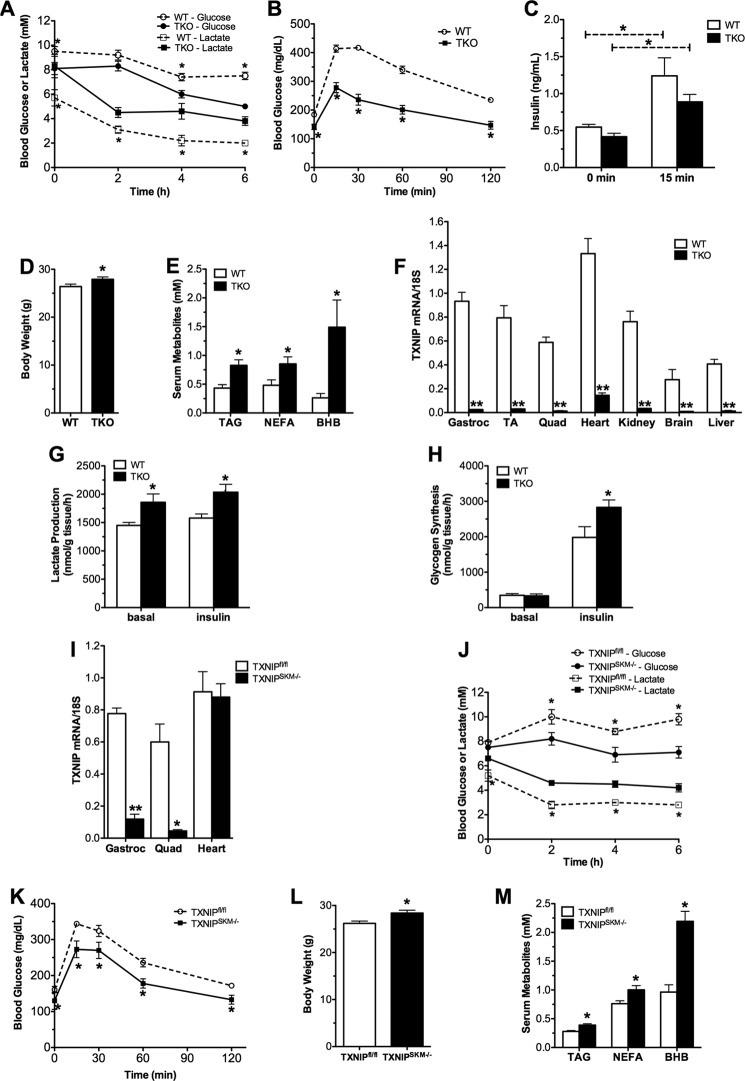
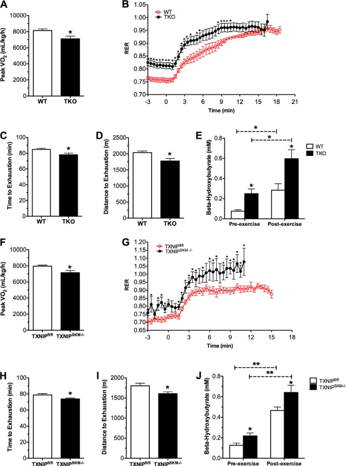
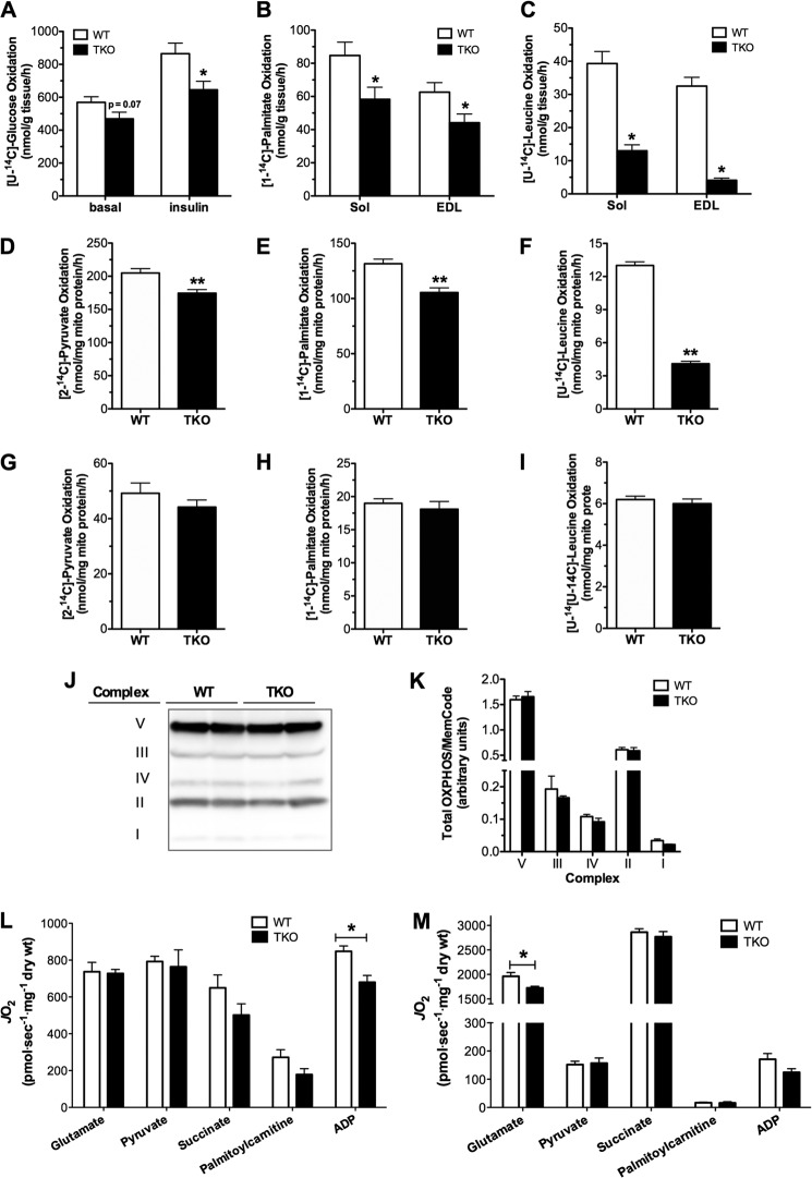
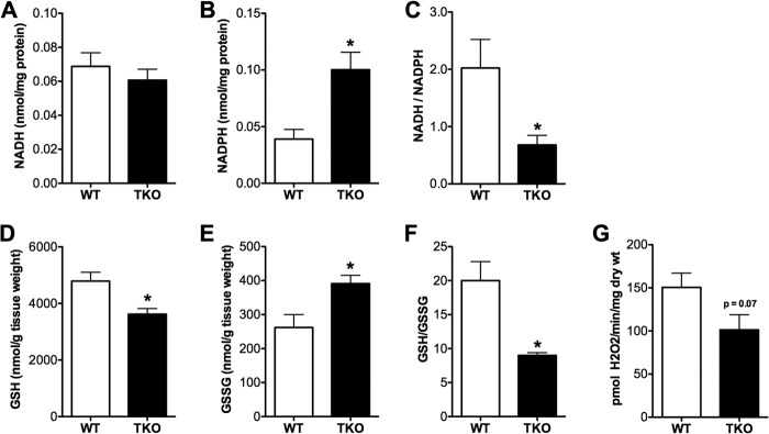
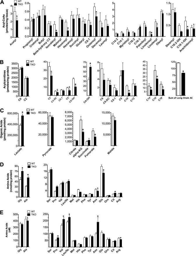
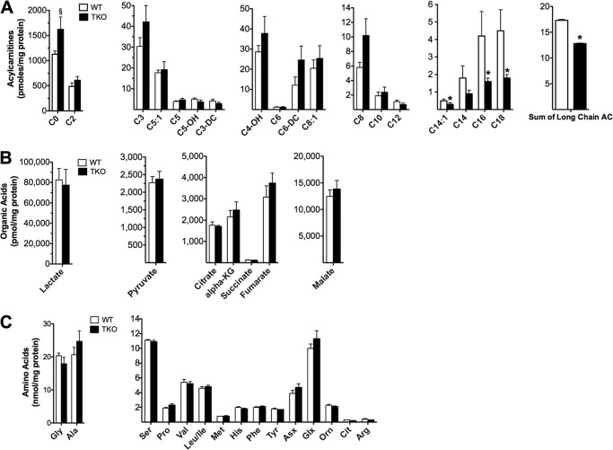
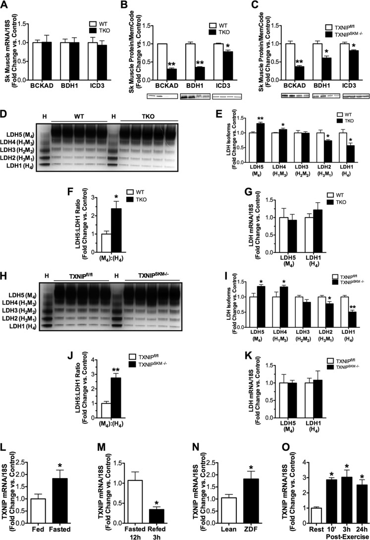
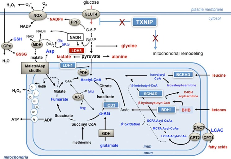
Thioredoxin-interacting protein (TXNIP) is an α-arrestin family member involved in redox sensing and metabolic control. Growing evidence links TXNIP to mitochondrial function, but the molecular nature of this relationship has remained poorly defined. Herein, we employed targeted metabolomics and comprehensive bioenergetic analyses to evaluate oxidative metabolism and respiratory kinetics in mouse models of total body (TKO) and skeletal muscle-specific (TXNIP(SKM-/-)) Txnip deficiency. Compared with littermate controls, both TKO and TXNIP(SKM-/-) mice had reduced exercise tolerance in association with muscle-specific impairments in substrate oxidation. Oxidative insufficiencies in TXNIP null muscles were not due to perturbations in mitochondrial mass, the electron transport chain, or emission of reactive oxygen species. Instead, metabolic profiling analyses led to the discovery that TXNIP deficiency causes marked deficits in enzymes required for catabolism of branched chain amino acids, ketones, and lactate, along with more modest reductions in enzymes of β-oxidation and the tricarboxylic acid cycle. The decrements in enzyme activity were accompanied by comparable deficits in protein abundance without changes in mRNA expression, implying dysregulation of protein synthesis or stability. Considering that TXNIP expression increases in response to starvation, diabetes, and exercise, these findings point to a novel role for TXNIP in coordinating mitochondrial fuel switching in response to nutrient availability.

Keywords: Branched Chain Amino Acids; Diabetes; Energy Metabolism; Ketone Body Metabolism; Mitochondrial Metabolism; Redox; Skeletal Muscle Metabolism; Thioredoxin Interacting Protein.


Figures

FIGURE 1.
FIGURE 1.
TXNIP deficiency alters whole body energy metabolism. TKO mice and WT littermates were fed a standard chow diet and metabolic parameters were measured at 18–22 weeks unless otherwise noted. A, blood glucose and lactate levels were measured 0–6 h following food withdrawal (n = 6). B, glucose tolerance tests were performed at 12 weeks of age using 1.75 g/kg of glucose administered 6 h after food withdrawal (n = 12). C, serum insulin was measured at 0 and 15 min. D, body weight at 22 weeks. E, plasma triglycerides (TAG), non-esterified fatty acids (NEFA), and β-hydroxybutyrate (BHB) were measured 5 h after food withdrawal at 9–12 weeks of age (n = 6–8). F, TXNIP mRNA tissue distribution was measured by real-time quantitative PCR and normalized to 18S as an endogenous control gene (n = 5). Lactate production (G) and rates of [14C]glucose (H) incorporation into glycogen were measured in isolated soleus muscles incubated 1 h ± 100 nm insulin. I, TXNIP mRNA expression in skeletal muscle and heart from TXNIPSKM−/− and TXNIPfl/fl mice (n = 3). Metabolic parameters of TXNIPSKM−/− mice and TXNIPfl/fl littermate controls were measured at 12–14 weeks of age. J, blood glucose and lactate levels (n = 5). K, glucose tolerance tests (n = 6). L, body weight at sacrifice. M, plasma metabolites (n = 9). Data are mean ± S.E. and results were analyzed by Student's t test (*, p ≤ 0.05 and **, p ≤ 0.001).
FIGURE 2.
FIGURE 2.
TXNIP deficiency decreases exercise tolerance. TKO mice and WT littermates were subjected to two different treadmill exercises at 12–15 weeks of age. Indirect calorimetry was used to measure peak VO2 (A) and the RER (VCO2/VO2) (B) as a function of workload during a graded, high intensity regimen. Exercise endurance was evaluated using an open-air treadmill and a mid-intensity regimen. Performance was assessed by measuring time (min) (C) and distance (meters) (D) to exhaustion. E, plasma β-hydroxybutyrate was measured prior to and immediately after endurance exercise. Data are expressed as mean ± S.E. from 7 to 8 animals per group. The same tests were administered to TXNIPSKM−/− mice and TXNIPfl/fl littermate controls at 22–24 weeks of age. F, peak VO2. G, RER. H, time; and I, distance to exhaustion. J, plasma β-hydroxybutyrate concentrations. Data are expressed as mean ± S.E. from 5 to 7 animals per group and results were analyzed by Student's t test (*, p ≤ 0.05 and **, p ≤ 0.001). Panels B and G were evaluated using a one-factor analysis of variance and post hoc analysis to determine differences between groups.
FIGURE 3.
FIGURE 3.
Substrate oxidation and respiratory function in skeletal muscle and liver mitochondria. Substrate oxidation assays were performed in tissues and mitochondria from TKO mice and WT littermates harvested 5–6 h after food withdrawal. A, oxidation of [U-14C]glucose to CO2 was measured in isolated soleus muscles during a 2-h incubation in modified KHB buffer containing 5 mm glucose ± 100 nm insulin. Oxidation of 500 μm [1-14C]palmitate (B) or 200 μm [U-14C]leucine (C) to CO2 was measured in isolated soleus and EDL muscles during a 2-h incubation in modified KHB (n = 6). Isolated mitochondria from gastrocnemius muscles were used to measure substrate oxidation to CO2 in the presence of 1 mm [2-14C]pyruvate (D), 100 μm [1-14C]palmitate (E), or 100 μm [U-14C]leucine (F) for 30 min (n = 11–12). Isolated mitochondria from liver was used to measure substrate oxidation to CO2 in the presence of 1 mm [2-14C]pyruvate (G), 100 μm [1-14C]palmitate (H), or 100 μm [U-14C]leucine (I) for 30 min (n = 6–12). Gastrocnemius muscles from TKO mice and WT littermate controls were used for Western blot of complexes I-V of the ETC (J) and quantified results (K) were normalized to MemCode staining to control for loading. Respiratory function in situ was measured using permeabilized fiber bundles under state 3 respiration conditions in the presence of saturating (L) or increasing (M) substrate concentrations to determine substrate sensitivity (Km). Data are expressed as mean ± S.E. and results were analyzed by Student's t test (*, p ≤ 0.05 and **, p ≤ 0.001).
FIGURE 4.
FIGURE 4.
Redox imbalance in TXNIP-deficient skeletal muscles. Skeletal muscles from TKO mice and WT littermate controls were harvested 5 h after food withdrawal and immediately flash frozen in liquid N2 for subsequent assessment of redox metabolites. Gastrocnemius muscles were used to measure NADH (A), NADPH (B), and the NADH/NADPH (C) ratio. Quadriceps muscles were used to measure reduced glutathione (GSH) (D), oxidized glutathione (GSSG) (E), and the GSH/GSSG (F) ratio. G, mitochondrial potential for producing reactive oxygen species was assessed in permeabilized fiber bundles by measuring succinate-supported H2O2 emission rates under state 4 conditions. Data are expressed as mean ± S.E. from 5 to 6 animals per group and results were analyzed by Student's t test (*, p ≤ 0.05).
FIGURE 5.
FIGURE 5.
Targeted metabolic profiling of skeletal muscle and serum. Gastrocnemius muscles and serum from TKO mice and WT littermate controls were harvested 5–6 h following food withdrawal at 18 weeks of age. Tissues were immediately flash frozen in liquid N2 and subsequently processed for mass spectrometry-based measurement of acyl-CoAs (A), acylcarnitines (B), organic acids (C), amino acids (D), and serum amino acids (E). Data are expressed as mean ± S.E. from 5 to 7 animals per group. Results were analyzed by Student's t test (*, p ≤ 0.05 and §, p ≤ 0.10).
FIGURE 6.
FIGURE 6.
Targeted metabolic profiling of liver. Liver from TKO mice and WT littermate controls were harvested 5–6 h following food withdrawal at 18 weeks of age. Specimens were immediately flash frozen in liquid N2 and subsequently processed for mass spectrometry-based measurement of acylcarnitines (A), organic acids (B), and amino acids (C). Data are mean ± S.E. from 6 animals per group and results were analyzed by Student's t test (*, p ≤ 0.05 and §, p ≤ 0.10).
FIGURE 7.
FIGURE 7.
Expression of mitochondrial dehydrogenase enzymes in skeletal muscle and liver. Tissues were harvested 5–6 h following food withdrawal at 18–22 weeks of age. Skeletal muscles from TKO mice and littermate controls were used for quantitative RT-PCR analysis of mRNAs encoding (A) bckad e1α, bdh1, and icd3 normalized to 18 S, and Western blot analysis of the BCKAD E1A, BDH1, and ICD3 proteins (B) normalized to Memcode staining. Expression of the same proteins was measured in gastrocnemius muscles (C) from TXNIPSKM−/− mice and TXNIPfl/fl controls. Skeletal muscles from TKO mice and littermate controls were used for: D, an LDH isozyme activity gel with WT heart shown as a control; E, quantification of results; F, calculation of the LDH5:LDH1 ratio; and G, quantitative RT-PCR analysis of mRNAs encoding ldh5 and ldh1. The same analyses were performed using gastrocnemius muscles from TXNIPSKM−/− mice and TXNIPfl/fl mice (H–K). Quantitative RT-PCR analysis of mRNA encoding txnip normalized to 18 S was measured in skeletal muscles from fed and fasted (12 h) C57BL/6J mice (L), fasted (12 h) and refed (3 h) C57BL/6J mice (M), Zucker Diabetic Fatty (ZDF) rats, and lean controls (O) C57BL/6J mice (N) at rest and 10 min, 3 h, or 24 h after a 90-min graded treadmill exercise bout (*, p ≤ 0.05 versus rest). Data are mean ± S.E. (n = 5 per group) and results were analyzed by Student's t test (*, p ≤ 0.05 and **, p ≤ 0.001).
FIGURE 8.
FIGURE 8.
TXNIP deficiency disrupts mitochondrial fuel selection. During periods of carbohydrate deprivation, up-regulation of TXNIP serves to limit glucose transport into skeletal muscle and promote mitochondrial oxidation of alternative fuels. TXNIP deficiency enhances glucose uptake and flux through both glycolysis and the pentose phosphate pathway. Despite this shift to glycolytic metabolism, respiratory function of the ETC remains intact, thereby permitting transfer of glucose-derived NADH to complex I via the malate/aspartate shuttle. Diminished protein expression and activities of specific mitochondrial dehydrogenase enzymes lowers muscle capacity for oxidizing lactate, fatty acid, amino acid, and ketones, thus disrupting adaptive mitochondrial fuel switching in response to starvation and exercise. Key: metabolites affected by TXNIP deficiency are indicated in red (increased) and blue (decreased). Abbreviations: AcAc, acetoacetate; α-KG, α-ketoglutarate dehydrogenase; AST, apartate aminotransferase; BHB, β-hydroxybutyrate; BDH1, β-hydroxybutyrate dehydrogenase; CACT, carnitine acylcarnitine translocase; CPT1 and CPT2, carnitine palmitoyltransferase 1 and 2; DHAP, dihydroxyacetone phosphate; G-6-P, glucose 6-phosphate; GDH, glutamate dehydrogenase; GPx, glutathione peroxidase; ICD3, NAD+-dependent isocitrate dehydrogenase; LCAC, long chain acylcarnitine; LCFA-CoA, long chain fatty acid acyl-CoA; LDH1, lactate dehydrogenase 1; LDH5, lactate dehydrogenase 5; MDH, malate dehydrogenase; NOX, NADPH oxidase; PDH, pyruvate dehydrogenase; PPP, pentose phosphate pathway.

References

    1. Chen K. S., DeLuca H. F. (1994) Isolation and characterization of a novel cDNA from HL-60 cells treated with 1,25-dihydroxyvitamin D3. Biochim. Biophys. Acta 1219, 26–32 - PubMed
    1. Nishiyama A., Matsui M., Iwata S., Hirota K., Masutani H., Nakamura H., Takagi Y., Sono H., Gon Y., Yodoi J. (1999) Identification of thioredoxin-binding protein-2/vitamin D3 up-regulated protein 1 as a negative regulator of thioredoxin function and expression. J. Biol. Chem. 274, 21645–21650 - PubMed
    1. Bodnar J. S., Chatterjee A., Castellani L. W., Ross D. A., Ohmen J., Cavalcoli J., Wu C., Dains K. M., Catanese J., Chu M., Sheth S. S., Charugundla K., Demant P., West D. B., de Jong P., Lusis A. J. (2002) Positional cloning of the combined hyperlipidemia gene Hyplip1. Nat. Genet. 30, 110–116 - PMC - PubMed
    1. Nordberg J., Arnér E. S. (2001) Reactive oxygen species, antioxidants, and the mammalian thioredoxin system. Free Radic. Biol. Med. 31, 1287–1312 - PubMed
    1. Alvarez C. E. (2008) On the origins of arrestin and rhodopsin. BMC Evol. Biol. 8, 222. - PMC - PubMed

Publication types

LinkOut - more resources