. 2014 Mar 7;289(10):6592-6603.
doi: 10.1074/jbc.M113.538991. Epub 2014 Jan 30.

Structural insights into the interaction between a potent anti-inflammatory protein, viral CC chemokine inhibitor (vCCI), and the human CC chemokine, Eotaxin-1

Affiliations

Structural insights into the interaction between a potent anti-inflammatory protein, viral CC chemokine inhibitor (vCCI), and the human CC chemokine, Eotaxin-1

Nai-Wei Kuo et al. J Biol Chem. .

Abstract

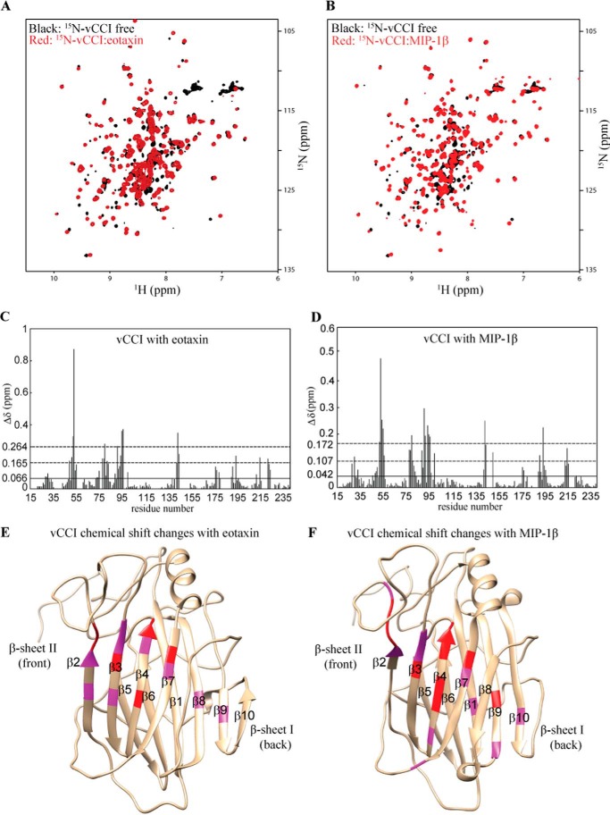
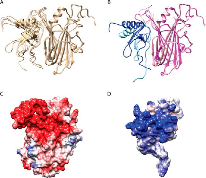
Chemokines play important roles in the immune system, not only recruiting leukocytes to the site of infection and inflammation but also guiding cell homing and cell development. The soluble poxvirus-encoded protein viral CC chemokine inhibitor (vCCI), a CC chemokine inhibitor, can bind to human CC chemokines tightly to impair the host immune defense. This protein has no known homologs in eukaryotes and may represent a potent method to stop inflammation. Previously, our structure of the vCCI·MIP-1β (macrophage inflammatory protein-1β) complex indicated that vCCI uses negatively charged residues in β-sheet II to interact with positively charged residues in the MIP-1β N terminus, 20s region and 40s loop. However, the interactions between vCCI and other CC chemokines have not yet been fully explored. Here, we used NMR and fluorescence anisotropy to study the interaction between vCCI and eotaxin-1 (CCL11), a CC chemokine that is an important factor in the asthma response. NMR results reveal that the binding pattern is very similar to the vCCI·MIP-1β complex and suggest that electrostatic interactions provide a major contribution to binding. Fluorescence anisotropy results on variants of eotaxin-1 further confirm the critical roles of the charged residues in eotaxin-1. In addition, the binding affinity between vCCI and other wild type CC chemokines, MCP-1 (monocyte chemoattractant protein-1), MIP-1β, and RANTES (regulated on activation normal T cell expressed and secreted), were determined as 1.1, 1.2, and 0.22 nm, respectively. To our knowledge, this is the first work quantitatively measuring the binding affinity between vCCI and multiple CC chemokines.

Keywords: Anti-inflammatory Protein; Biophysics; Chemokine-binding Protein; Chemokines; Eotaxin; Fluorescence Anisotropy; Molecular Docking; NMR; Protein-Protein Interactions; vCCI.


Figures

FIGURE 1.
FIGURE 1.
Sequence comparison of CC chemokines (eotaxin, MCP-1, MIP-1α, MIP-1β, RANTES, and I-309) (A) and CXC chemokines (IL-8 and SDF-1α) (B). Conserved cysteine residues are highlighted in yellow. The positively charged residues putatively critical to vCCI binding are highlighted in red. The hydrophobic residue putatively important in vCCI binding is highlighted in pink. The 47th position has been shown to modulate binding such that mutation to Ala can enhance binding to vCCI (cyan). In A, all of the chemokines are high affinity ligands for vCCI except for I-309 (for comparison, see Ref. 24). The sequence numbers in A are according to eotaxin, and those in B are according to IL-8.
FIGURE 2.
FIGURE 2.
Mapping the contact surface of eotaxin and MIP-1β on vCCI. A, overlay of the 1H-15N HSQC spectra of free vCCI (black) and eotaxin-bound vCCI (red). In these spectra, only the vCCI is isotopically labeled with 15N. B, overlay of the 1H-15N HSQC spectra of free vCCI (black) and MIP-1β-bound vCCI (red). C, chemical shift changes of vCCI versus its residue number upon binding to eotaxin. 15N-Labeled vCCI was combined with unlabeled eotaxin to a final molar ratio of 1:1. The average chemical shift change (Δδ) is indicated with a solid horizontal line. The dashed lines indicate the 1 and 2 S.D. values from the average. Residues with chemical shift changes above 0.165 ppm (1 S.D.) are considered significantly involved in contact with eotaxin. D, as in C, except that 15N-labeled vCCI was combined with unlabeled MIP-1β. Residues with the chemical shift changes above 0.107 ppm (1 S.D.) are considered significantly involved in contact with MIP-1β. E, mapping the significant eotaxin contact residues on the vCCI structure. Pink, chemical shift changes above 1 S.D. greater than average; red, chemical shift changes above 2 S.D. values greater than average. The vCCI structure is from cluster 1 of the docked structure. F, as in E, except mapping the significant MIP-1β contact residues on the vCCI structure (Protein Data Bank code for vCCI: 2FFK). B, D, and F, all MIP-1β data shown here are from Ref. and used for comparison.
FIGURE 3.
FIGURE 3.
Mapping the contact surface of vCCI on eotaxin. A, overlay of the 1H-15N HSQC spectra of free 15N eotaxin (black) and vCCI-bound 15N eotaxin (red). B, chemical shift changes of eotaxin versus its residue number upon binding vCCI. 15N-Labeled eotaxin was combined with unlabeled vCCI to a final molar ratio of 1:1. The average chemical shift change (Δδ) is indicated with a solid horizontal line. Residues with the chemical shift changes above 0.21 ppm are considered significantly involved in contact with vCCI. C, mapping the significant vCCI contact residues on the eotaxin structure. Orange, chemical shift changes above the average; pink, chemical shift changes above 1 S.D. greater than average; red, chemical shift changes above 2 S.D. values greater than average. Yellow, disulfide bond; the other disulfide bond is C10:C50. The eotaxin structure is from cluster 1 of the docked structure.
FIGURE 4.
FIGURE 4.
Fluorescence anisotropy studies of binding of eotaxin, eotaxin variants, and other CC chemokines to vCCI. A, left, fluorescence anisotropy of eotaxin-fluor binding to vCCI. Right, fluorescence anisotropy competition assay between unlabeled eotaxin and a complex of eotaxin-fluor and vCCI. B, selected fluorescence anisotropy competition assays between unlabeled eotaxin variants and a complex of eotaxin-fluor and vCCI. C, fluorescence anisotropy competition assays between unlabeled CC chemokines and a complex of eotaxin-fluor and vCCI. Three independent experiments were performed for each assay. The graphs show a representative experiment. Error bar, S.D. of multiple measurements of that point. The Kd was calculated as described under “Experimental Procedures.”
FIGURE 5.
FIGURE 5.
Structural model of the vCCI·eotaxin complex. A, overlay of four docking structures from the vCCI·eotaxin cluster 1. Eotaxin is shown on the left, and vCCI is shown on the right. B, overlay of the vCCI·eotaxin (pink and blue) complex and the vCCI·MIP-1β (light pink and light blue) complex for comparison. The vCCI·MIP-1β complex is from Ref. . C and D, electrostatic potential maps are shown for the binding interface of vCCI (C) and eotaxin (D). Negatively charged residues are shown in red, and positively charged residues are shown in blue. The vCCI and eotaxin surface is from cluster 1 of the docked structure.

References

    1. Charo I. F., Ransohoff R. M. (2006) The many roles of chemokines and chemokine receptors in inflammation. N. Engl. J. Med. 354, 610–621 - PubMed
    1. Rot A., von Andrian U. H. (2004) Chemokines in innate and adaptive host defense. Basic chemokinese grammar for immune cells. Annu. Rev. Immunol. 22, 891–928 - PubMed
    1. Fernandez E. J., Lolis E. (2002) Structure, function, and inhibition of chemokines. Annu. Rev. Pharmacol. Toxicol. 42, 469–499 - PubMed
    1. Lau E. K., Allen S., Hsu A. R., Handel T. M. (2004) Chemokine-receptor interactions. GPCRs, glycosaminoglycans and viral chemokine binding proteins. Adv. Protein Chem. 68, 351–391 - PubMed
    1. Luster A. D. (1998) Chemokines. Chemotactic cytokines that mediate inflammation. N. Engl. J. Med. 338, 436–445 - PubMed

Publication types

MeSH terms

LinkOut - more resources