. 2014 Mar 14;289(11):7589-98.
doi: 10.1074/jbc.M113.517243. Epub 2014 Jan 30.

Sry HMG box protein 9-positive (Sox9+) epithelial cell adhesion molecule-negative (EpCAM-) biphenotypic cells derived from hepatocytes are involved in mouse liver regeneration

Affiliations

Sry HMG box protein 9-positive (Sox9+) epithelial cell adhesion molecule-negative (EpCAM-) biphenotypic cells derived from hepatocytes are involved in mouse liver regeneration

Naoki Tanimizu et al. J Biol Chem. .

Abstract

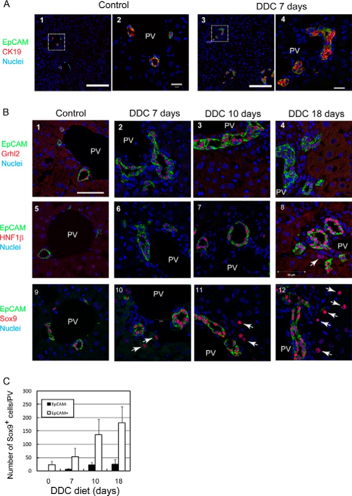
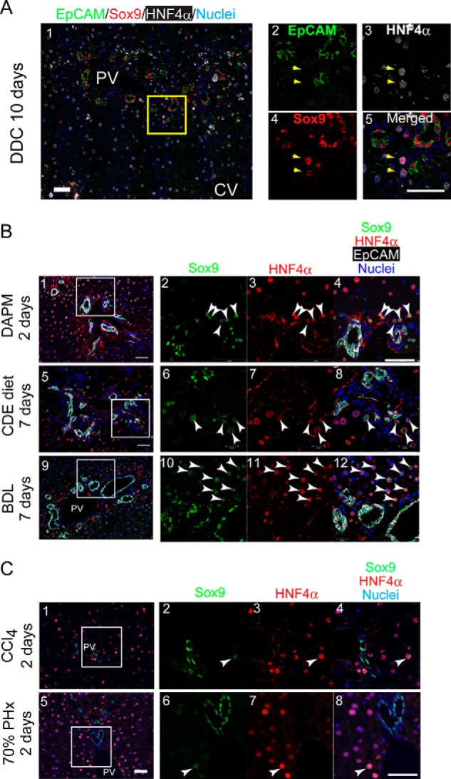
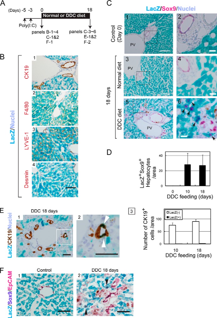
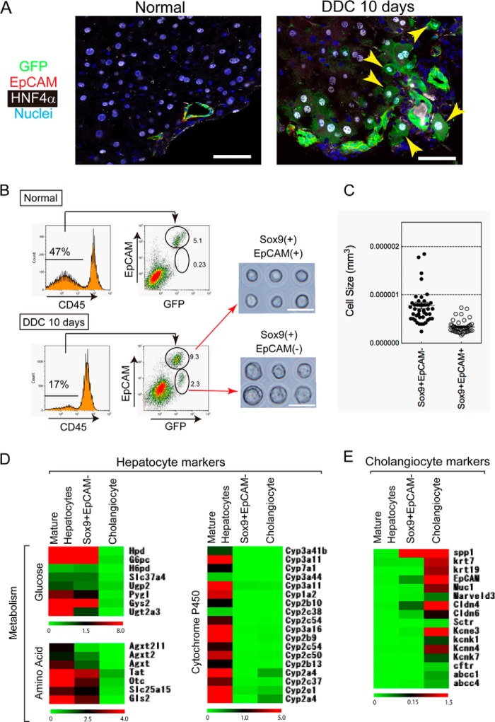
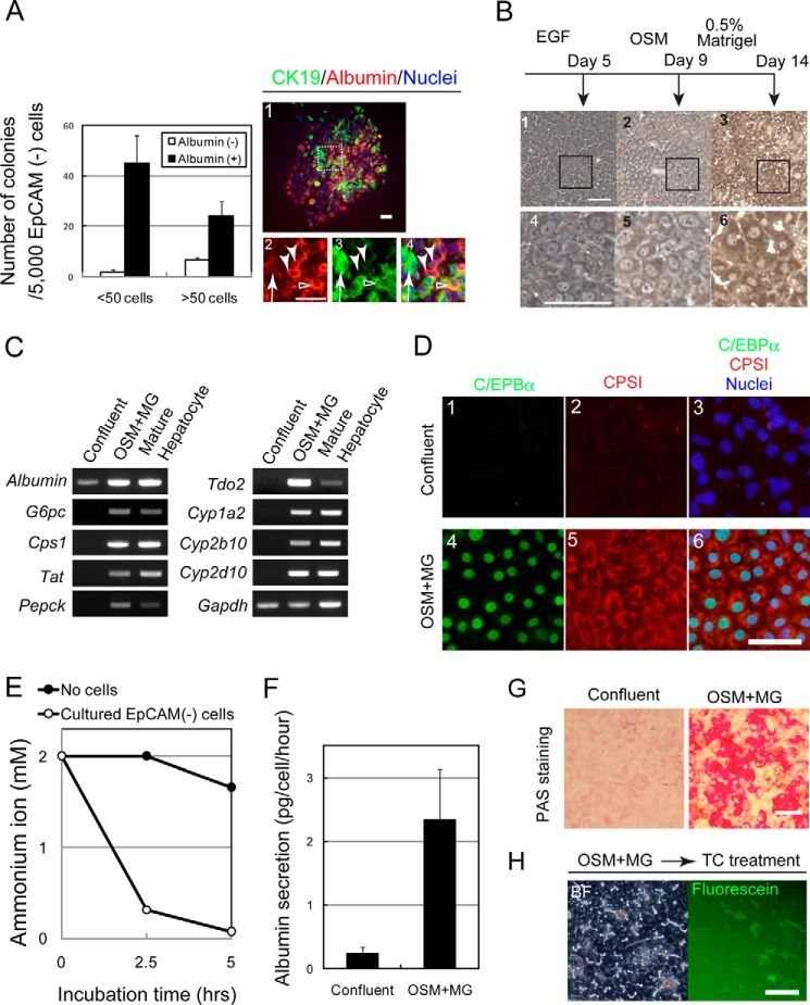
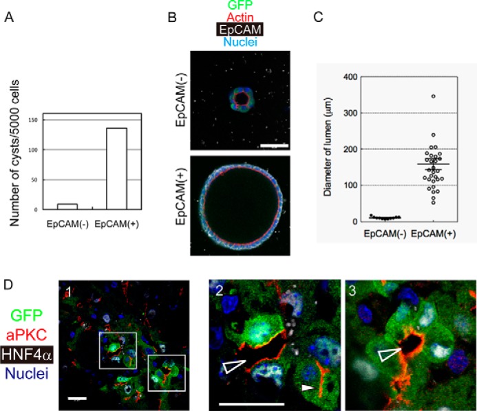
It has been shown that mature hepatocytes compensate tissue damages not only by proliferation and/or hypertrophy but also by conversion into cholangiocyte-like cells. We found that Sry HMG box protein 9-positive (Sox9(+)) epithelial cell adhesion molecule-negative (EpCAM(-)) hepatocyte nuclear factor 4α-positive (HNF4α(+)) biphenotypic cells showing hepatocytic morphology appeared near EpCAM(+) ductular structures in the livers of mice fed 3,5-diethoxycarbonyl-1,4-dihydrocollidine (DDC)-containing diet. When Mx1-Cre:ROSA mice, which were injected with poly(I:C) to label mature hepatocytes, were fed with the DDC diet, we found LacZ(+)Sox9(+) cells near ductular structures. Although Sox9(+)EpCAM(-) cells adjacent to expanding ducts likely further converted into ductular cells, the incidence was rare. To know the cellular characteristics of Sox9(+)EpCAM(-) cells, we isolated them as GFP(+)EpCAM(-) cells from DDC-injured livers of Sox9-EGFP mice. Sox9(+)EpCAM(-) cells proliferated and could differentiate to functional hepatocytes in vitro. In addition, Sox9(+)EpCAM(-) cells formed cysts with a small central lumen in collagen gels containing Matrigel® without expressing EpCAM. These results suggest that Sox9(+)EpCAM(-) cells maintaining biphenotypic status can establish cholangiocyte-type polarity. Interestingly, we found that some of the Sox9(+) cells surrounded luminal spaces in DDC-injured liver while they expressed HNF4α. Taken together, we consider that in addition to converting to cholangiocyte-like cells, Sox9(+)EpCAM(-) cells provide luminal space near expanded ductular structures to prevent deterioration of the injuries and potentially supply new hepatocytes to repair damaged tissues.

Keywords: Biphenotypic Cells; Cell Differentiation; Cell Polarity; Ductular Reaction; Hepatocyte; Liver; Liver Stem/Progenitor Cells; Regeneration.


Figures

FIGURE 1.
FIGURE 1.
Expression of cholangiocyte specific transcription factors in DDC-injured liver. A, ductular reactions are induced by DDC diet. Ductular structures consisting of CK19+EpCAM+ cells are expanded in the liver of DDC diet-fed mice. The boxes in panels 1 and 3 are enlarged in panels 2 and 4, respectively. Bars in panels 1 and 3 and panels 2 and 4 are 100 μm and 20 μm, respectively. PV, portal vein. B, expression of Grhl2, HNF1β, and Sox9 in DDC-injured livers. In the control, Grhl2, HNF1β, and Sox9 are exclusively expressed in EpCAM+ bile ducts around the portal veins (PV) (panels 1, 5, and 9). In DDC-injured livers, Grhl2 is expressed only in EpCAM+ ductular structures (panels 2–4). HNF1β is mostly expressed in EpCAM+ cells (panels 6–8), although it is occasionally detected in EpCAM cells (arrow in panel 8). On the other hand, Sox9 is apparently expressed in EpCAM cells in addition to EpCAM+ ductular cells (arrows in panels 10–12). Bar represents 50 μm. C, the number of Sox9+EpCAM and Sox9+EpCAM+ cells at different time points during DDC feeding. Numbers of Sox9+EpCAM cells are much less than EpCAM+ cells but increase in livers of DDC diet fed-mice. At each time point, livers of 3–4 mice were analyzed. Bars represent means ± S.E.
FIGURE 2.
FIGURE 2.
Sox9+ biphenotypic cells appear in injured livers associated with ductular reactions. A, expression of HNF4α in non-ductular Sox9+ cells. Non-ductular Sox9+ cells (arrows) are also positive for HNF4α, a hepatocyte marker. The box in panel 1 is enlarged in panels 2–5. Bars represent 50 μm. B, expression of Sox9 and HNF4α in injured livers including ductular reactions. Sox9+HNF4α+ cells (arrowheads) emerge in DAPM-, CDE-, and BDL-injured livers. The boxes in panels 1, 5, and 9 are enlarged in panels 2–4, 6–8, and 10–12, respectively. Bars represent 50 μm. C, expression of Sox9 and HNF4α in regenerating livers after 70% partial hepatectomy and carbon tetrachloride (CCl4) injection. Sox9+HNF4α+ cells were occasionally observed in carbon tetrachloride-injured livers and in regenerating liver after 70% partial hepatectomy (PHx) (white arrowheads). The boxes in panels 1 and 5 are enlarged in panels 2–4 and 6–8, respectively. Bars represent 50 μm.
FIGURE 3.
FIGURE 3.
Mature hepatocytes are the origin of Sox9+ biphenotypic cells. A, the time course of the labeling of MHs in Mx1-Cre:ROSA mice and the induction of liver injury. B, specificity of hepatocytes labeling in Mx1-Cre:ROSA mice. Hepatocytes were labeled with LacZ after poly(I:C) injection. On the other hand, CK19+ cholangiocytes, LYVE-1+ sinusoidal endothelial cells, F4/80+ Kupffer cells, and Desmin+ stellate cells were not labeled with LacZ in Mx1-Cre:ROSA mice injected with poly(I:C). Bar represents 50 μm. C, LacZ+ hepatocytes convert to Sox9+ cells. Before DDC injury, LacZ staining is limited to MHs (panels 1 and 2). After feeding mice with the DDC diet for 10 and 18 days, LacZ+Sox9+ cells appear near the portal vein (PV) (arrows in panels 4 and 6). Bars in panels 1, 3, and 5 represent 100 μm, while those in panels 2, 4, and 6 represent 20 μm. D, the number of LacZ+Sox9+ cells with hepatocyte morphology. The number of LacZ+Sox9+ cells showing hepatocyte morphology was counted on liver sections of normal and DDC diet-fed mice. Error bars represent S.E. E, CK19+LacZ+ cells emerge in DDC-injured livers of Mx1-Cre:ROSA mice. After 18 days of DDC injury, some of LacZ+ cells are incorporated to ductular structures as CK19+ cells (white arrowheads). The number of CK19+LacZ+ cell is slightly increased between 10 and 18 days of DDC injury. At day 18, it represents about 2% of CK19+ cells. Bars in panels 1 and 2 are 50 μm. F, LacZ+Sox9+ cells emerging in DDC-injured liver of Mx1-Cre mice are negative for EpCAM. In the control liver, LacZ+ cells express neither Sox9 nor EpCAM (panel 1). After DDC injury, LacZ+Sox9+ biphenotypic cells, which are negative for EpCAM, are observed (arrowheads in panel 2). EpCAM and Sox9 were visualized with Permanent Red and 5-bromo-4-chloro-3-indolyl phosphate, respectively. Bars represent 50 μm.
FIGURE 4.
FIGURE 4.
Isolation of Sox9+EpCAM cells from DDC-injured liver of Sox9-EGFP mice. A, expression of GFP, HNF4α, and EpCAM in the liver of a DDC diet-fed Sox9-EGFP mouse. Expression of GFP and HNF4α is mutually exclusive in the normal liver (panel 1). In DDC-injured liver, some of the GFP+ cells are positive for HNF4α (arrowheads in panel 2). The GFP+HNF4α+ cells are negative for EpCAM. Bars represent 50 μm. B, flow cytometric analysis of CD45 non-parenchymal cells isolated from normal and DDC-injured livers. In the control, GFP+ (Sox9+) cells are mostly EpCAM+, indicating that they are ductular cholangiocytes. After mice were fed with the DDC diet for 10 days, GFP+EpCAM cells, in addition to GFP+EpCAM+ cells, become evident. Purified GFP+EpCAM and GFP+EpCAM+ cells are shown in the right panels. EpCAM cells are larger and show high granularity in their cytoplasm when compared with EpCAM+ cells. C, Sox9+EpCAM cells are larger than Sox9+EpCAM+ cells. Cells were isolated from livers after feeding mice with the DDC diet for 10 days. Pictures of purified cells were used for acquiring cell size. Cell size was calculated from the diameter of each cell. D, expression of hepatocyte markers in Sox9+EpCAM cells. The expression profile of hepatocyte markers is shown. The data suggested that genes related to glucose and amino acid metabolism are weakly expressed in Sox9+EpCAM biphenotypic cells, whereas Cyps are not expressed. Hierarchical clustering analysis was performed on the MultiExperiment Viewer. E, cholangiocyte markers are barely expressed in Sox9+EpCAM cells. The expression profile of cholangiocyte markers is shown. Cholangiocyte markers including CK7, CK19, and cystic fibrosis transmembrane conductance regulator (cftr) are not expressed in Sox9+EpCAM biphenotypic cells. On the other hand, the data suggested that osteopontin (OPN) is significantly expressed in biphenotypic cells.
FIGURE 5.
FIGURE 5.
Sox9+EpCAM cells have the potential to differentiate into mature hepatocytes. A, colony-forming ability of Sox9+EpCAM cells. In the low density culture, Sox9+EpCAM cells form small and large colonies containing albumin+ and CK19+ cells (closed bars) and colonies containing only CK19+ cells (open bars). A typical colony derived from a Sox9+EpCAM cell is shown in the right panels. It consists of albumin+CK19 (arrowheads), albumin+CK19+ (open arrowhead), and albuminCK19+ (arrow) cells. The box in panel 1 is enlarged in panels 2–4. Cells were isolated from 2–3 mice, and experiments were repeated four times. The average values of the number of colonies with S.E. are shown in the graph. Bars represent 50 μm. B, Sox9+EpCAM cells show hepatocyte-like morphology in the culture. In the presence of oncostatin M (OSM), Sox9+EpCAM cells show hepatocyte-like morphology (panels 2 and 3), which becomes clearer after the overlay with Matrigel (panels 3 and 6). The boxes in panels 1–3 are enlarged in panels 4–6. Bars represent 50 μm. C, Sox9+EpCAM cells are induced to express metabolic enzymes and cytochrome P450s. EpCAM cells differentiate to express markers of MHs in the presence of oncostatin M and Matrigel. Expressions of albumin, CPSI, and tyrosine aminotransferase (Tat) are enhanced during the culture, whereas those of glucose 6-phosphatase (G6pc), phosphoenolpyruvate carboxykinase (Pepck), Tdo2, and Cyps are induced. D, Sox9+EpCAM cells are induced to express CCAAT-enhancer-binding protein α (C/EBPα) and CPSI proteins. Bar represents 50 μm. E, hepatocytes derived from Sox9+EpCAM cells eliminate ammonium ions from the medium. Two mm ammonium chloride was added to EpCAM cells treated with Matrigel. The concentration of ammonium ion was examined by using an ammonia test Wako kit. F, hepatocytes derived from Sox9+EpCAM cells secrete albumin into the medium. When cells became confluent, at which about 2 × 10⁵ cells were in each well of a 24-well plate, or after they were incubated with Matrigel (MG) for 4 days, medium was changed to fresh medium and then kept for 24 h. The concentration of albumin was measured by a sandwich ELIZA. G, hepatocytes derived from Sox9+EpCAM cells store glycogen in cytosol. Sox9+EpCAM cells before and after hepatocyte differentiation were fixed, and their glycogen storage was examined by periodic acid-Schiff (PAS) staining. Bar represents 50 μm. H, hepatocytes derived from Sox9+EpCAM cells form BC-like structures. After Matrigel overlay, Sox9+EpCAM cells were further treated with 100 μm taurocholate (TC). When fluorescein diacetate is added to the medium, fluorescein is accumulated into BC-like structures. Bar represents 50 μm.
FIGURE 6.
FIGURE 6.
Sox9+EpCAM cells show the ability to establish cholangiocyte-type epithelial polarity both in vitro and in vivo. A, number of cysts derived from Sox9+EpCAM and Sox9+EpCAM+ cells. Cellular structures associated with the apical lumen were counted after 2 weeks of three-dimensional culture. Sox9+EpCAM and Sox9+EpCAM+ cells were isolated from DDC-injured Sox9-EGFP mice by FACS and seeded onto Matrigel. After being overlaid with 5% Matrigel, cells were incubated for 2 weeks in the presence of EGF, hepatocyte growth factor, and Y-27632. Cells were isolated from three mice and plated into 3 wells of an 8-well coverglass chamber. Experiments were repeated twice. The average of the number of cysts is shown in the graph. B, Sox9+EpCAM cells form small cysts in three-dimensional culture. A typical cystic structure that emerged in three-dimensional culture of EpCAM cells is shown with that derived from an EpCAM+ cell. A Sox9+EpCAM cyst consists of GFP+EpCAM cells and is associated with a tiny lumen. Bar represents 50 μm. C, the lumen size of cysts derived from EpCAM and EpCAM+ cells. The lumen size of EpCAM cysts is much smaller than that of EpCAM+ cells. Data points are shown with means ± S.E. D, Sox9+ biphenotypic cells surround luminal space in DDC-injured liver. GFP+ (Sox9+)HNF4α+ cells emerge in DDC-injured livers. BCs between hepatocytes are recognized by atypical PKC (aPKC) staining (closed arrowhead in panel 2). Additionally, luminal spaces that are obviously larger than BCs are evident (open arrowheads in panels 2 and 3). The boxes in panel 1 are enlarged in panels 2 and 3. Bars represent 20 μm.

References

    1. Duncan A. W., Dorrell C., Grompe M. (2009) Stem cells and liver regeneration. Gastroenterology 137, 466–481 - PMC - PubMed
    1. Tanaka M., Itoh T., Tanimizu N., Miyajima A. (2011) Liver stem/progenitor cells: their characteristics and regulatory mechanisms. J. Biochem. 149, 231–239 - PubMed
    1. Cardinale V., Wang Y., Carpino G., Mendel G., Alpini G., Gaudio E., Reid L. M., Alvaro D. (2012) The biliary tree–a reservoir of multipotent stem cells. Nat. Rev. Gastroenterol. Hepatol. 9, 231–240 - PubMed
    1. Okabe M., Tsukahara Y., Tanaka M., Suzuki K., Saito S., Kamiya Y., Tsujimura T., Nakamura K., Miyajima A. (2009) Potential hepatic stem cells reside in EpCAM+ cells of normal and injured mouse liver. Development 136, 1951–1960 - PubMed
    1. Suzuki A., Sekiya S., Onishi M., Oshima N., Kiyonari H., Nakauchi H., Taniguchi H. (2008) Flow cytometric isolation and clonal identification of self-renewing bipotent hepatic progenitor cells in adult mouse liver. Hepatology 48, 1964–1978 - PubMed

MeSH terms