

doi: 10.1091/mbc.E12-12-0863.
Conventional transmission electron microscopy
Affiliations
- PMID: 24482357
- PMCID: PMC3907272
- DOI: 10.1091/mbc.E12-12-0863
Item in Clipboard
Conventional transmission electron microscopy
Mol Biol Cell.
2014 Feb.
Abstract
Researchers have used transmission electron microscopy (TEM) to make contributions to cell biology for well over 50 years, and TEM continues to be an important technology in our field. We briefly present for the neophyte the components of a TEM-based study, beginning with sample preparation through imaging of the samples. We point out the limitations of TEM and issues to be considered during experimental design. Advanced electron microscopy techniques are listed as well. Finally, we point potential new users of TEM to resources to help launch their project.
Figures
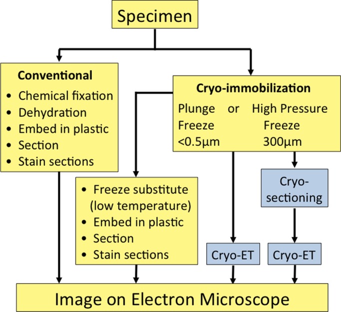
A brief flowchart showing the work to be done with different types of sample preparation
for conventional electron microscopy (yellow background). The advanced cryo-EM techniques
are shown with a blue background. For immuno-EM, the samples can be stained before
embedding (pre-embedding staining) or the sections can be stained (post-embedding
staining).
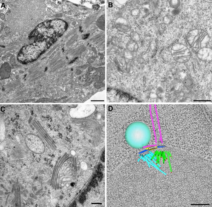
Cell structure as visualized by transmission electron microscopy. (A) Actin-myosin
cytoskeleton revealed in a cultured cardiomyocyte prepared by conventional chemical
fixation. Bar, 1 µm (B) Cytoplasmic organelles in a mouse macrophage prepared by
conventional chemical fixation. Bar, 700 nm (C) Golgi membranes in a cultured 3T3 cell
prepared by high-pressure freezing and freeze substitution. Bar, 200 nm. (D)
Three-dimensional tomographic model of a forming mitotic spindle from budding yeast.
Bar, 200 nm.
References
-
- Bouchet-Marquis C, Hoenger A. Cryo-electron tomography on vitrified sections: a critical analysis of benefits and limitations for structural cell biology. Micron. 2011;42:152–162. - PubMed
-
- Bozzola JJ, Russell LD. Electron Biology Principles and Techniques for Biologists. Sudbury, MA: Jones & Bartlett Learning; 1999.
-
- Dahl R, Staehelin LA. High-pressure freezing for the preservation of biological structure: theory and practice. J Electron Microsc Tech. 1989;13:165–174. - PubMed
-
- Gilkey JC, Staehelin LA. Advances in ultrarapid freezing for the preservation of cellular ultrastructure. J Electron Microsc Tech. 1986;3:177–210.
Publication types
MeSH terms
Substances
Grants and funding
LinkOut - more resources
Full Text Sources
Other Literature Sources
