. 2014 Sep;38(5):823-64.
doi: 10.1111/1574-6976.12063. Epub 2014 Feb 24.

S-layers: principles and applications

Affiliations

S-layers: principles and applications

Uwe B Sleytr et al. FEMS Microbiol Rev. 2014 Sep.

Abstract

Monomolecular arrays of protein or glycoprotein subunits forming surface layers (S-layers) are one of the most commonly observed prokaryotic cell envelope components. S-layers are generally the most abundantly expressed proteins, have been observed in species of nearly every taxonomical group of walled bacteria, and represent an almost universal feature of archaeal envelopes. The isoporous lattices completely covering the cell surface provide organisms with various selection advantages including functioning as protective coats, molecular sieves and ion traps, as structures involved in surface recognition and cell adhesion, and as antifouling layers. S-layers are also identified to contribute to virulence when present as a structural component of pathogens. In Archaea, most of which possess S-layers as exclusive wall component, they are involved in determining cell shape and cell division. Studies on structure, chemistry, genetics, assembly, function, and evolutionary relationship of S-layers revealed considerable application potential in (nano)biotechnology, biomimetics, biomedicine, and synthetic biology.

Keywords: bacterial surface layers; biomimetics; crystalline cell surface layers (S-layers); nanobiotechnology; self-assembly; synthetic biology.


Figures

Figure 1
Figure 1
TEM image of a freeze‐etched and metal shadowed preparation of (a) an archaeal cell (from Methanocorpusuculum sinense), and (b) a bacterial cell (from Desulfotomaculum nigrificans). Bars, 200 nm.
Figure 2
Figure 2
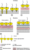
Schematic illustration of the supramolecular architecture of the major classes of prokaryotic cell envelopes containing surface (S) layers. S‐layers in archaea with glycoprotein lattices as exclusive wall component are composed either of mushroom‐like subunits with pillar‐like, hydrophobic trans‐membrane domains (a), or lipid‐modified glycoprotein subunits (b). Individual S‐layers can be composed of glycoproteins possessing both types of membrane anchoring mechanisms. Few archaea possess a rigid wall layer (e.g. pseudomurein in methanogenic organisms) as intermediate layer between the plasma membrane and the S‐layer (c). In Gram‐positive bacteria, (d) the S‐layer (glyco)proteins are bound to the rigid peptidoglycan‐containing layer via secondary cell wall polymers. In Gram‐negative bacteria, (e) the S‐layer is closely associated with the lipopolysaccharide of the outer membrane.
Figure 3
Figure 3
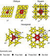
Schematic drawing of the different S‐layer lattice types, their base vectors, the unit cell (shaded in gray), and the corresponding symmetry axis. The proteins at one morphological unit are shown in red.
Figure 4
Figure 4
Schematic drawing of the reassembly of isolated S‐layer (glyco)proteins in suspension, at the air–water interface, on solid supports, on lipid films, on liposomes, emulsomes, polyelectrolyte nanocapsules, and (magnetic) beads.
Figure 5
Figure 5
Schematic drawings of mono‐ and oligomeric S‐layer fusion proteins: (a) fusion protein with single function, (b) fusion protein acting as template for oligomeric assemblies, (c) functional domains bound via flexible linkers to S‐layer proteins assemble on the surface of the S‐layer lattice. (d) Cartoon illustrating self‐assembled S‐layer fusion proteins (see Table 2) carrying functional domains (represented as knights) in defined position and orientation (Sleytr et al., 2007a, b).
Figure 6
Figure 6
Schematic drawing of a lipid membrane on S‐layer (yellow) covered solid (black; left) or porous (black/white; right) supports. Some head groups of the lipid molecules within the membrane (gray) interact electrostatically with certain domains on the S‐layer lattice. A further (glyco)protein S‐layer lattice can be recrystallized on the outer leaflet of the lipid membrane (left). In analogy, some head groups of the lipid molecules within the membrane (gray) interact electrostatically with certain domains of the S‐layer proteins. The lipid molecules on the left side depict schematically phospholipids, whereas the lipid molecules on the right side indicate ether lipids.

References

    1. Abdul Halim MF, Pfeiffer F, Zou J et al (2013) Haloferax volcanii archaeosortase is required for motility, mating, and C‐terminal processing of the S‐layer glycoprotein. Mol Microbiol 88: 1164–1175. - PubMed
    1. Akca E, Claus H, Schultz N, Karbach G, Schlott B, Debaerdemaeker T, Declercq JP & König H (2002) Genes and derived amino acid sequences of S‐layer proteins from mesophilic, thermophilic, and extremely thermophilic methanococci. Extremophiles 6: 351–358. - PubMed
    1. Albers SV & Meyer BH (2011) The archaeal cell envelope. Nat Rev Microbiol 9: 414–426. - PubMed
    1. Amselem S, Yogev A, Zawoznik E & Friedman D (1994) Emulsomes, a novel drug delivery technology. Proc Contr Rel Soc 21: 668–669.
    1. Andresen TL, Jensen SS & Jørgensen K (2005) Advanced strategies in liposomal cancer therapy: problems and prospects of active and tumor specific drug release. Prog Lipid Res 44: 68–97. - PubMed

Publication types

MeSH terms