Review
. 2014 Mar;38(2):254-99.
doi: 10.1111/1574-6976.12065. Epub 2014 Mar 3.

Nutrient sensing and signaling in the yeast Saccharomyces cerevisiae

Affiliations
Review

Nutrient sensing and signaling in the yeast Saccharomyces cerevisiae

Michaela Conrad et al. FEMS Microbiol Rev. 2014 Mar.

Abstract

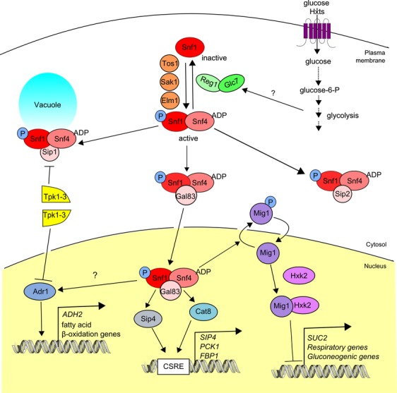
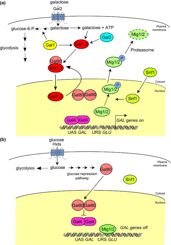
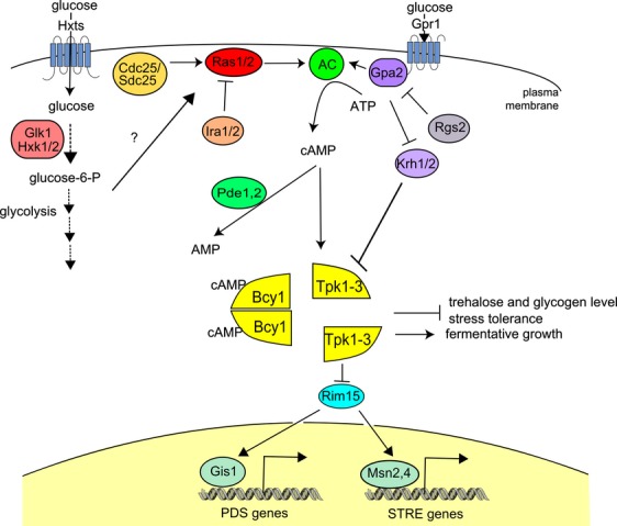
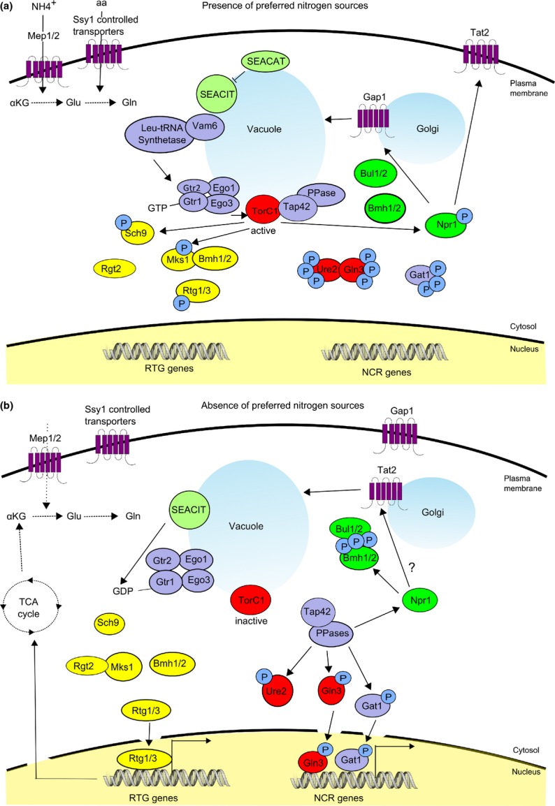
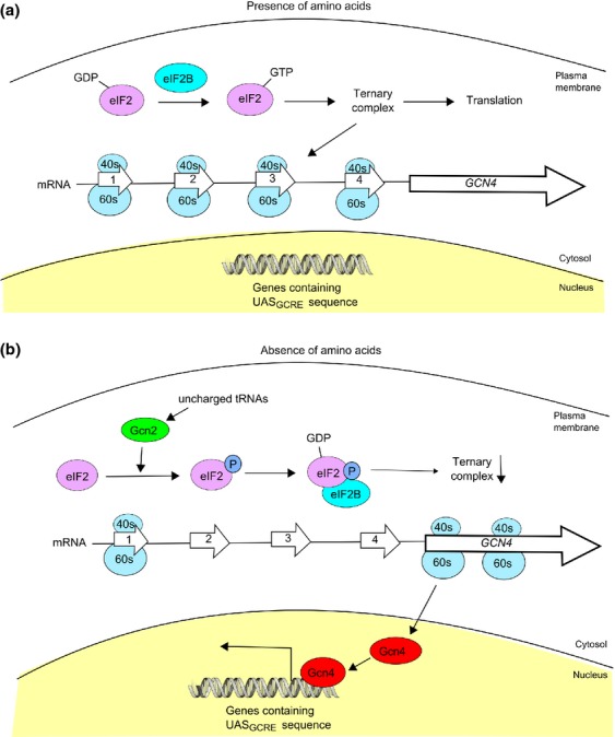
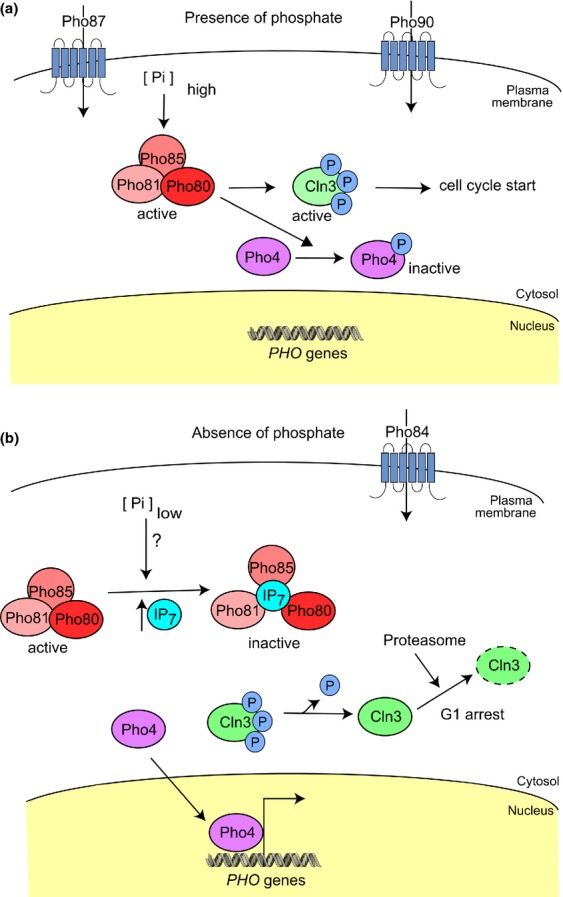
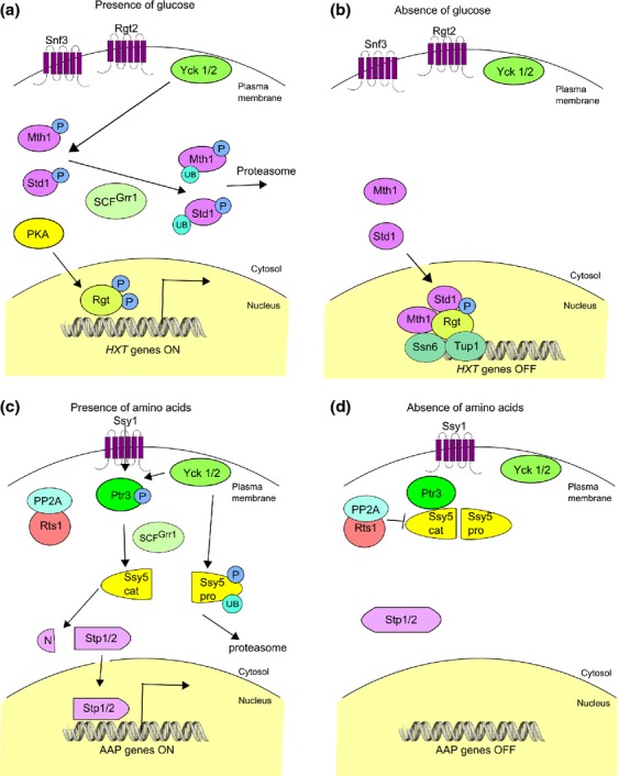
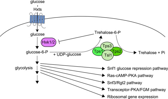
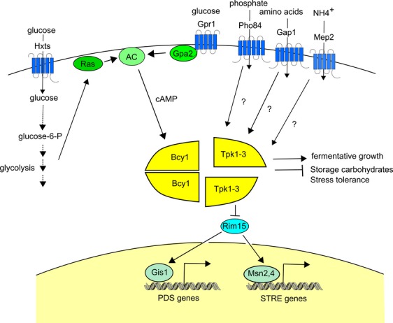
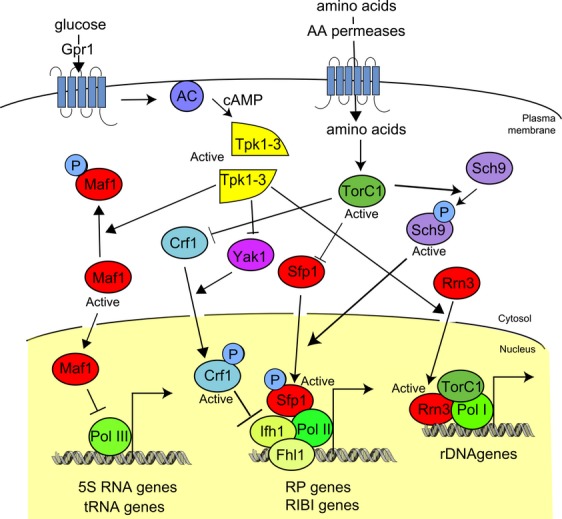
The yeast Saccharomyces cerevisiae has been a favorite organism for pioneering studies on nutrient-sensing and signaling mechanisms. Many specific nutrient responses have been elucidated in great detail. This has led to important new concepts and insight into nutrient-controlled cellular regulation. Major highlights include the central role of the Snf1 protein kinase in the glucose repression pathway, galactose induction, the discovery of a G-protein-coupled receptor system, and role of Ras in glucose-induced cAMP signaling, the role of the protein synthesis initiation machinery in general control of nitrogen metabolism, the cyclin-controlled protein kinase Pho85 in phosphate regulation, nitrogen catabolite repression and the nitrogen-sensing target of rapamycin pathway, and the discovery of transporter-like proteins acting as nutrient sensors. In addition, a number of cellular targets, like carbohydrate stores, stress tolerance, and ribosomal gene expression, are controlled by the presence of multiple nutrients. The protein kinase A signaling pathway plays a major role in this general nutrient response. It has led to the discovery of nutrient transceptors (transporter receptors) as nutrient sensors. Major shortcomings in our knowledge are the relationship between rapid and steady-state nutrient signaling, the role of metabolic intermediates in intracellular nutrient sensing, and the identity of the nutrient sensors controlling cellular growth.

Keywords: G-protein-coupled receptor; Pho85 protein kinase; Ras; Snf1 protein kinase; target of rapamycin; transceptor.


Figures

Figure 1
Figure 1
The Snf1 protein kinase as a central player in the main glucose repression pathway. The Snf1 protein kinase orchestrates glucose repression of alternative carbon source utilization, and genes involved in respiration and gluconeogenesis. The Snf1 heterotrimeric complex consists of the catalytic subunit Snf1, the stimulatory subunit, Snf4, and one of the three β-subunits: Gal83, Sip1, or Sip2. Snf1 is active in phosphorylated form and the phosphorylation is performed by the three upstream protein kinases Sak1, Tos3, and Elm1, while the phosphatase Glc7 in conjunction with its regulatory subunit Reg1 is responsible for its dephosphorylation. The actual glucose-sensing mechanism, in which Hxk2 appears to play an important role, possibly activates the Glc7-Reg1 protein phosphatase to trigger dephosphorylation of Snf1. In its active form, Snf1-Snf4 binds to each of the three β-subunits, acquiring differential specificity for localization and target phosphorylation. Upon glucose exhaustion, a major role is played by the Snf1–Gal83 complex, which enters the nucleus to trigger derepression. This is accomplished by activation of the transcription factors Adr1, Sip4, and Cat8 and inactivation of Mig1 by dislodging its interaction with Hxk2 and promoting its cytosolic localization by phosphorylation. This leads to the expression of a wide range of carbon source-responsive element (CSRE) containing genes involved in the use of alternative carbon sources, gluconeogenesis, ethanol, and fatty acid metabolism. Metabolic reactions are depicted by dotted arrows; regulatory and signaling interactions by full arrows.
Figure 2
Figure 2
Induction of the GAL regulon for galactose utilization. (a) In the absence of glucose and presence of galactose, the GAL genes are induced. Galactose enters the cells via its transporter, Gal2, which is present at a low basal level under this condition. Trace amounts of the intracellular sensor protein, Gal3, bind galactose and ATP in the cytosol, which promotes binding of Gal3 to the GAL-specific transcriptional repressor, Gal80. This prevents the accumulation of Gal80 in the nucleus, which reduces its inhibition of the transcriptional activator Gal4. A tripartite interaction between Gal3, Gal80, and Gal4 may also occur in the nucleus to facilitate Gal4 release from Gal-80-mediated inhibition. In later stages of galactose induction, the bifunctional protein Gal1 replaces Gal3 in its signaling role. The Snf1 protein kinase complex, which is active under this condition, phosphorylates the Mig1/2 repressor proteins, which causes their dissociation from upstream repressor sequences (URSGLU) and subsequent export to the cytosol. Gal4 activation facilitates the association of chromatin remodeling complexes and the basal transcriptional machinery leading to induction of the GAL genes. (b) In the presence of glucose (irrespective of the absence or presence of galactose), expression of the GAL genes is repressed. Glucose enters the cells via the multiple hexose transporters (HXT). Once the levels of intracellular glucose increase, Gal80 is relieved from inhibition by Gal1,3 and enters the nucleus where it inhibits Gal4. Glucose also causes inactivation of the Snf1 protein kinase, which favors Mig1/2 nuclear import and thus downregulation of the GAL genes by these transcriptional repressors. Metabolic reactions are depicted by dotted arrows; regulatory and signaling interactions by full arrows.
Figure 3
Figure 3
Glucose activation of the cAMP-PKA pathway. AC is activated by glucose through two different G-protein-coupled systems. The Gpr1-Gpa2-Rgs2 GPCR system senses extracellular glucose, while the Cdc25,Sdc25-Ras1,2-Ira1,Ira2 system senses intracellular glucose through glucose catabolism in glycolysis in a way that is not yet understood. The glucose-sensing GPCR, Gpr1, and the Cdc25,Sdc25 proteins stimulate guanine nucleotide exchange on Gpa2 and Ras1,2, respectively. Rgs2 and Ira1,2 act as GAPs on Gpa2 and Ras1,2, respectively. cAMP binds to the Bcy1 regulatory subunits of PKA causing dissociation and activation of the catalytic subunits, Tpk1-3. The Krh1,2 kelch repeat proteins mediate a cAMP-independent pathway triggered by the glucose-sensing GPCR system for direct activation of PKA, by lowering the affinity between catalytic and regulatory subunits. Metabolic reactions are depicted by dotted arrows; regulatory and signaling interactions by full arrows.
Figure 4
Figure 4
Role of TORC1 in the NCR and RTG pathways. (a) Preferred nitrogen sources for yeast are these that can easily be converted into glutamate (Glu) and glutamine (Gln), major precursors for amino acid biosynthesis. Their presence in the medium results in increased levels of intracellular glutamate and glutamine. This causes repression of genes involved in the metabolism of less preferred nitrogen sources, nitrogen catabolite repression (NCR). This transcriptional repression is achieved mainly by hyperphosphorylation of Ure2 and Gln3, causing their association and preventing nuclear localization of the transcription factor Gln3. The Gat1 transcription factor is regulated in a similar way. High glutamine levels as well as other amino acids stimulate the vacuolar/endosome membrane-located, EGO complex. This complex is composed of the two Ras-like GTPases, Gtr1, Gtr2, and the Ragulator-like, Ego3 and Ego1. Activation of EGO is stimulated by GTP-bound Gtr1 and GDP-bound Gtr2. GTP loading of Gtr1 is stimulated by the guanine nucleotide-exchange factor (GEF) activities of Vam6/Vps39 and the L-Leu-tRNA synthetase. SEACAT prevents GAP activity of SEACIT on Gtr1. Activated EGO stimulates in turn the vacuolar membrane-associated fraction of the TORC1 complex. TORC1 also phosphorylates Sch9 and Tap42, the latter leading to the inhibition of several protein phosphatases (PPA2, Sit4, etc.). As a result, the protein phosphatases can no longer dephosphorylate the Ure2 complexes with Gln3 and Gat1, reinforcing their hyperphosphorylation. Synthesis of glutamine and glutamate occurring via anaplerotic reactions shared with the TCA cycle is also downregulated. In this case, TORC1 phosphorylated Mks1 bound to Bmh1,2 proteins prevents nuclear localization of the RTG transcription factors, Rtg1 and Rtg3. TORC1-dependent phosphorylation of Npr1 causes Npr1 inactivation, which in a yet not completely understood manner increases plasma membrane stabilization of specific AAPs like Tat2, while stimulating endocytosis of the alternative general AAP, Gap1. (b) Under poor nitrogen conditions, intracellular glutamate and glutamine levels drop. GAPs like the SEACIT increase GDP loading of Gtr1, which inactivates the EGO complex. An inactive EGO complex can no longer stimulate TORC1, which leads to release into the cytosol and activation of Tap42–protein phosphatase complexes. They reduce phosphorylation of Ure2, Gln3, and Gat1 causing nuclear localization of the latter two and subsequent stimulation of NCR gene expression. The phosphatases also dephosphorylate Mks1, which then complexes with Rtg2. This allows Rtg1,3 nuclear localization resulting in stimulation of the expression of RTG genes, sustaining amino acid biosynthesis through the synthesis of glutamate and glutamine. The phosphatases also dephosphorylate Npr1, which then phosphorylates the Rsp5-associated arrestins Bul1 and Bul2 provoking their association with Bmh1/2 proteins, which in turn leads to the stabilization of Gap1 at the plasma membrane. Metabolic reactions are depicted by dotted arrows; regulatory and signaling interactions by full arrows.
Figure 5
Figure 5
The GAAC pathway. (a) In the presence of amino acids, eukaryotic translation initiation factor, eIF2, is mainly in the GTP-bound state as a result of stimulation by its GEF, eIF2B. GTP-bound eIF2 forms a TC with initiator Met-tRNA. TC along with the 40S ribosomal unit scans mRNA and recruits the 60S ribosomal unit to form the functional ribosome. The latter starts translating mRNA into protein once it encounters the start codon. This is also true for the GCN4 mRNA, but the ORF is preceded by multiple μORFs, which largely prevent the ribosomes from reaching the GCN4 ORF. Hence, under these conditions, production of the Gcn4 transcription factor is very limited. (b) Under amino acid starvation, the levels of uncharged tRNA increase, which activates the Gcn2 protein kinase. This enhances the level of phosphorylated eIF2, which as a result binds too tightly to eIF2B, preventing the stimulation of the exchange of GDP for GTP on eIF2. Hence, eIF2 is largely in the GDP-bound state and the level of TC drops, which causes a strong reduction in the level of general protein synthesis. However, paradoxically, the μORFs in front of the GCN4 ORF are now largely read through by the ribosomes because of the lack of TC to initiate translation. As a result, the ribosomes are now able to reach the main GCN4 ORF, causing its translation into Gcn4 protein. The enhanced level of the Gcn4 transcription factor stimulates the expression of genes involved in amino acid biosynthesis, resulting in a strong increase in the endogenous synthesis of amino acids when amino acids are absent in the medium.
Figure 6
Figure 6
Central role of Pho85 in phosphate regulation of the PHO pathway. (a) When present in high levels, external phosphate is imported by the low-affinity phosphate carriers Pho87 and Pho90, which raises the intracellular phosphate level. This activates the Pho85 complex, which phosphorylates the transcriptional activator Pho4, causing its sequestration in the cytosol. As a result, the PHO genes, encoding, for example, the secreted phosphatases Pho3 and Pho5, are not expressed. Active Pho85 also phosphorylates cyclin Cln3, which aids in progression over start in G1 of the cell cycle. (b) Upon intracellular phosphate limitation, inositol pyrophosphate (IP7) levels increase. IP7 promotes Pho81-dependent inhibition of the Pho85–cyclin Pho80 complex. This inhibition results in dephosphorylation of Pho4, which then migrates to the nucleus where it activates transcription of the PHO and VTC genes. Inactivation of the Pho85 complex also dephosphorylates Cln3, which is then degraded by the proteasome leading to cell cycle arrest.
Figure 7
Figure 7
Nutrient sensing by transporter-like plasma membrane sensors. The Snf3-Rgt2 glucose-sensing pathway. (a) Glucose binding to the Snf3/Rgt2 sensors recruits Mth1 and Std1 to the plasma membrane, where they are phosphorylated by Yck1,2. This phosphorylation targets them for ubiquitination by Grr1 and degradation by the proteasome, exposing Rgt1 to phosphorylation by PKA. This turns Rgt1 into a transcriptional activator for the expression of HXT (hexose transporter) genes. (b) In absence of glucose, Yck1,2 fail to phosphorylate Mth1 and Std1, which are no longer degraded and enter the nucleus to repress the expression of HXT genes. The SPS amino acid-sensing pathway. (c) In the presence of external amino acids, the amino acids bind to Ssy1, causing recruitment of the Yck1,2 protein kinases. They hyperphosphorylate Ptr3 and the Ssy5 prodomain, making it sensitive to ubiquitination by Grr1, after which it is broken down by the proteasome. Subsequently, the enhanced Ssy5 protease activity toward Stp1,2 leads to the removal of the N-terminal part of Stp1,2, which enables it to enter the nucleus and activate transcription of the target AAP genes. (d) In the absence of extracellular amino acids, phosphorylation of Ssy5 is counteracted by the phosphatase PP2A and its subunit Rts1, which keeps Ssy5 inactive. This prevents migration of Stp1,2 from the cytosol to the nucleus, and the expression of AAP genes is thus kept down.
Figure 8
Figure 8
Function of trehalose-6-phosphate as allosteric regulator of Hxk2 activity. Glucose is taken up by the hexose transporters (Hxt) and subsequently phosphorylated predominantly by hexokinase I (Hxk1) and II (Hxk2) to glucose-6-P, which is then further converted in glycolysis. Glucose-6-P and UDP-glucose are converted to trehalose-6-phosphate by the Tps1 enzyme and further to trehalose by the Tps2 enzyme in the trehalose synthase complex, which also contains the regulatory subunits Tps3 and Tsl1. Trehalose-6-phosphate is a potent allosteric inhibitor of Hxk1 and Hxk2, causing feedback inhibition on the influx of glucose into glycolysis. Although the precise mechanisms are generally unclear, the early steps of glucose catabolism are in some way important for the activation of most glucose signaling pathways. Metabolic reactions are depicted by dotted arrows; regulatory and signaling interactions by full arrows.
Figure 9
Figure 9
Activation of the PKA pathway in the presence of glucose by different essential nutrients through multiple transceptors. Glucose-fermenting cells of Saccharomyces cerevisiae that are starved for another essential nutrient, like nitrogen or phosphate, enter stationary phase and develop a low-PKA phenotype, that is, accumulation of trehalose and glycogen, acquirement of high stress tolerance, and downregulation of stress-responsive genes. Re-addition of nitrogen or phosphate triggers rapid activation of PKA targets, which is not mediated by cAMP as a second messenger. The nutrient sensors or ‘transceptors’ involved are specific transporters that were induced in the starvation period and that act as nutrient receptors for activation of the PKA pathway. Gap1 senses amino acids, Mep2 senses ammonium, and Pho84 senses phosphate in appropriately starved cells. The presence of glucose is essential for nutrient transceptor activation. It can be detected either by the GPCR (Gpr1-Gpa2) system for extracellular glucose sensing or by the Ras system, which is activated by intracellular glucose catabolism. Metabolic reactions are depicted by dotted arrows; regulatory and signaling interactions by full arrows.
None
Nutrient control of ribosomal gene expression. Ribosomal gene expression involves RNA polymerase (Pol) I for expression of rRNA, Pol II for expression of RPs and ribosome biogenesis (RiBi) factors, and Pol III for tRNA and small nuclear (5S) RNA. Different nutrient-sensing pathways are involved. Glucose sensing by Gpr1 activates the cAMP-PKA pathway, which stimulates ribosome biogenesis through effects on the three polymerases. The presence of preferred nitrogen sources impinges on this pathway via TORC1 and its downstream kinase Sch9. Pol I-dependent expression is positively regulated via interaction of Rrn3 with Pol I. TORC1 stabilizes this interaction. Glucose may stimulate Pol I-dependent transcription through PKA control of the level of Rrn3. Pol II-dependent expression is positively regulated by Sfp1 and Fhl1. Upon nutrient deprivation, the Crf1 inhibitor competes with the co-activator Ifh1 for binding to Fhl1, which causes inactivation of the latter. TORC1 prevents Crf1 nuclear shuttling, whereas Yak1 favors it. Sfp1 stimulates Pol II-dependent expression and is positively regulated by TorC1 via Sch9. Pol III-dependent expression is negatively regulated by Maf1. PKA phosphorylates Maf1, which prevents entrance of Maf1 into the nucleus and thus allows Pol III-directed gene expression.

References

    1. Abate G, Bastonini E, Braun KA, Verdone L, Young ET. Caserta M. Snf1/AMPK regulates Gcn5 occupancy, H3 acetylation and chromatin remodelling at S. cerevisiae ADY2 promoter. Biochim Biophys Acta. 2012;1819:419–427. - PMC - PubMed
    1. Abdel-Sater F, El Bakkoury M, Urrestarazu A, Vissers S. Andre B. Amino acid signaling in yeast: casein kinase I and the Ssy5 endoprotease are key determinants of endoproteolytic activation of the membrane-bound Stp1 transcription factor. Mol Cell Biol. 2004;24:9771–9785. - PMC - PubMed
    1. Abdel-Sater F, Jean C, Merhi A, Vissers S. Andre B. Amino acid signaling in yeast: activation of Ssy5 protease is associated with its phosphorylation-induced ubiquitylation. J Biol Chem. 2011;286:12006–12015. - PMC - PubMed
    1. Abe F. Iida H. Pressure-induced differential regulation of the two tryptophan permeases Tat1 and Tat2 by ubiquitin ligase Rsp5 and its binding proteins, Bul1 and Bul2. Mol Cell Biol. 2003;23:7566–7584. - PMC - PubMed
    1. Abramczyk D, Holden S, Page CJ. Reece RJ. Interplay of a ligand sensor and an enzyme in controlling expression of the Saccharomyces cerevisiae GAL genes. Eukaryot Cell. 2012;11:334–342. - PMC - PubMed

Publication types

MeSH terms