. 2014 May;15(5):488-99.
doi: 10.1111/tra.12158. Epub 2014 Mar 12.

Legionella pneumophila subversion of host vesicular transport by SidC effector proteins

Affiliations

Legionella pneumophila subversion of host vesicular transport by SidC effector proteins

Florian A Horenkamp et al. Traffic. 2014 May.

Abstract

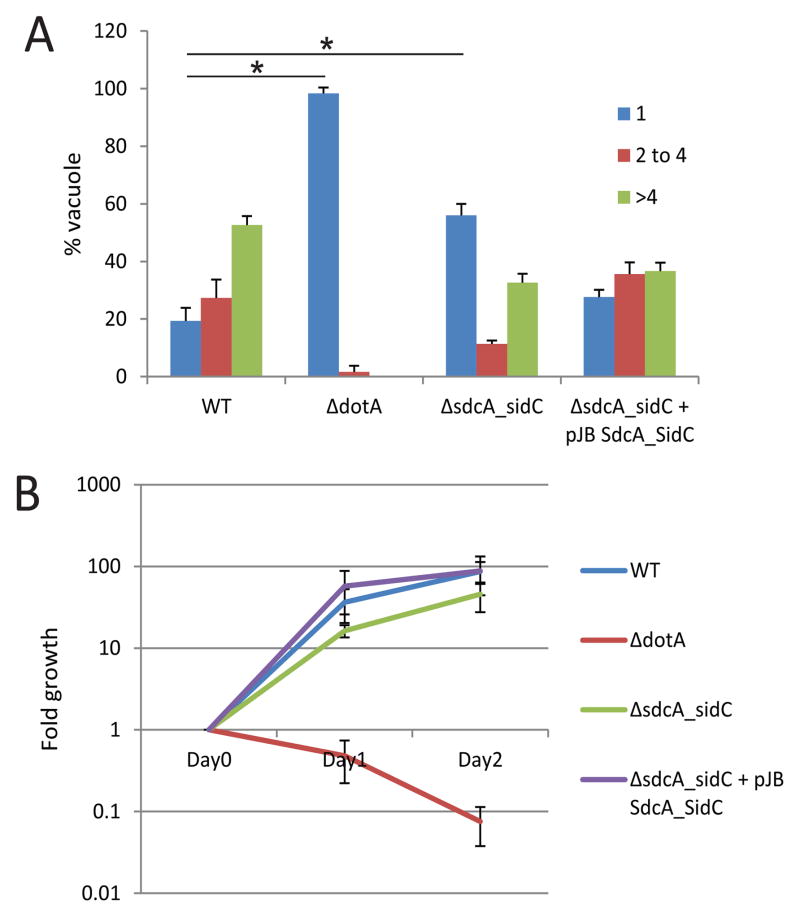
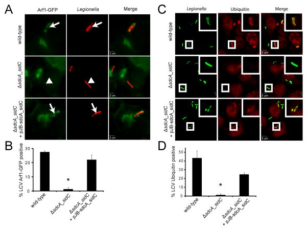
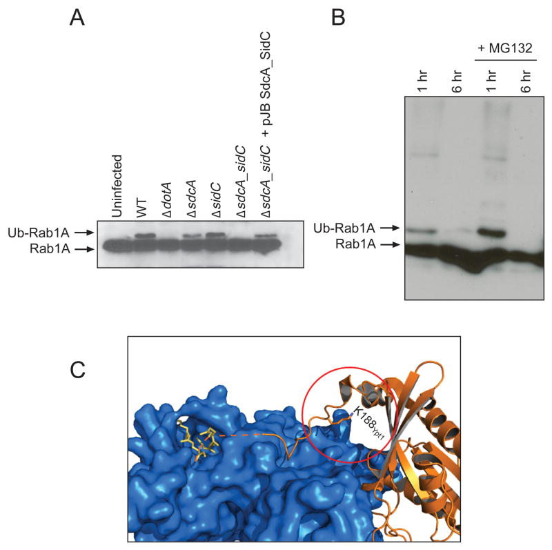
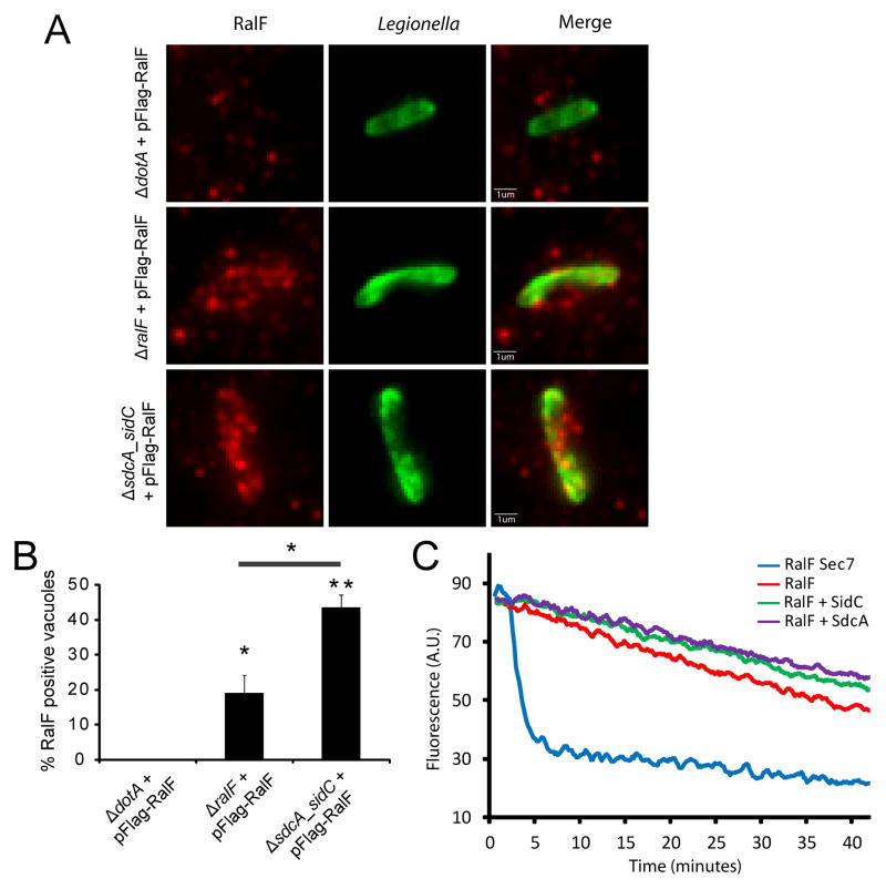
Tethering proteins play a key role in vesicular transport, ensuring that cargo arrives at a specific destination. The bacterial effector protein SidC and its paralog SdcA have been described as tethering factors encoded by the intracellular pathogen Legionella pneumophila. Here, we demonstrate that SidC proteins are important for early events unique to maturation of vacuoles containing Legionella and discover monoubiquitination of Rab1 as a new SidC-dependent activity. The crystal structure of the SidC N-terminus revealed a novel fold that is important for function and could be involved in Legionella adaptations to evolutionarily divergent host cells it encounters in natural environments.

Keywords: Rab1 modification; Tethering.


Conflict of interest statement

Conflict of Interest: The authors declare NO conflict of interest.

Figures

Figure 1
Figure 1. SdcA/SidC are required for optimal Legionella growth
A and B) NLRC4 −/− bone marrow derived macrophages were infected with L. pneumophila wild-type, ΔdotA, ΔsdcA-sidC and ΔsdcA-sidC complemented with a plasmid-encoded sdcA-sidC operon. A) Cells were fixed 8 h after infection. Intracellular replication of L. pneumophila was assessed by counting the number of bacteria residing in a single vacuole in infected cells. The data are presented as vacuoles containing 1 (blue), 2–4 (red) or >4 bacteria (green). Average ± standard deviations were obtained from 3 independent experiments in which 300 vacuoles were scored for each strain. Statistical significance was obtained by comparing the number of cells showing a single bacteria between WT and ΔdotA or WT and ΔsdcA-sidC (* P<0.01). B) Intracellular replication of L. pneumophila strains in NLRC4−/− cells were determined by counting the number of colony forming units (cfus) on charcoal yeast extract agar plates. Values are the mean ± SEM of four independent experiments.
Figure 2
Figure 2. SdcA/SidC are required for early recruitment of specific LCV markers
A) and B) HEK293 cells stably expressing GFP-tagged Arf1 were infected with L. pneumophila wild-type, ΔsdcA-sidC and ΔsdcA-sidC complemented with a plasmid-encoded sdcA-sidC operon. A) Cells were fixed 1 h post-infection. Intracellular bacteria are shown in red. Arrows show Arf1-positive vacuoles, arrowheads show Arf1-negative vacuoles. B) Quantification of the proportion of Arf1 positive vacuoles. Average ± standard deviations were obtained from 3 independent experiments (* P<0.01). C) and D) HEK293 cells were infected with L. pneumophila wild-type, ΔsdcA-sidC and ΔsdcA-sidC complemented with a plasmid-encoded sdcA-sidC operon. C) Cells were fixed 1 h post-infection, and stained for ubiquitin with FK2 antibodies. Intracellular bacteria are shown in green. D) Quantification of the proportion of ubiquitin positive vacuoles. Average ± standard deviations were obtained from 3 independent experiments (* P<0.01).
Figure 3
Figure 3. Rab1 is ubiquitinated during infection.<
br>A) HEK293 cells stably expressing Flag-tagged Rab1 were either uninfected or infected with L. pneumophila wild-type, ΔdotA, ΔsdcA, ΔsidC, ΔsdcA-sidC and ΔsdcA-sidC complemented with a plasmid-encoded sdcA-sidC operon. Cell lysates were prepared 1 h post-infection, and Rab1A was probed with anti-Rab1A antibody. B) HEK293 cells stably expressing Flag-tagged Rab1 were infected with L. pneumophila wild-type for 1 h or 6 h with or without the proteasome inhibitor MG132 and probed with anti-Rab1A antibody. C) Structure of the doubly prenylated (yellow) yeast Rab1 homologue Ypt1 (orange) in complex with GDI (blue) (PDB: 2BCG) (18). K188Ypt1 is the Ypt1 residue homologous to the Rab1A ubiquitination site K187. Addition of a ubiquitin moiety at this position would interfere with the Rab1:GDI interaction. The radius of the red circle around K188Ypt1 is 10 Å.
Figure 4
Figure 4. SidC does not alter RalF function
A) and B) RalF is recruited at the surface of the L pneumophila ΔsdcA-sidC LCV. HEK293 cells were infected with L. pneumophila ΔdotA, ΔralF and ΔsdcA-sidC expressing 3*Flag-tagged RalF from a plasmid. A) Cells were fixed 2 h post-infection, and RalF was probed with anti-Flag antibodies (red). Intracellular bacteria are shown in green. B) Quantification of the proportion of RalF positive vacuoles. Average ± standard deviations were obtained from one experiment done in triplicate and are representative of 3 independent experiments (* P=0.01; ** P=0.001). C) SidC or SdcA do not relieve full length RalF ArfGEF activity autoinhibition. His-ΔN17Arf1 nucleotide exchange was measured by mant-GDP release fluorescence assay in the presence of indicated MBP-RalF and GST-SdcA or SidC proteins. The MBP-RalF Sec7 domain was used as a positive control for ArfGEF activity.
Figure 5
Figure 5. Structure of SidCNT (residue 1-608)
A) SidCNT, colored from blue at the N-terminus to red at the C-terminus. The domains are labeled A–C. B) Secondary structure topology of SidCNT. C) Sequence alignment of SidC and SdcA. D) HEK293 cells stably expressing 3XFlag-tagged Rab1 were transfected with the cDNAs encoding 3XFLAG-tagged SdcA full-length (3XFLAG-SdcAFL) or SdcA lacking residues 221-329 (3XFLAG-SdcAΔC). 14 hours after transfection, cells were infected with a L. pneumophila wild-type strain or ΔsdcA-sidC strain for 1 h, following which they were lysed. Rab1A was immunoprecipitated using FLAG-antibody coated beads. After incubation, beads were boiled and subjected to SDS-PAGE analysis and subsequently probed with either a Rab1A specific antibody or a FLAG antibody. E) Model of SdcA/SidC-mediated vesicle tethering at the LCV.

References

    1. Yu IM, Hughson FM. Tethering factors as organizers of intracellular vesicular traffic. Annu Rev Cell Dev Biol. 2010;26:137–156. - PubMed
    1. Brocker C, Engelbrecht-Vandre S, Ungermann C. Multisubunit tethering complexes and their role in membrane fusion. Current biology: CB. 2010;20(21):R943–952. - PubMed
    1. Rowbotham TJ. Preliminary report on the pathogenicity of Legionella pneumophila for freshwater and soil amoebae. Journal of clinical pathology. 1980;33(12):1179–1183. - PMC - PubMed
    1. Horwitz MA, Silverstein SC. Legionnaires’ disease bacterium (Legionella pneumophila) multiples intracellularly in human monocytes. The Journal of clinical investigation. 1980;66(3):441–450. - PMC - PubMed
    1. Horwitz MA. Formation of a novel phagosome by the Legionnaires’ disease bacterium (Legionella pneumophila) in human monocytes. The Journal of experimental medicine. 1983;158(4):1319–1331. - PMC - PubMed

Publication types

MeSH terms

Substances

Associated data