. 2014 May;25(5):461-74.
doi: 10.1089/hum.2013.200. Epub 2014 Mar 21.

Silencing mutant huntingtin by adeno-associated virus-mediated RNA interference ameliorates disease manifestations in the YAC128 mouse model of Huntington's disease

Affiliations

Silencing mutant huntingtin by adeno-associated virus-mediated RNA interference ameliorates disease manifestations in the YAC128 mouse model of Huntington's disease

Lisa M Stanek et al. Hum Gene Ther. 2014 May.

Abstract

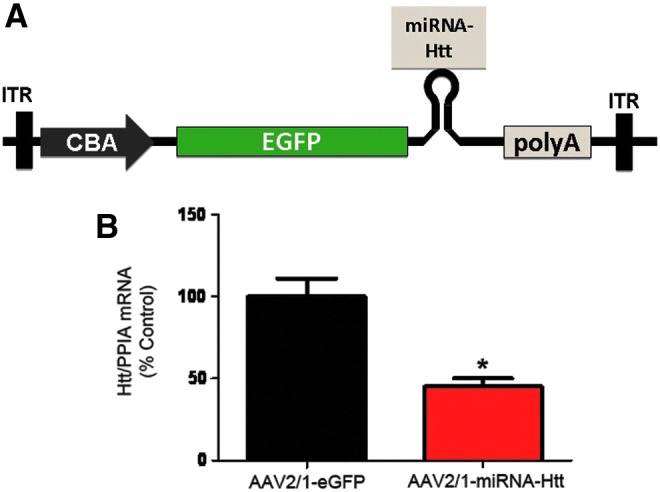
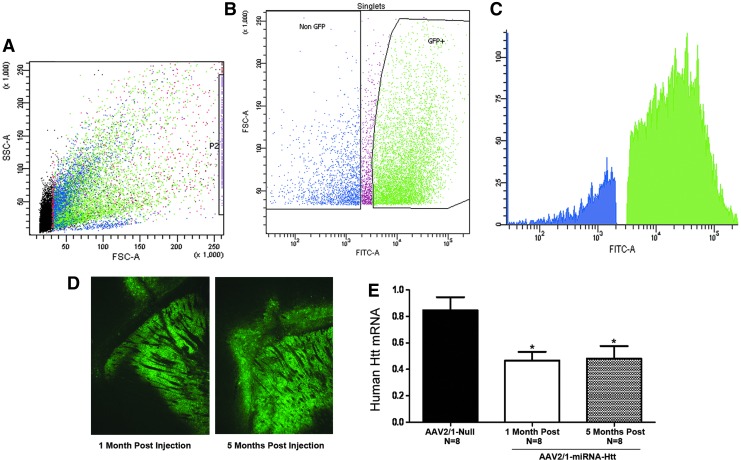
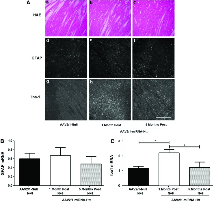
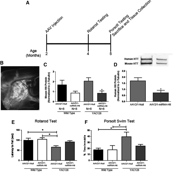
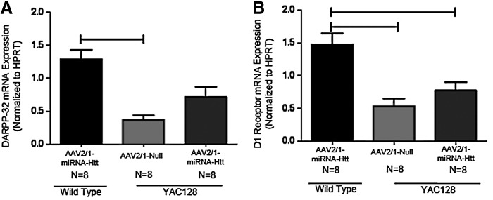
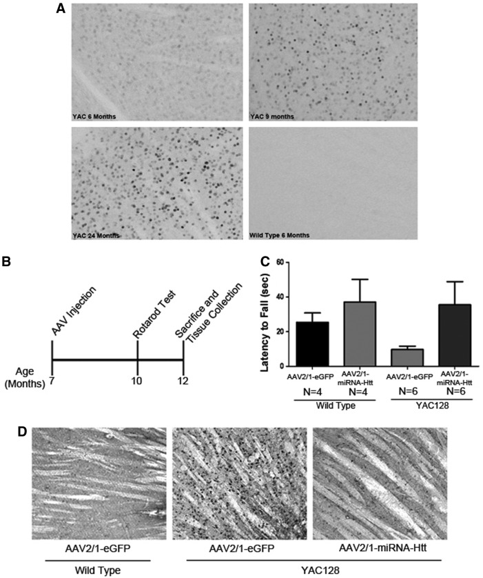
Huntington's disease (HD) is a fatal autosomal dominant neurodegenerative disease caused by an increase in the number of polyglutamine residues in the huntingtin (Htt) protein. With the identification of the underlying basis of HD, therapies are being developed that reduce expression of the causative mutant Htt. RNA interference (RNAi) that seeks to selectively reduce the expression of such disease-causing agents is emerging as a potential therapeutic strategy for this and similar disorders. This study examines the merits of administering a recombinant adeno-associated viral (AAV) vector designed to deliver small interfering RNA (siRNA) that targets the degradation of the Htt transcript. The aim was to lower Htt levels and to correct the behavioral, biochemical, and neuropathological deficits shown to be associated with the YAC128 mouse model of HD. Our data demonstrate that AAV-mediated RNAi is effective at transducing greater than 80% of the cells in the striatum and partially reducing the levels (~40%) of both wild-type and mutant Htt in this region. Concomitant with these reductions are significant improvements in behavioral deficits, reduction of striatal Htt aggregates, and partial correction of the aberrant striatal transcriptional profile observed in YAC128 mice. Importantly, a partial reduction of both the mutant and wild-type Htt levels is not associated with any notable overt neurotoxicity. Collectively, these results support the continued development of AAV-mediated RNAi as a therapeutic strategy for HD.


Figures

<b>FIG. 1.</b>
FIG. 1.
AAV2/1-miRNA-Htt mediates the reduction of Htt levels in vitro. (A) Schematic of the previral construct used to generate AAV2/1-miRNA-Htt. The plasmid was designed to express GFP and an miRNA sequence against Htt under the transcriptional control of the chicken β-actin (CBA) promoter. ITR, inverted terminal repeat; eGFP, enhanced green fluorescent protein. (B) Quantitative PCR analysis evaluating Htt mRNA levels in HEK293 cells 48 hr after AAV-2/1-miRNA-Htt treatment. PPIA served as a normalization control gene. Values are given as means±SEM. *p<0.05. Color images available online at www.liebertpub.com/hum
<b>FIG. 2.</b>
FIG. 2.
Widespread striatal transduction and Htt reduction after intrastriatal injection of AAV2/1-miRNA-Htt into YAC128 mice. (A) Flow cytometric scatter profile of striatal cells with eGFP. Forward light scatter A (FSC-A) represents relative cell size, area and SSC-A represents relative cell complexity, area with each dot representing one cell. (B) Dot plot based on FSC-A versus FITC-A analysis. Dead cells were sorted out and eGFP+ and eGFP cells were selected. GFP expression from these cells was evaluated, quantified, and shown in (C), a fluorescence plot of eGFP fluorescence intensity collected with a 530/30BP filter 505LP. (D) Fluorescence microscopy showing widespread eGFP expression throughout the striatum after intracranial administration of AAV2/1-eGFP-miRNA-Htt. (E) Quantitative PCR analysis evaluating Htt mRNA levels in the striatum 1 and 5 moths after injection of AAV2/1-miRNA-Htt or AAV2/1-null control vector. PPIA served as a normalization control gene. Values are given as means±SEM. *p<0.05. AAV2/1-miRNA-Htt-treated YAC128 mice (n=8) showed an approximately 50% reduction in Htt mRNA levels in the striatum when compared with AAV2/1-Null-injected mice (n=8 per time point) at 1 and 5 months posttreatment.
<b>FIG. 3.</b>
FIG. 3.
Sustained lowering of Htt levels in YAC128 mice by AAV2/1-miRNA-Htt does not cause overt neuroinflammation. (A) Panels a–c: Hematoxylin and eosin (H&E) staining of striatal tissue sections 1 or 5 months after AAV-miRNA-Htt injection. Panels d–i: GFAP and Iba-1 immunohistochemical staining of sections from YAC128 mice treated with AAV2/1-miRNA-Htt or AAV2/1-Null vector. All photographs were exposure-matched for accurate comparisons. Scale bar: 0.25 mm. (B) Striatal levels of GFAP mRNA levels by qPCR 1 or 5 months after the injection of AAV2/1-miRNA-Htt. (C) Iba-1 mRNA levels by qPCR 1 or 5 months after the injection of AAV2/1-miRNA-Htt. Values are given as means±SEM. *Significant difference, p<0.05; ANOVA. Color images available online at www.liebertpub.com/hum
<b>FIG. 4.</b>
FIG. 4.
Striatal administration of AAV2/1-miRNA-Htt reduces behavioral deficits in YAC128 mice. (A) Illustration of experimental timeline. Two-month-old YAC128 (YAC) and wild-type (WT) mice received bilateral striatal injections of either AAV2/1-miRNA-Htt (YAC, n=8; WT, n=8) or AAV2/1-Null control (YAC, n=8; WT, n=8) and were subjected to a Rotarod test and the Porsolt swim test at 4 and 5 months of age, respectively. All mice were killed at 5 months of age, and tissues were then collected for biochemical and histological analyses. (B) Fluorescence microscopy showing eGFP expression in the striatum 3 months posttreatment. (C and D) Mouse and human Htt protein levels by Western blot 3 months after AAV2/1-miRNA-Htt treatment. (E) Accelerating Rotarod test 2 months after the injection of AAV2/1-miRNA-Htt. (F) Time spent immobile in the Porsolt swim test 3 months after the injection of AAV2/1-miRNA-Htt. Values are given as means±SEM. *Significant difference, p<0.05; ANOVA followed by Tukey post-hoc test.
<b>FIG. 5.</b>
FIG. 5.
Treatment with AAV2/1-miRNA-Htt partially corrects the transcriptional dysregulation of DARPP-32 and D1 receptor in YAC128 mice. DARPP-32 and D1 receptor mRNA levels in the striatum of YAC128 and wild-type (WT) mice were assessed by qPCR 3 months after the injection of either AAV2/1-miRNA-Htt (YAC, n=8; WT, n=8) or AAV2/1-Null control (YAC, n=8; WT, n=8). (A) Striatal DARPP-32 mRNA levels in YAC128 and FVB wild-type littermate mice after AAV2/1-Null or AAV2/1-miRNA-Htt treatment. (B) Striatal D1 receptor mRNA levels in YAC128 and FVB wild-type littermate mice after AAV2/1-Null or AAV2/1-miRNA-Htt treatment. Values are given as means±SEM. *Significant difference, p<0.05; ANOVA followed by Tukey post-hoc test.
<b>FIG. 6.</b>
FIG. 6.
Intracranial administration of AAV2/1-miRNA-Htt ameliorates motor deficits and reduces mutant Htt aggregates in the striatum of aged YAC128 mice. (A) Immunohistochemical staining of YAC128 mouse brain sections showing mutant Htt aggregates in the striatum. Aggregates were observed in 6, 9, 12 (data not shown), and 24-month-old YAC128 mice. Wild-type mice exhibited no aggregates at all ages tested. (B) An illustration of the experimental timeline for testing AAV2/1-miRNA-HTT in aged YAC128 and wild-type mice. Seven-month-old mice received bilateral intrastriatal injections of AAV2/1-miRNA-Htt (YAC, n=6; WT, n=4) or AAV2/1-GFP control (YAC, n=6; WT, n=4) and were then subjected to behavioral testing at 10 months of age. Brains were harvested 5 months postinjection (when the mice were 12 months old). (C) Performance of aged YAC128 mice on the Rotarod test 3 months after injection of AAV-miRNA-Htt. (D) EM48 immunohistochemical analysis of brain sections of AAV2/1-miRNA-Htt- or AAV2/1-eGFP-treated YAC128 mice 5 months posttreatment.

References

    1. Ambros V. (2004). The functions of animal microRNAs. Nature 431, 350–355 - PubMed
    1. Arrasate M., Mitra S., Schweitzer E.S., et al. (2004). Inclusion body formation reduces levels of mutant huntingtin and the risk of neuronal death. Nature 431, 805–810 - PubMed
    1. Augood S.J., Faull R.L., and Emson P.C. (1997). Dopamine D1 and D2 receptor gene expression in the striatum in Huntington's disease. Ann. Neurol. 42, 215–221 - PubMed
    1. Bankiewicz K.S., Forsayeth J., Eberling J.L., et al. (2006). Long-term clinical improvement in MPTP-lesioned primates after gene therapy with AAV-hAADC. Mol. Ther. 14, 564–570 - PubMed
    1. Bates G. (2003). Huntingtin aggregation and toxicity in Huntington's disease. Lancet 361, 1642–1644 - PubMed

MeSH terms