

Fiber tractography of the axonal pathways linking the basal ganglia and cerebellum in Parkinson disease: implications for targeting in deep brain stimulation
- PMID: 24484226
- PMCID: PMC4708079
- DOI: 10.3171/2013.12.JNS131537
Fiber tractography of the axonal pathways linking the basal ganglia and cerebellum in Parkinson disease: implications for targeting in deep brain stimulation
Abstract
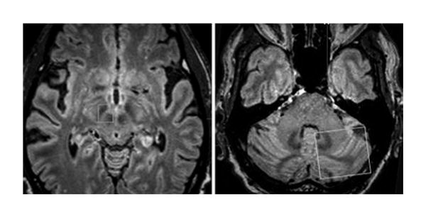
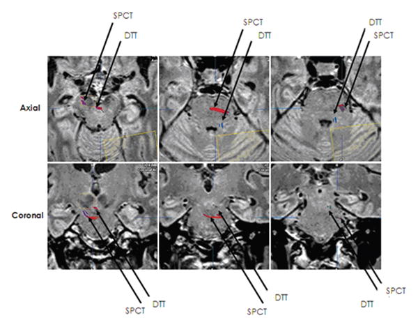
Object: Stimulation of white matter pathways near targeted structures may contribute to therapeutic effects of deep brain stimulation (DBS) for patients with Parkinson disease (PD). Two tracts linking the basal ganglia and cerebellum have been described in primates: the subthalamopontocerebellar tract (SPCT) and the dentatothalamic tract (DTT). The authors used fiber tractography to evaluate white matter tracts that connect the cerebellum to the region of the basal ganglia in patients with PD who were candidates for DBS.
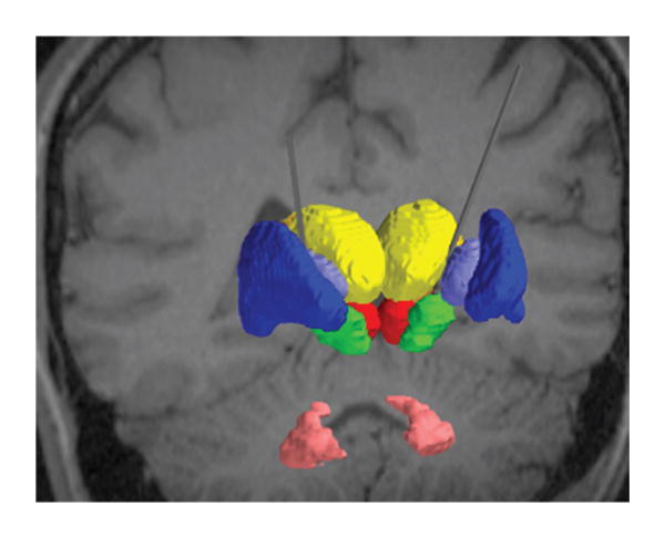
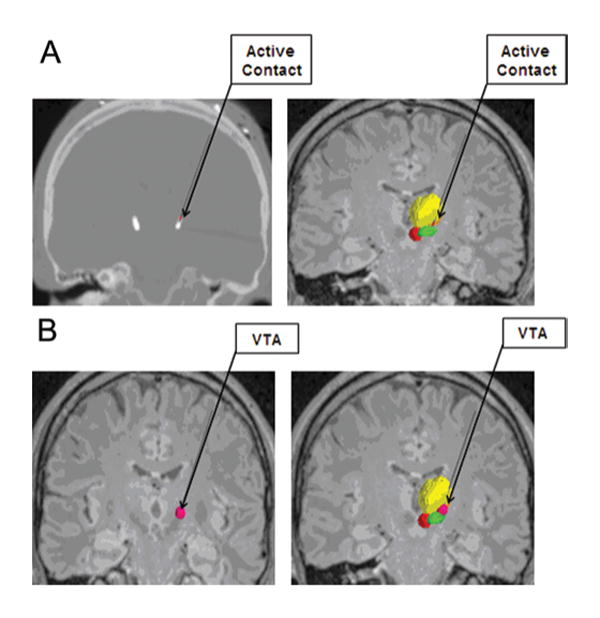
Methods: Fourteen patients with advanced PD underwent 3-T MRI, including 30-directional diffusion-weighted imaging sequences. Diffusion tensor tractography was performed using 2 regions of interest: ipsilateral subthalamic and red nuclei, and contralateral cerebellar hemisphere. Nine patients underwent subthalamic DBS, and the course of each tract was observed relative to the location of the most effective stimulation contact and the volume of tissue activated.
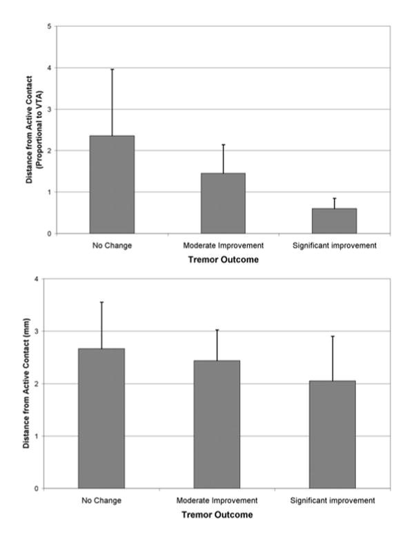
Results: In all patients 2 distinct tracts were identified that corresponded closely to the described anatomical features of the SPCT and DTT, respectively. The mean overall distance from the active contact to the DTT was 2.18 ± 0.35 mm, and the mean proportional distance relative to the volume of tissue activated was 1.35 ± 0.48. There was a nonsignificant trend toward better postoperative tremor control in patients with electrodes closer to the DTT.
Conclusions: The SPCT and the DTT may be related to the expression of symptoms in PD, and this may have implications for DBS targeting. The use of tractography to identify the DTT might assist with DBS targeting in the future.
Conflict of interest statement
Figures
Comment in
-
Letters to the editor: the cerebellum and Parkinson's disease.J Neurosurg. 2014 Aug;121(2):494-5. doi: 10.3171/2014.3.JNS14544. Epub 2014 Jun 27. J Neurosurg. 2014. PMID: 24972127 No abstract available.
-
Response.J Neurosurg. 2014 Aug;121(2):495. J Neurosurg. 2014. PMID: 25221808 No abstract available.
References
-
- Amtage F, Henschel K, Schelter B, Vesper J, Timmer J, Lucking CH, et al. Tremor-correlated neuronal activity in the subthalamic nucleus of Parkinsonian patients. Neurosci Lett. 2008;442:195–199. - PubMed
-
- Coenen VA, Mädler B, Schiffbauer H, Urbach H, Allert N. Individual fiber anatomy of the subthalamic region revealed with diffusion tensor imaging: a concept to identify the deep brain stimulation target for tremor suppression. Neurosurgery. 2011;68:1069–1076. - PubMed
MeSH terms
Grants and funding
LinkOut - more resources
Full Text Sources
Other Literature Sources
Medical
