. 2014 Mar;11(3):232-8.
doi: 10.2174/1567205011666140131120922.

Myelin injury and degraded myelin vesicles in Alzheimer's disease

Affiliations

Myelin injury and degraded myelin vesicles in Alzheimer's disease

Xinhua Zhan et al. Curr Alzheimer Res. 2014 Mar.

Abstract

Objective: Myelin disruption is an important feature of Alzheimer's disease (AD) that contributes to impairment of neuronal circuitry and cognition. In this study we characterize myelin degradation in the brains of patients with Alzheimer's disease compared with normal aged controls.

Methods: Myelin from patients with AD (n=13) was compared to matched controls (n=6). Myelin degradation was examined by immunohistochemistry in frontal white matter (WM) for intact myelin basic protein (MBP), degraded MBP, the presence of myelin lipid and for PAS staining. The relationship of myelin degradation and axonal injury was also assessed.

Results: Brains from patients with AD had significant loss of intact MBP, and an increase in degraded MBP in periventricular WM adjacent to a denuded ependymal layer. In regions of myelin degradation, vesicles were identified that stained positive for degraded MBP, myelin lipid, and neurofilament but not for intact MBP. Most vesicles stained for PAS, a corpora amylacea marker. The vesicles were significantly more abundant in the periventricular WM of AD patients compared to controls (44.5 ± 11.0 versus 1.7 ± 1.1, p=0.02).

Conclusion: In AD patients degraded MBP is associated in part with vesicles particularly in periventricular WM that is adjacent to areas of ependymal injury.


Figures
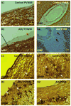
Fig. 1
Fig. 1. Myelin loss and degraded myelin vesicles in the white matter of Alzheimer’s disease (AD) brains
In controls, intact MBP staining was observed in the PVWM (Periventricular White Matter) (A1) and around the white matter of PVSR (Perivascular Region of white matter) (A2). In PVWM of AD brains, there was loss of ependymal cells adjacent to the ventricle (B1). There was marked decrease of staining for intact MBP in the PVWM adjacent to the denuded ependyma (B1, red arrow). Note more intact ependymal cells to the left of the black line along with intact MBP staining being closer to the ependymal layer (B1). There was decreased MBP staining in the PVSR of AD brains (B2) and vesicles in these regions did not stain for intact MBP (B2, arrows). An antibody specific for degraded MBP (dMBP) was used to detect degraded myelin in AD and control brains. In areas of decreased overall MBP staining, dMBP immunostaining was detected mainly in different types of vesicles: solid (D1 and D2, white arrows), clear (D1 and D2, black arrows), and a complex morphology (D1 and D2, red arrows). In control, few dMBP+ vesicles were detected (C1 and C2, arrows) in PVWM. These “degraded myelin vesicles” varied in sizes from 5 μm to 25 μm. Brown = positive staining, MBP = intact myelin basic protein, dMBP = degraded MBP, AD= Alzheimer’s disease. Bars = 25μm.
Fig. 2
Fig. 2. Identification of “degraded myelin vesicles” in regions of myelin loss in AD brains
Double labeling showed that in the area absent of intact-MBP (A1, arrows), degraded MBP+ vesicles (A2 and A3, arrows) were observed. In addition, in the area absent of intact-MBP (B1 and C1, arrows), a specific axonal marker, NF was stained in the vesicles (B2 and B3, PVWM; C2 and C3, PVSR) were observed. MBP = intact myelin basic protein, dMBP = degraded MBP, NF = neurofilament, PVWM = periventricular white matter, PVSR = perivascular region of white matter. Bars = 25μm.
Fig 3
Fig 3. Co-localization of NF and dMBP in the vesicles of AD brains
Double labeling showed that NF (A1, B1 and C1) and dMBP (A2, B2 and C2) were co-localized in the same vesicles (A3, B3 and C3). dMBP = degraded MBP, NF = neurofilament, PVWM = periventricular white matter, DWM = deep white matter, PVSR = perivascular region of white matter. Bars = 25μm.
Fig 4
Fig 4. Localization of myelin lipid in the vesicles in AD brains
Galactocerebroside (GALC), a myelin lipid, was localized in the vesicles of PVWM (A), vesicles in DWM (B) and vesicles in PVSR (C) in AD brains. In age matched control brains there were fewer GALC+ vesicles in PVWM (D), DWM (E) and PVSR (F). Note that the GALC+ ependymal cells were lost in the AD brain (A) but GALC+ ependymal cells were present in control (D). Brown = positive staining, AD = Alzheimer’s disease, PVWM = periventricular white matter, DWM = deep white matter, PVSR = perivascular region of white matter. Bars = 25μm.
Fig 5
Fig 5. Localization of myelin lipid in the degraded myelin vesicles in AD brains
Galactocerebroside (GALC) (A1 and B1), a myelin lipid, was localized in the dMBP+ vesicles (A2, A3, B2 and B3) of PVWM (A1, A2,A3) and PVSR (B1, B2, B3) in AD brains. PVWM = periventricular white matter. PVSR = perivascular region of white matter, Bars = 25μm.
Fig 6
Fig 6. Double staining of dMBP and PAS in AD brains
dMBP+ vesicles (A1, B1, white arrows) often stained for PAS, a corpora amylacea marker (A2, B2, black arrows) that appeared to be co-localized (A3, B3). Occasional dMBP stained vesicles (A1, yellow arrow) did not stain for PAS (A2, A3 yellow arrows). Note that in some large and intensely-stained dMBP+ vesicles (B1, gray arrow), PAS staining was weak (B2, gray arrow) but did co-localize with dMBP (B3, gray arrow). Occasional PAS stained vesicles (B2, B3, orange arrows) did not stain for dMBP (B1, orange arrow). PAS not only stained vesicles, but also stained vessels (B2 and B3, blue arrows), which were negative for dMBP (B1). PVWM = periventricular white matter; PVSR = perivascular regions of white matter; dMBP, degraded myelin basic protein; PAS, periodic acid-Schiff. Bars = 25μm.

References

    1. Wallin A, Gottfries CG, Karlsson I, Svennerholm L. Decreased myelin lipids in Alzheimer’s disease and vascular dementia. Acta Neurol Scand. 1989;80:319–23. - PubMed
    1. Bartzokis G, Cummings JL, Sultzer D, Henderson VW, Nuechterlein KH, Mintz J. White matter structural integrity in healthy aging adults and patients with Alzheimer disease: a magnetic resonance imaging study. Arch Neurol. 2003;60:393–8. - PubMed
    1. Chia LS, Thompson JE, Moscarello MA. X-ray diffraction evidence for myelin disorder in brain from humans with Alzheimer’s disease. Biochim Biophys Acta. 1984;775:308–12. - PubMed
    1. de la Monte SM. Quantitation of cerebral atrophy in preclinical and end-stage Alzheimer’s disease. Ann Neurol. 1989;25:450–9. - PubMed
    1. Englund E, Brun A, Alling C. White matter changes in dementia of Alzheimer’s type. Biochemical and neuropathological correlates. Brain. 1988;111 (Pt 6):1425–39. - PubMed

Publication types

Substances