. 2014 Jul;51(1):114-24.
doi: 10.1165/rcmb.2012-0179OC.

Dihydrotestosterone potentiates EGF-induced ERK activation by inducing SRC in fetal lung fibroblasts

Affiliations

Dihydrotestosterone potentiates EGF-induced ERK activation by inducing SRC in fetal lung fibroblasts

Matt K Lee et al. Am J Respir Cell Mol Biol. 2014 Jul.

Abstract

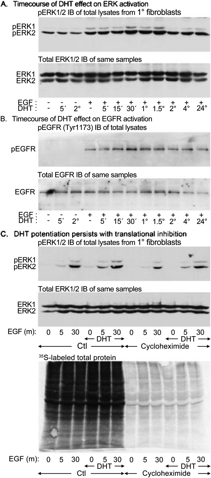
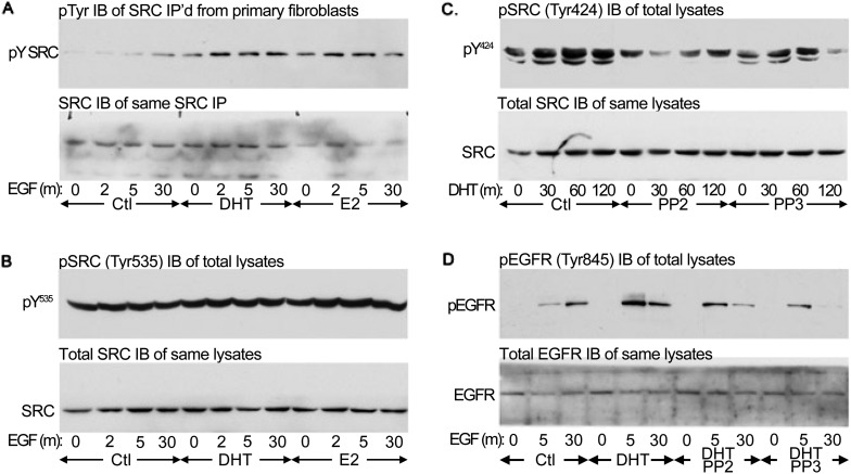
Lung maturation is regulated by interactions between mesenchymal and epithelial cells, and is delayed by androgens. Fibroblast-Type II cell communications are dependent on extracellular signal-regulated kinases (ERK) 1/2 activation by the ErbB receptor ligands epidermal growth factor (EGF), transforming growth factor (TGF)-α, and neuregulin (Nrg). In other tissues, dihydrotestosterone (DHT) has been shown to activate SRC by a novel nontranscriptional mechanism, which phosphorylates EGF receptors to potentiate EGF-induced ERK1/2 activation. This study sought to determine if DHT potentiates EGFR signaling by a nontranscriptional mechanism. Embryonic day (E)17 fetal lung cells were isolated from dams treated with or without DHT since E12. Cells were exposed to 30 ng/ml DHT for periods of 30 minutes to 3 days before being stimulated with 100 ng/ml EGF, TGF-α, or Nrg for up to 30 minutes. Lysates were immunoblotted for ErbB and SRC pathway signaling intermediates. DHT increased ERK1/2 activation by EGF, TGF-α, and Nrg in fibroblasts and Type II cells. Characterization in fibroblasts showed that potentiation of the EGF pathway was significant after 60 minutes of DHT exposure and persisted in the presence of the translational inhibitor cycloheximide. SRC and EGF receptor phosphorylation was increased by DHT, as was EGF-induced SHC1 phosphorylation and subsequent association with GRB2. Finally, SRC silencing, SRC inhibition with PP2, and overexpression of a dominant-negative SRC each prevented DHT from increasing EGF-induced ERK1/2 phosphorylation. These results suggest that DHT activates SRC to potentiate the signaling pathway leading from the EGF receptor to ERK activation in primary fetal lung fibroblasts.

Keywords: ErbB; MAP kinases; androgens; fibroblasts.


Figures

Figure 1.
Figure 1.
Dihydrotestosterone (DHT) increases ErbB-induced extracellular signal-regulated (ERK) activation in primary canalicular stage Type II cells and fibroblasts. Fibroblasts and Type II cells continuously exposed to DHT since embryonic day (E)12 were isolated from E17 lungs, cultured in the presence of DHT until 80% confluent, and then stimulated with epidermal growth factor (EGF), transforming growth factor (TGF)-α, or neuregulin (Nrg) at 100 ng/ml for the indicated times. The resulting phosphorylation of ERK1 and ERK2 was detected by immunoblotting and normalized against total ERK1 and ERK 2 within the same samples. Images shown are paired phosphoERK1/2 and total ERK1/2 immunoblots of the same samples but not necessarily of the same membrane. EGF induced fibroblast ERK1/2 phosphorylation regardless of the presence or absence of DHT (A). However, DHT significantly increased EGF responsiveness (densitometric quantitation and statistical comparisons are provided as Figure E1). (B) TGF-α also activated fibroblast ERK1 and ERK2. This activation was significantly increased by DHT. (C) Nrg had nonsignificant effects on fibroblast ERK1/2 induction in the absence of DHT. In its presence, Nrg responsiveness increased and became significant. DHT had similar effects on primary fetal Type II cells. In the absence of DHT, EGF significantly activated ERK1 but not ERK2 (D). DHT treatment increased EGF-induced phosphorylation of ERK1 and ERK2 so that in its presence both were induced by EGF. (E) TGF-α conversely phosphorylated ERK2 but not ERK1. DHT treatment also increased TGF-α–induced phosphorylation of ERK1 and ERK2. (F) Nrg had insignificant effects on ERK1 and ERK2 phosphorylation in Type II cells. DHT treatment increased responsiveness to Nrg so that in its presence both ERKs were activated.
Figure 2.
Figure 2.
DHT potentiation of EGF-induced ERK induction in fibroblasts requires relatively short DHT exposure and is independent of protein translation. DHT exposure potentiates ErbB responsiveness, but the mechanism is unclear. To begin characterization, the minimum duration of DHT exposure necessary to potentiate ERK activation was determined. Pulmonary fibroblasts were isolated from untreated E17 mice and exposed to 30 ng/ml DHT for up to 24 hours before uniform stimulation with 100 ng/ml EGF for 5 minutes. Phosphorylated ERK1/2 (pERK1 and pERK2) was detected by immunoblotting and normalized to total ERK1 and ERK2 content. (A) DHT potentiated EGF-induced ERK phosphorylation within 1 hour. Densitometric and statistical analyses are provided in Figure E2. (B) phosphorylated and total EGF receptor (pEGFR and EGFR) immunoblots of the same experiment. Normalized EGF-induced EGFR phosphorylation was also increased by 1 hour of DHT, consistent with receptor-level potentiation of EGF signaling. (C) The requirement for de novo protein synthesis in ERK potentiation by DHT was evaluated. Previously untreated primary pulmonary fibroblasts were exposed to the translational inhibitor cycloheximide (1 μg/ml) for 1 hour before the addition of 30 ng/ml DHT. After 1 hour, cells were stimulated with EGF, 100 ng/ml for 5 or 30 minutes. Top: pERK1/2 immunoblot of lysates showing that DHT potentiation of EGF-induced ERK phosphorylation persists in the presence of cycloheximide. The total ERK1/2 reprobe of the same blot is underneath. Bottom: Autoradiograph of resolved E17 fetal lung mesenchymal cell lysates treated identically with cycloheximide and DHT in the presence of 35S-methionine.
Figure 3.
Figure 3.
DHT increases SRC phosphorylation and potentiates the ligand-dependent activation of the EGFR. EGF-induced SRC and EGFR pathway activation is potentiated by DHT. Previously untreated primary fetal lung fibroblasts were stimulated with 100 mg/ml EGF for 0, 2, 5, or 30 minutes with or without a prior 1-hour exposure to 30 ng/ml DHT or 30 ng/ml estradiol (E2). Phosphoprotein blots are paired with total protein immunoblots of the same samples but not necessarily of the same membrane. (A, top) Immunoblot of SRC phosphorylated at Tyr424 in the kinase domain (Y424). Phosphorylation at this site correlates with SRC activation. (A, bottom) Total SRC immunoblot of the same lysates. Densitometric analyses are provided in Figure E3. EGF-induced SRC Tyr424 phosphorylation is significantly higher in DHT-treated cells than in controls (Ctl) or E2-treated cells. (B) EGF-induced EGFR phosphorylation is potentiated by DHT. Primary fibroblasts were similarly stimulated with EGF in the presence or absence of DHT or E2. Top: Immunoblot of EGFR phosphorylated at the Tyr1173 SHC1 binding site. Phosphorylation of SHC1 by EGFR mediates ERK1/2 activation. Bottom: Total EGFR immunoblot of the same lysates. EGF-induced EGFR phosphorylation is significantly higher in DHT-treated cells than in controls or E2-treated cells. (C) EGF-induced SHC1 phosphorylation and GRB2 association are increased by DHT. Fetal lung fibroblasts were treated with DHT and stimulated with EGF as previously described. The lysates were then equalized by protein content before SHC1 immunoprecipitation. The activation of SHC1, a substrate of EGFR and initiator of the ERK1/2 pathway, was then assessed by probing the precipitates for GRB2 and for phosphotyrosine. Top: Phosphotyrosine immunoblot showing phosphorylation of the p66, p52, and p46 SHC1 isoforms. Middle: GRB2 immunoblot of SHC1 immunoprecipitated from the same experiment. Bottom: SHC1 immunoblot of the same precipitates. EGF-induced p52SHC1 phosphorylation was significantly higher in DHT-treated cells relative to untreated or E2-treated cells. EGF-induced GRB2 coimmunoprecipitation, normalized to immunoprecipitated SHC1, was also increased by DHT relative to controls. (D) EGF-induced ERK1/2 phosphorylation is more potently potentiated by DHT than by E2. Fibroblasts were again stimulated with EGF with or without a prior 1-hour exposure to 30 ng/ml DHT or E2. ERK1 and ERK2 phosphorylation were assessed as previously described. Top: pERK1/2 immunoblot. Bottom: Total ERK1/2 immunoblot of the same samples. EGF-induced ERK1 phosphorylation was significantly higher in DHT-treated cells than in control or E2-treated cells. EGF-induced ERK2 phosphorylation was significantly higher in DHT-treated cells than in controls and approached significance compared with E2-treated cells (P = 0.055).
Figure 4.
Figure 4.
SRC activation by DHT is consistent with direct binding. SRC may be activated by dephosphorylating Tyr535 or by disrupting its intramolecular bonding. Androgen receptors putatively bind the SH3 domain to unmask the kinase domain. Releasing this inhibition enables SRC to autophosphorylate on multiple residues. Of these, Tyr424 is necessary for full activation. Activated SRC then associates with the EGFR and, in the presence of EGF ligand, induces its phosphorylation at Tyr845. To assess the mechanism of activation, specific and global tyrosine phosphorylation of SRC and its EGFR substrate were assessed. The corresponding densitometric analyses are presented in Figure E4. Primary fibroblasts were exposed to DHT or E2 for 1 hour before stimulation with up to 30 minutes EGF as previously described. The SRC was immunoprecipitated, and the isolates were immunoblotted for phosphotyrosine. (A) Phosphotyrosine blot of SRC immunoprecipitates (IP) (top) and SRC immunoblot (IB) of the same samples (bottom). EGF-induced SRC tyrosine phosphorylation, and, hence, activation is increased by DHT and, to a lesser extent, by E2. Global tyrosine phosphorylation parallels Tyr424 phosphorylation. (B) Immunodetection of SRC Tyr535 (top) and SRC immunoblot of the same samples (bottom). Phosphorylation of Tyr535 is not decreased by DHT or E2. (C) PhosphoTyr424 immunoblot (top) and SRC immunoblot (below) of cells exposed to DHT in the absence of EGF and in the presence of the SRC inhibitor PP2 or its control, PP3. DHT significantly increases Tyr424 phosphorylation independently of EGF in control cells and in cells exposed to PP3 but not in cells treated with PP2. This suggests that SRC is activated by DHT in a manner that requires SRC kinase activity. (D) EGFR Tyr845 immunoblot of cells exposed to DHT in the presence of PP2 or PP3 and then stimulated with EGF. EGF-induced EGFR Tyr845 phosphorylation is potentiated by DHT only in the absence of PP2 or PP3. Because PP3 attenuates EGFR activity, this result confirms that SRC and EGFR kinase activities are both necessary for DHT potentiation.
Figure 5.
Figure 5.
Src inhibition prevents DHT from enhancing the activity of the EGFR. (AC) PP2 inhibition of SRC prevents DHT from potentiating EGF signaling. Previously untreated primary fetal lung fibroblasts were exposed to PP2 before DHT treatment and EGF stimulation as previously described. Lysates were immunoblotted for phosphorylated and total signaling intermediates; densitometric analyses are presented in Figure E5. (A) Phosphorylated ERK1/2 and total ERK1/2 immunoblots of the same samples. ERK activation is potentiated by DHT in the absence but not in the presence of PP2. (B) EGFR phosphoTyr1173 and total EGFR immunoblots of the same samples. EGFR activation is potentiated by DHT in the absence but not in the presence of PP2. (C) SRC phosphoTyr424 and total SRC immunoblots of the same samples. SRC activation is potentiated by DHT in the absence but not in the presence of PP2. (D) Src silencing prevents DHT from enhancing EGF signaling. Primary fetal lung fibroblasts were transfected with small interfering RNA (siRNA) against Src or Gapdh before DHT treatment and EGF stimulation. Lysates were immunoblotted for phosphorylated and total ERK1/2 (top pair of images) and for phosphorylated EGFR Tyr1173 and total EGFR (bottom pair of images). EGF-induced ERK phosphorylation was potentiated by DHT only in cells transfected with Gapdh siRNA, and DHT-associated ERK phosphorylation was significantly lower in cells transfected with Src siRNA. Similarly, EGF-induced receptor phosphorylation was potentiated by DHT only in the presence of Gapdh silencing and was significantly lower when Src was silenced. The efficiency of Src silencing was confirmed by immunoblotting the same samples for SRC (bottom). By densitometry, SRC content was reduced by 58% (SD = 9%). (E) overexpression of dominant–negative (DN) SRC prevents DHT from potentiating EGF signaling. Primary fibroblasts were infected with adenoviruses encoding green fluorescent protein (GFP) or a DN Src. Cells were inoculated 2 days before exposure to DHT and EGF as previously described. DN Src expression was confirmed by SRC immunoblotting (bottom). DHT potentiated EGF-induced ERK activation in cells expressing GFP but not in cells expressing DN Src. EGF-induced ERK2 phosphorylation in the presence of DHT was significantly lower in cells expressing DN Src than in GFP controls. ERK1 phosphorylation was also decreased by DN Src but did not achieve significance (P = 0.062).

References

    1. Torday JS, Nielsen HC, Fencl MM, Avery ME. Sex differences in fetal lung maturation. Am Rev Respir Dis. 1981;123:205–208. - PubMed
    1. Miller HC, Futrakul P. Birth weight, gestational age, and sex as determining factors in the incidence of respiratory distress syndrome of prematurely born infants. J Pediatr. 1968;72:628–635. - PubMed
    1. Farrell PM, Avery ME. Hyaline membrane disease. Am Rev Respir Dis. 1975;111:657–688. - PubMed
    1. Nielsen HC, Zinman HM, Torday JS. Dihydrotestosterone inhibits fetal rabbit pulmonary surfactant production. J Clin Invest. 1982;69:611–616. - PMC - PubMed
    1. Gross I, Wilson CM, Ingleson LD, Brehier A, Rooney SA. The influence of hormones on the biochemical development of fetal rat lung in organ culture: I. Estrogen. Biochim Biophys Acta. 1979;575:375–383. - PubMed

Publication types

MeSH terms

Substances