

Inter-study reproducibility of arterial spin labelling magnetic resonance imaging for measurement of renal perfusion in healthy volunteers at 3 Tesla
- PMID: 24484613
- PMCID: PMC3909760
- DOI: 10.1186/1471-2369-15-23
Inter-study reproducibility of arterial spin labelling magnetic resonance imaging for measurement of renal perfusion in healthy volunteers at 3 Tesla
Abstract
Background: Measurement of renal perfusion is a crucial part of measuring kidney function. Arterial spin labelling magnetic resonance imaging (ASL MRI) is a non-invasive method of measuring renal perfusion using magnetised blood as endogenous contrast. We studied the reproducibility of ASL MRI in normal volunteers.
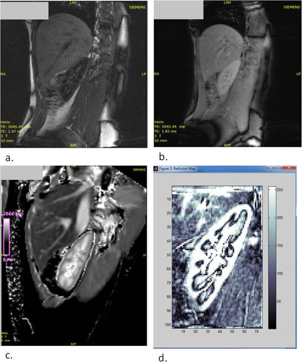
Methods: ASL MRI was performed in healthy volunteers on 2 occasions using a 3.0 Tesla MRI scanner with flow-sensitive alternating inversion recovery (FAIR) perfusion preparation with a steady state free precession (True-FISP) pulse sequence. Kidney volume was measured from the scanned images. Routine serum and urine biochemistry were measured prior to MRI scanning.
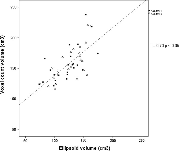
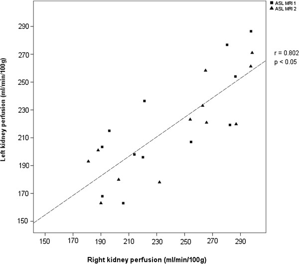
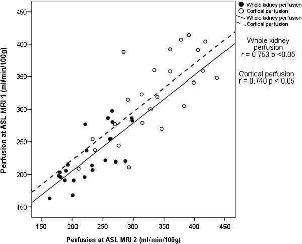
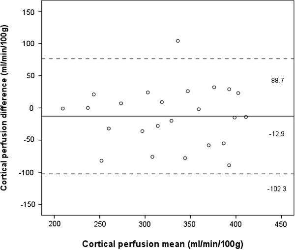
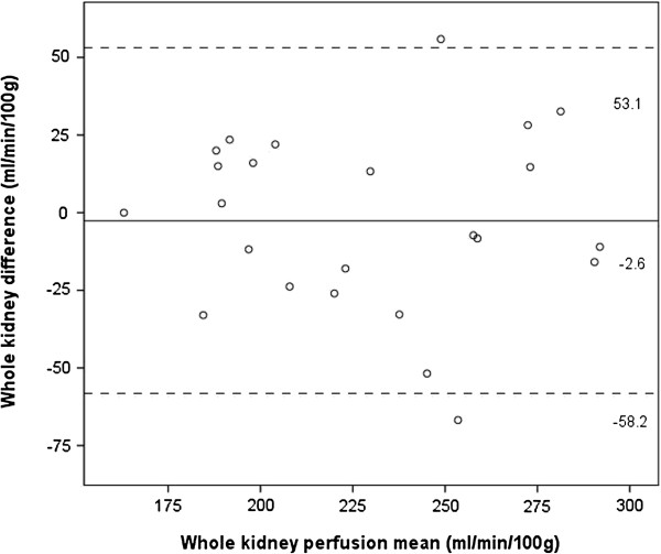
Results: 12 volunteers were recruited yielding 24 kidneys, with a mean participant age of 44.1 ± 14.6 years, blood pressure of 136/82 mmHg and chronic kidney disease epidemiology formula estimated glomerular filtration rate (CKD EPI eGFR) of 98.3 ± 15.1 ml/min/1.73 m2. Mean kidney volumes measured using the ellipsoid formula and voxel count method were 123.5 ± 25.5 cm3, and 156.7 ± 28.9 cm3 respectively. Mean kidney perfusion was 229 ± 41 ml/min/100 g and mean cortical perfusion was 327 ± 63 ml/min/100 g, with no significant differences between ASL MRIs. Mean absolute kidney perfusion calculated from kidney volume measured during the scan was 373 ± 71 ml/min. Bland Altman plots were constructed of the cortical and whole kidney perfusion measurements made at ASL MRIs 1 and 2. These showed good agreement between measurements, with a random distribution of means plotted against differences observed. The intra class correlation for cortical perfusion was 0.85, whilst the within subject coefficient of variance was 9.2%. The intra class correlation for whole kidney perfusion was 0.86, whilst the within subject coefficient of variance was 7.1%.
Conclusions: ASL MRI at 3.0 Tesla provides a repeatable method of measuring renal perfusion in healthy subjects without the need for administration of exogenous compounds. We have established normal values for renal perfusion using ASL MRI in a cohort of healthy volunteers.
Figures
References
-
- Cole BR, Giangiacomo J, Ingelfinger JR, Robson AM. Measurement of renal function without urine collection. A critical evaluation of the constant-infusion technic for determination of inulin and para-aminohippurate. N Engl J Med. 1972;287(22):1109–1114. doi: 10.1056/NEJM197211302872202. - DOI - PubMed
-
- Unlicensed Medicinal Products for Human Use (Transmissable Spongiform Encephalopathies)(Safety) Regulations. UK; 2003.
Publication types
MeSH terms
Substances
LinkOut - more resources
Full Text Sources
Other Literature Sources
Research Materials
Miscellaneous
