

Quantitative characterization of mineralized silk film remodeling during long-term osteoblast-osteoclast co-culture
- PMID: 24484674
- PMCID: PMC3964259
- DOI: 10.1016/j.biomaterials.2014.01.034
Quantitative characterization of mineralized silk film remodeling during long-term osteoblast-osteoclast co-culture
Abstract
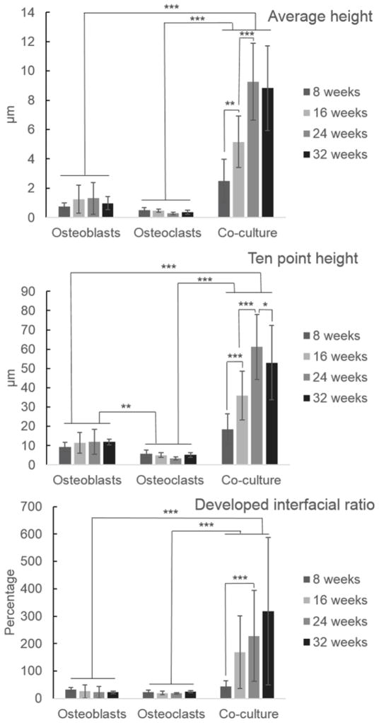
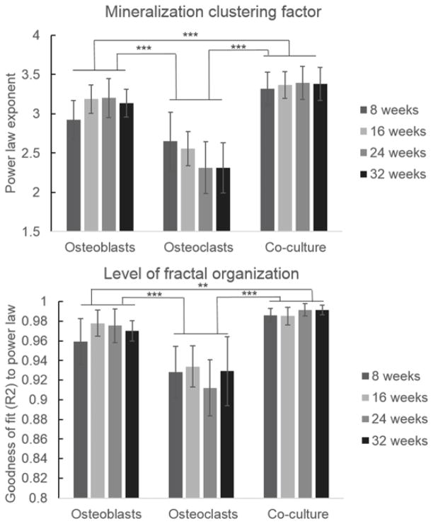
The goal of this study was to explore quantitative assessments of mineralized silk protein biomaterial films by co-cultures of human mesenchymal stem cell-derived osteoblasts and human acute monocytic leukemia cell line-derived osteoclasts during long-term culture (8-32 weeks). The remodeled films were quantitatively assessed using three different techniques during this extended cultivation to provide more comprehensive insight into the impact of co-cultures on surface remodeling. Scanning electron microscopy (SEM) with three dimensional surface reconstructions was used to quantitatively determine various surface morphological features and measures of roughness indicative of remodeling by the cells. Additionally, reconstructed surfaces were converted to depth images for Fourier analysis to quantify the potential fractal organization of biomineralization. The long-term remodeled films were also imaged using confocal reflectance microscopy and micro-computed tomography (micro-CT) to further quantify morphological changes. Films remodeled in co-culture demonstrated increased roughness parameters, fractal organization, and volume compared to films remodeled by osteoblasts alone. The combination of these techniques to quantify remodeling of mineralized protein films shows promise for quantifying processes related to mineralized surfaces.
Keywords: Bone remodeling; Bone tissue engineering; Confocal microscopy; Scanning electron microscopy; Silk; Surface topography.
Copyright © 2014 Elsevier Ltd. All rights reserved.
Figures


Similar articles
-
Effects of clodronate and alendronate on osteoclast and osteoblast co-cultures on silk-hydroxyapatite films.Acta Biomater. 2014 Jan;10(1):486-93. doi: 10.1016/j.actbio.2013.09.028. Epub 2013 Oct 1. Acta Biomater. 2014. PMID: 24096150 Free PMC article.
-
Osteoblast: osteoclast co-cultures on silk fibroin, chitosan and PLLA films.Biomaterials. 2009 Oct;30(29):5376-84. doi: 10.1016/j.biomaterials.2009.07.028. Epub 2009 Aug 3. Biomaterials. 2009. PMID: 19647869
-
Cell-tethered ligands modulate bone remodeling by osteoblasts and osteoclasts.Adv Funct Mater. 2014 Jan 29;24(4):472-479. doi: 10.1002/adfm.201302210. Adv Funct Mater. 2014. PMID: 25419210 Free PMC article.
-
[Research propress of co-culture system of osteoblast with osteoclast and its applications].Zhongguo Gu Shang. 2013 Apr;26(4):349-53. Zhongguo Gu Shang. 2013. PMID: 23844502 Review. Chinese.
-
From the Clinical Problem to the Basic Research-Co-Culture Models of Osteoblasts and Osteoclasts.Int J Mol Sci. 2018 Aug 3;19(8):2284. doi: 10.3390/ijms19082284. Int J Mol Sci. 2018. PMID: 30081523 Free PMC article. Review.
Cited by
-
Zinc Sulfate Stimulates Osteogenic Phenotypes in Periosteum-Derived Cells and Co-Cultures of Periosteum-Derived Cells and THP-1 Cells.Life (Basel). 2021 Apr 30;11(5):410. doi: 10.3390/life11050410. Life (Basel). 2021. PMID: 33946199 Free PMC article.
-
Tuning the resorption-formation balance in an in vitro 3D osteoblast-osteoclast co-culture model of bone.Bone Rep. 2022 Dec 12;18:101646. doi: 10.1016/j.bonr.2022.101646. eCollection 2023 Jun. Bone Rep. 2022. PMID: 36578830 Free PMC article.
-
A Comparison of Osteoblast and Osteoclast In Vitro Co-Culture Models and Their Translation for Preclinical Drug Testing Applications.Int J Mol Sci. 2020 Jan 30;21(3):912. doi: 10.3390/ijms21030912. Int J Mol Sci. 2020. PMID: 32019244 Free PMC article. Review.
-
Biomaterial Cues for Regulation of Osteoclast Differentiation and Function in Bone Regeneration.Adv Ther (Weinh). 2025 Jan;8(1):2400296. doi: 10.1002/adtp.202400296. Epub 2024 Nov 15. Adv Ther (Weinh). 2025. PMID: 39867107
-
Optimization of the Static Human Osteoblast/Osteoclast Co-culture System.Iran J Med Sci. 2018 Mar;43(2):208-213. Iran J Med Sci. 2018. PMID: 29749990 Free PMC article.
References
-
- Cuijpers VMJI, Walboomers XF, Jansen JA. Scanning electron microscopy stereoimaging for three-dimensional visualization and analysis of cells in tissue-engineered constructs: technical note. Tissue Eng Part C. 2011;17(6):663–8. - PubMed
-
- Dunn AK, Smithpeter C, Welch AJ, Richards-Kortum R. Sources of contrast in confocal reflectance imaging. Appl Opt. 1996;35(19):3441–6. - PubMed
Publication types
MeSH terms
Substances
Grants and funding
LinkOut - more resources
Full Text Sources
Other Literature Sources
