

A tunable silk-alginate hydrogel scaffold for stem cell culture and transplantation
- PMID: 24484675
- PMCID: PMC3971886
- DOI: 10.1016/j.biomaterials.2014.01.029
A tunable silk-alginate hydrogel scaffold for stem cell culture and transplantation
Abstract
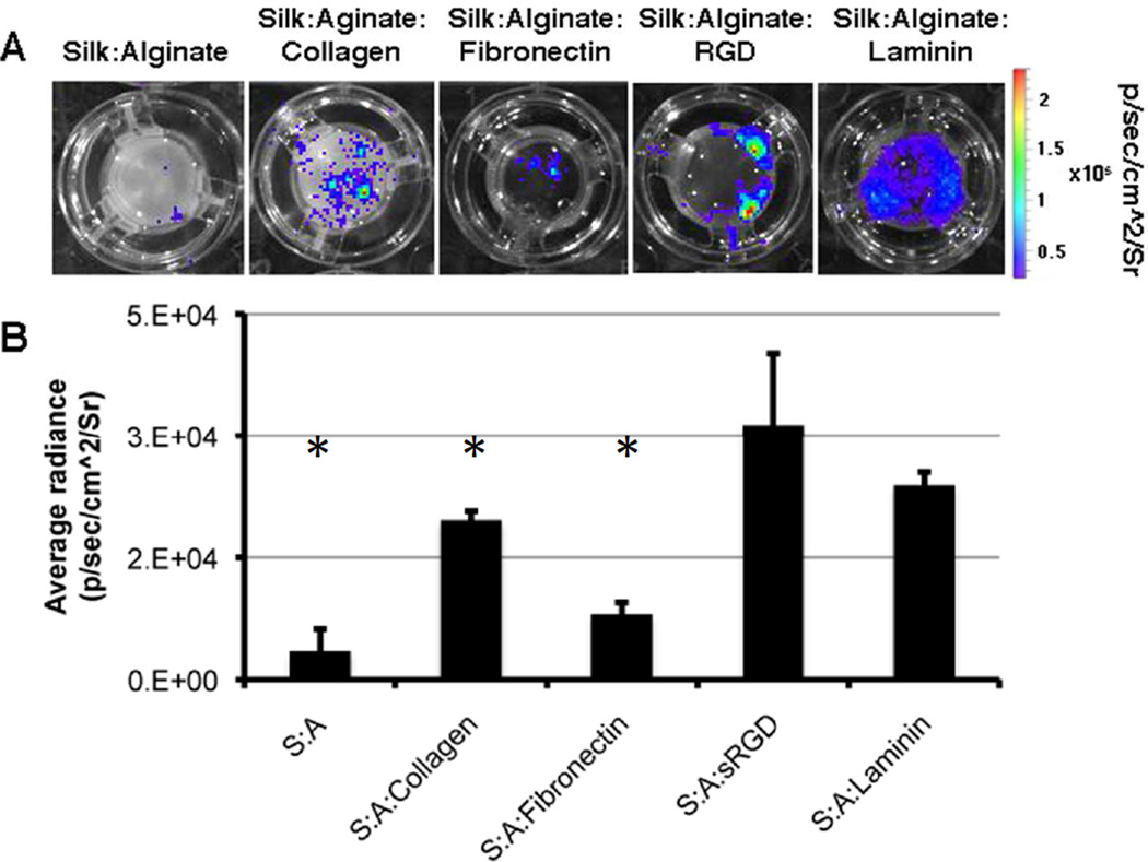
One of the major challenges in regenerative medicine is the ability to recreate the stem cell niche, which is defined by its signaling molecules, the creation of cytokine gradients, and the modulation of matrix stiffness. A wide range of scaffolds has been developed in order to recapitulate the stem cell niche, among them hydrogels. This paper reports the development of a new silk-alginate based hydrogel with a focus on stem cell culture. This biocomposite allows to fine tune its elasticity during cell culture, addressing the importance of mechanotransduction during stem cell differentiation. The silk-alginate scaffold promotes adherence of mouse embryonic stem cells and cell survival upon transplantation. In addition, it has tunable stiffness as function of the silk-alginate ratio and the concentration of crosslinker--a characteristic that is very hard to accomplish in current hydrogels. The hydrogel and the presented results represents key steps on the way of creating artificial stem cell niche, opening up new paths in regenerative medicine.
Keywords: Alginate; Elasticity; Laminin; Scaffold; Silk; Stem cells.
Copyright © 2014 Elsevier Ltd. All rights reserved.
Figures


Similar articles
-
The type and composition of alginate and hyaluronic-based hydrogels influence the viability of stem cells of the apical papilla.Dent Mater. 2014 Dec;30(12):e349-61. doi: 10.1016/j.dental.2014.08.369. Epub 2014 Aug 31. Dent Mater. 2014. PMID: 25182372
-
Evaluation of alginate hydrogel cytotoxicity on three-dimensional culture of type A spermatogonial stem cells.Int J Biol Macromol. 2017 Feb;95:888-894. doi: 10.1016/j.ijbiomac.2016.10.074. Epub 2016 Oct 27. Int J Biol Macromol. 2017. PMID: 27984148
-
Silk-Reinforced Collagen Hydrogels with Raised Multiscale Stiffness for Mesenchymal Cells 3D Culture.Tissue Eng Part A. 2020 Mar;26(5-6):358-370. doi: 10.1089/ten.TEA.2019.0199. Tissue Eng Part A. 2020. PMID: 32085691
-
Chondroinductive Alginate-Based Hydrogels Having Graphene Oxide for 3D Printed Scaffold Fabrication.ACS Appl Mater Interfaces. 2020 Jan 29;12(4):4343-4357. doi: 10.1021/acsami.9b22062. Epub 2020 Jan 17. ACS Appl Mater Interfaces. 2020. PMID: 31909967
-
Processing silk hydrogel and its applications in biomedical materials.Biotechnol Prog. 2015 May-Jun;31(3):630-40. doi: 10.1002/btpr.2058. Epub 2015 Mar 4. Biotechnol Prog. 2015. PMID: 25740113 Review.
Cited by
-
Enzymatically crosslinked silk and silk-gelatin hydrogels with tunable gelation kinetics, mechanical properties and bioactivity for cell culture and encapsulation.Biomaterials. 2020 Feb;232:119720. doi: 10.1016/j.biomaterials.2019.119720. Epub 2019 Dec 23. Biomaterials. 2020. PMID: 31896515 Free PMC article.
-
Easy Manipulation of Architectures in Protein-based Hydrogels for Cell Culture Applications.J Vis Exp. 2017 Aug 4;(126):55813. doi: 10.3791/55813. J Vis Exp. 2017. PMID: 28809838 Free PMC article.
-
Nanostructured platforms for the sustained and local delivery of antibiotics in the treatment of osteomyelitis.Crit Rev Ther Drug Carrier Syst. 2015;32(1):1-59. doi: 10.1615/critrevtherdrugcarriersyst.2014010920. Crit Rev Ther Drug Carrier Syst. 2015. PMID: 25746204 Free PMC article. Review.
-
Mechanical Characterization for Cellular Mechanobiology: Current Trends and Future Prospects.Front Bioeng Biotechnol. 2020 Nov 12;8:595978. doi: 10.3389/fbioe.2020.595978. eCollection 2020. Front Bioeng Biotechnol. 2020. PMID: 33282852 Free PMC article. Review.
-
Cytoprotection of Human Progenitor and Stem Cells through Encapsulation in Alginate Templated, Dual Crosslinked Silk and Silk-Gelatin Composite Hydrogel Microbeads.Adv Healthc Mater. 2022 Sep;11(17):e2200293. doi: 10.1002/adhm.202200293. Epub 2022 Jun 22. Adv Healthc Mater. 2022. PMID: 35686928 Free PMC article.
References
-
- Berthiaume F, Maguire TJ, Yarmush ML. Tissue engineering and regenerative medicine: history, progress, and challenges. Annu Rev Chem Biomol Eng. 2011;2:403–430. - PubMed
-
- Iglesias-Garcia O, Pelacho B, Prosper F. Induced pluripotent stem cells as a new strategy for cardiac regeneration and disease modeling. J Mol Cell Cardiol. 2013;62:43–50. - PubMed
Publication types
MeSH terms
Substances
Grants and funding
LinkOut - more resources
Full Text Sources
Other Literature Sources
