. 2014 May;8(3):581-95.
doi: 10.1016/j.molonc.2014.01.005. Epub 2014 Jan 16.

miR181b is induced by the chemopreventive polyphenol curcumin and inhibits breast cancer metastasis via down-regulation of the inflammatory cytokines CXCL1 and -2

Affiliations

miR181b is induced by the chemopreventive polyphenol curcumin and inhibits breast cancer metastasis via down-regulation of the inflammatory cytokines CXCL1 and -2

Emanuel Kronski et al. Mol Oncol. 2014 May.

Abstract

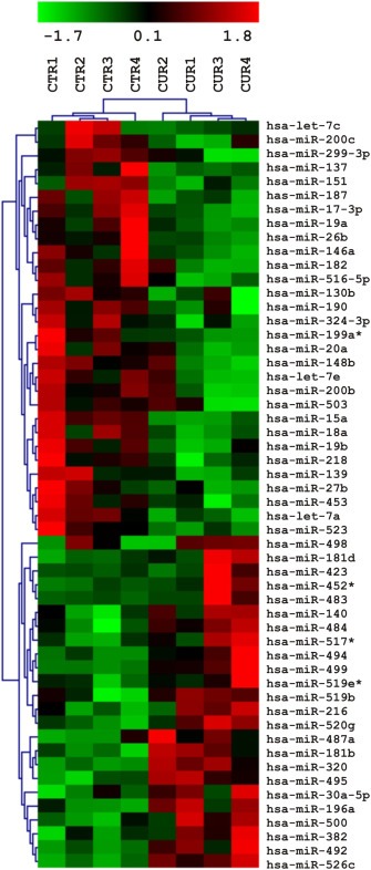
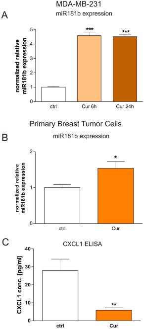
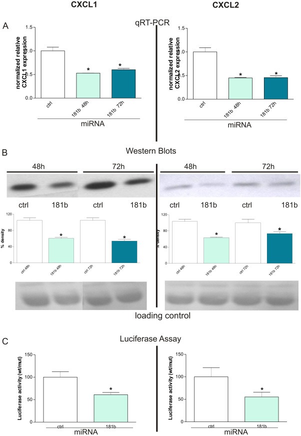
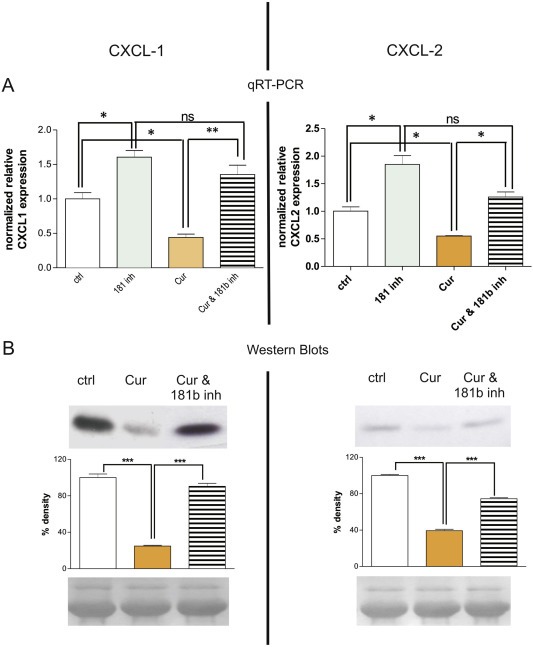
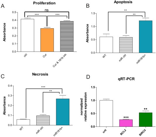
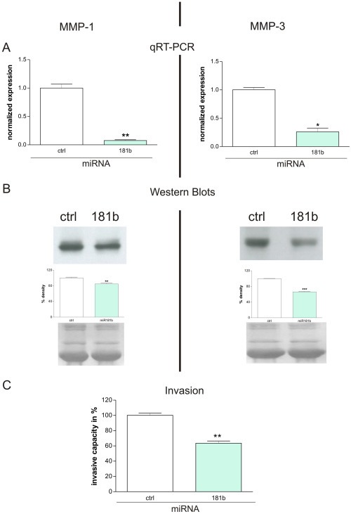
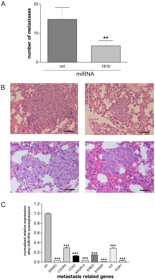
Chronic inflammation is a major risk factor for the development and metastatic progression of cancer. We have previously reported that the chemopreventive polyphenol Curcumin inhibits the expression of the proinflammatory cytokines CXCL1 and -2 leading to diminished formation of breast and prostate cancer metastases. In the present study, we have analyzed the effects of Curcumin on miRNA expression and its correlation to the anti-tumorigenic properties of this natural occurring polyphenol. Using microarray miRNA expression analyses, we show here that Curcumin modulates the expression of a series of miRNAs, including miR181b, in metastatic breast cancer cells. Interestingly, we found that miR181b down-modulates CXCL1 and -2 through a direct binding to their 3'-UTR. Overexpression or inhibition of miR181b in metastatic breast cancer cells has a significant impact on CXCL1 and -2 and is required for the effect of Curcumin on these two cytokines. miR181b also mediates the effects of Curcumin on inhibition of proliferation and invasion as well as induction of apoptosis. Importantly, over-expression of miR181b in metastatic breast cancer cells inhibits metastasis formation in vivo in immunodeficient mice. Finally, we demonstrated that Curcumin up-regulates miR181b and down-regulates CXCL1 and -2 in cells isolated from several primary human breast cancers. Taken together, these data show that Curcumin provides a simple bridge to bring metastamir modulation into the clinic, placing it in a primary and tertiary preventive, as well as a therapeutic, setting.

Keywords: Breast cancer; Curcumin; Inflammatory cytokines; Metastases prevention; microRNAs.


Figures

Figure 1
Figure 1
Curcumin modulates miRNA expression in human breast cancer cells. Microarray miRNA expression analysis. Four samples each of Curcumin treated (6 h) MDA‐MB‐231 cells and carrier‐treated controls were analyzed by hierarchical clustering applying euclidean distance and average linkage. Expression levels are indicated by a color code, green = expression below, red = expression above mean levels of expression, black = mean level.
Figure 2
Figure 2
Curcumin modulates miR181b expression in human breast cancer cells. A: Quantitative RT‐PCR reveals that human metastatic breast cancer cells (MDA‐MB‐231) treated with Curcumin for 6 h (“Cur”) express four fold higher amounts of miRNA181b in respect to carrier‐treated cells (“ctrl”). ***P < 0.001, student's t‐test. B: miR181b expression was analyzed in breast cancer cells from human primary tumor samples, cultured in vitro and treated for 24 h with Curcumin with respect to carrier‐treated control cells from the same origin. For each case, we observed up‐regulation of miR181b after Curcumin treatment (data not shown) and by pooling the results from all patient data we obtained a mean miR181b up‐regulation rate of 50%, which was statistically significant (*p < 0.05; student's t‐test). Mean + SD from 3 different patients are shown. C: CXCL1 protein expression secreted from breast cancer cells isolated from primary human tumors that were treated with Curcumin in vitro was analyzed by ELISA. CXCL1 concentrations were statistically significantly down‐regulated about 3.5‐fold in Curcumin treated tumor cells (**p < 0.01; student's t‐test). Mean + SD from 3 different patients are shown.
Figure 3
Figure 3
miR181b regulates CXCL1 expression through a direct binding to its 3′ UTR. A: Transient overexpression of miR181b in MDA‐MB‐231 breast cancer cells leads to a ∼50% down‐regulation of CXCL1 (left side) and CXCL2 (right side) transcripts as evidenced by qRT‐PCR (*P < 0.05, student's t‐test). Mean + SD from 3 independent experiments normalized to the respective controls are shown. B: On the corresponding protein level the same effect could be seen as indicated by Western Blot (upper panel) analysis 48 h and 72 h after transient introduction of miR181b into the tumor cells. Densitometric analysis of the bands (middle panel) reveals that CXCL1 (left side) and CXCL2 (right side) protein syntheses were statistically significantly (*P < 0.05, student's t‐test) impaired by about 50% 48 h after transfection. Ponceau staining was used as loading control to evaluate that equal amounts of proteins were applied (lower panel). Mean + SD from 3 independent experiments are shown. C: Luciferase assays demonstrated that regulation of CXCL1 and ‐2 occurs through direct binding of miR181b to their corresponding 3′UTRs. Co‐transfection of pGL3‐cont‐CXCL1 UTR‐wt or pGL3‐cont‐CXCL2 UTR‐wt together with miR‐181b, but not with a scrambled miR181b oligonucleotide, caused a decrease of the luciferase activity in He–La cells. Conversely, miR181b oligonucleotide did not inhibit luciferase activity of pGL3‐cont‐CXCL1 UTR‐mut or pGL3‐cont‐CXCL2 UTR‐mut. Mean + SD from 3 independent experiments are shown.
Figure 4
Figure 4
miR181b mediates Curcumin‐related down‐modulation of CXCL1 and CXCL2. A: qRT‐PCR results reveal that treatment of MDA‐MB‐231 cells with Curcumin for 24 h leads to inhibition of CXCL1 expression (left side, “Cur”) that can be reverted to original expression levels (“ctrl”) by concomitant application of a miR181b hairpin inhibitor (“Cur &181b inh”). Conversely, CXCL2 expression (right side) was only partially restored in cells treated with Curcumin plus the miR181b hairpin inhibitor, indicating that additional regulation mechanisms may exist. Inhibition of mir181b by specific small hairpin inhibitors leads to induction of CXCL1 and ‐2 expression compared to control cells transfected with scrambled unspecific hairpin inhibitors (*P < 0.05; **P < 0.001; ANOVA with Bonferroni's post‐test). Mean + SD from 3 independent experiments are shown. "ns”: not significant. B: The corresponding analysis on protein level (Western Blots, upper panel) confirm the data obtained from qRT‐PCR, strengthening the evidence that miR181b mediates down‐modulation of CXCL1 (left side) and CXCL2 (right side) by Curcumin. Densitometric analysis of the bands (middle panel) reveals that differences in expression levels of CXCL1 and ‐2 after Curcumin treatment were statistically significant (***P < 0.001; ANOVA with Bonferroni's post‐test). Loading controls (bottom panel) indicate that equal amounts of total protein were subjected to each lane of the gel. Mean + SD from 3 independent experiments are shown.
Figure 5
Figure 5
Involvement of miR181b in tumor cell proliferation, apoptosis and invasion. A: Curcumin inhibits proliferation via miR181b. Treatment of MDA‐MB‐231 breast cancer cells for 24 h with Curcumin (“Cur”) inhibits cell proliferation. However, treatment with Curcumin together with a specific miR181b hairpin‐inhibitor (“Cur & 181b inh”) abolishes the anti‐proliferative effect of Curcumin, returning cell proliferation to control‐levels. The effect of miR181b expression on proliferation was statistically highly significant as evidenced by student's t‐test (***P < 0.001). "ns”: not significant. B, C: miR181b overexpression induces apoptosis and necrosis in breast cancer cells. Functional apoptosis/necrosis assays revealed that miR181b over‐expression led to enhanced apoptosis (left panel) as well as necrosis (middle panel). 24 h after transfection with miR181b oligos apoptosis rate in MDA‐MB‐231 cells was doubled as compared to wildtype MDA‐MB‐231 cells or to MDA‐MB‐231 cells transfected with an appropriate control oligo (left panel). Likewise, necrosis rate was increased approximately 5 fold in miR181b over‐expressing MDA‐MB‐231 cells as compared to wildtype cells and over 2.5 fold as compared to MDA‐MB‐231 cells expressing an appropriate control oligo (**P < 0.01; ***P < 0.001; ANOVA with Bonferroni's post test). Mean + SD from 3 independent experiments are shown. D: Using quantitative RT‐PCR, expression of the apoptosis related factors BCL2 and survivin/BIRC5 in MDA‐MB‐231 over‐expressing miR181b showed a statistically significant down‐regulation achieved 72 h after transfection with a double stranded miR181b oligo as compared to the appropriate controls (right panel) (**P < 0.01 and ***P < 0.001; student's t‐test). Mean + SD (SEM?) from 3 independent experiments are shown.
Figure 6
Figure 6
miR181b impairs the expression of MMP‐1 and MMP‐3. A: qRT‐PCR shows that MMP‐1 (left side) and MMP‐3 (right side) transcripts were downregulated 93% (**P = 0.0059; student's t‐test) and 75% (*P = 0.0109; student's t‐test) respectively in MDA‐MB‐231 cells stably transfected with a miR181b overexpression vector (lanes indicated with 181b) when compared to cells stably transfected with an appropriate control vector (lanes indicated with ctrl). Mean + SD from 3 independent experiments are shown. B: Western blots (upper panel) of conditioned media from MDA‐MB‐231 cells stably transfected with a miR181b overexpression vector (lanes indicated with 181b), reveal a downregulation of MMP‐1 and ‐3 protein when compared to cells stably transfected with an appropriate control vector (lanes indicated with ctrl). This effect was quantified by subsequent densitometry (middle panel) showing that the differences in expression levels were about 15% (**P = 0.0068; student's t‐test) for MMP‐1 and about 34% (***P < 0.0001; student's t‐test) for MMP‐3. Equal amounts of total protein were loaded to each lane of the gels and to verify this, we visualized the protein bands blotted onto the nitrocellulose membranes (after the gel run) by Ponceau staining (lower panel). Mean + SD from 3 independent experiments are shown. C: miR181b over‐expression impairs the invasive capacity of breast cancer cells. Invasion of MDA‐MB‐231 breast cancer cells stably over‐expressing miR181b through a reconstituted basement membrane (Matrigel) was significantly reduced (**P < 0.01; student's t‐test) by about 50% after an incubation period of 6 h. Mean + SD from 3 independent experiments are shown.
Figure 7
Figure 7
Breast cancer cells over‐expressing miR181b have a lower capacity to metastasize in vivo. Breast cancer cells over‐expressing miR181b had a statistically significant (Mann–Whitney test, p = 0.0070) reduction in their capacity to metastasize into the lung of the animals (A). While the average number of metastasis in mice belonging to the control group was approximately 15 per animal, animals carrying MDA‐MB‐231miR1871b cells developed an average of only 6 metastases per animal. The tumor cell morphology showed characteristic atypia, significant expression of human p53 protein was observed only in human tumor cells. Vitality of the tumor cells was confirmed by a high number of proliferating Ki‐67 positive cells thus excluding tumor cell dormancy (B). Bars in the right lower corners of all photos are equivalent to 50 μm. Expression analysis (qRT‐PCR) of a series of metastasis‐related genes in MDA‐MB‐231 cells transiently over‐expressing miR181b in comparison to corresponding MDA‐MB‐231 control cells showed that expression of the metastasis‐related genes SPARC, CXCR4, COX2, ANGPL4, EFEMP, IL‐6, and EGR1 to be statistically highly significantly down‐regulated (C) 72 h after transfection (***p < 0.001; student's t‐test). Mean + SD from 3 independent experiments are shown.

References

    1. Albini, A., Indraccolo, S., Noonan, D.M., Pfeffer, U., Functional genomics of endothelial cells treated with anti-angiogenic or angiopreventive drugs. Clin. Exp. Metastasis 27, 419–439. - PubMed
    1. Albini, A. , Mirisola, V. , Pfeffer, U. , 2008. Metastasis signatures: genes regulating tumor-microenvironment interactions predict metastatic behavior. Cancer Metastasis Rev.. 27, 75–83. - PubMed
    1. Albini, A. , Sporn, M.B. , 2007. The tumour microenvironment as a target for chemoprevention. Nat.Rev. Cancer. 7, 139–147. - PubMed
    1. Albini, A. , Tosetti, F. , Benelli, R. , Noonan, D.M. , 2005. Tumor inflammatory angiogenesis and its chemoprevention. Cancer Res.. 65, 10637–10641. - PubMed
    1. Albini, A. , Tosetti, F. , Li, V.W. , Noonan, D.M. , Li, W.W. , 2012. Cancer prevention by targeting angiogenesis. Nat. Rev. Clin. Oncol.. 9, 498–509. - PubMed

MeSH terms