
Exploring necrotizing autoimmune myopathies with a novel immunoassay for anti-3-hydroxy-3-methyl-glutaryl-CoA reductase autoantibodies

Laurent Drouot et al. Arthritis Res Ther. .

Abstract

Introduction: Necrotizing autoimmune myopathies (NAM) have recently been defined as a distinct group of severe acquired myopathies, characterized by prominent myofiber necrosis without significant muscle inflammation. Because of the lack of appropriate biomarkers, these diseases have been long misdiagnosed as atypical forms of myositis. NAM may be associated to autoantibodies directed against signal recognition particle (SRP) or 3-hydroxy-3-methyl-glutaryl-CoA reductase (HMGCR). The objective of this work was to quantify anti-HMGCR autoantibodies in patients with suspicion of NAM through the development of a new addressable laser bead immunoassay (ALBIA).

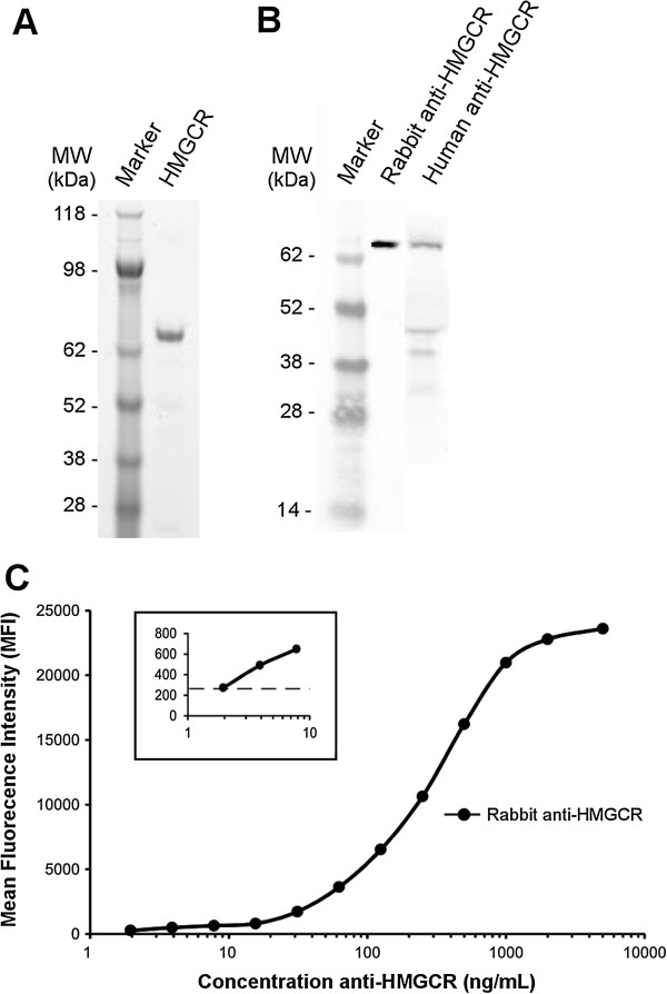
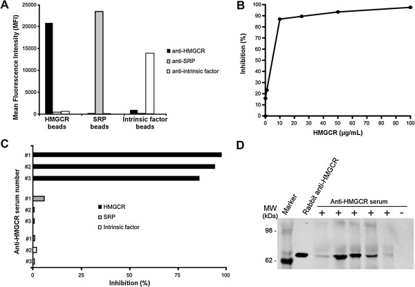
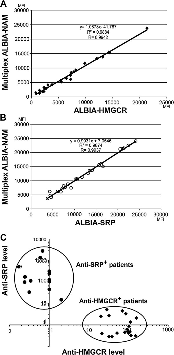
Methods: Recombinant HMGCR C-domain was bound to fluorescent beads. After incubation with serum, autoantibodies were revealed using class- or subclass-specific anti-human immunoglobulin G (IgG) antibodies. Anti-HMGCR levels were assayed in 150 patients with suspicion of NAM, 142 controls with different inflammatory/autoimmune diseases and 100 healthy donors. Inhibition with free recombinant HMGCR and immunoprecipitation experiments confirmed test specificity. Reproducibility and repeatability were determined from sera with various levels of anti-HMGCR autoantibodies. A multiplex assay (ALBIA-NAM) was also developed to permit the simultaneous quantification of anti-HMGCR and anti-signal recognition particle autoantibodies.

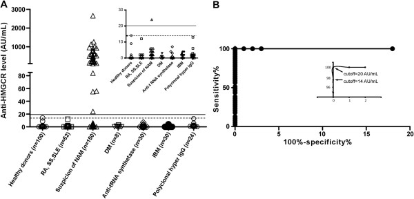
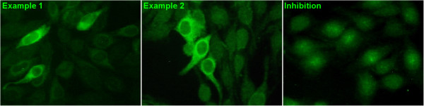
Results: No controls scored positive. Of 150 patients with suspicion of NAM, 24% were positive for anti-HMGCR autoantibodies with levels ranging from 24 to 2,656 AU/mL. Anti-HMGCR positivity could be associated to a cytoplasmic pattern in immunofluorescence assay on HEp-2 cells. Anti-HMGCR-positive patients had high creatine kinase (CK) levels (mean 6,630 IU/L) and only 40% of them had been exposed to statins. Multiplex ALBIA-NAM was equally as effective as monoplex anti-HMGCR and anti-SRP ALBIA.

Conclusions: Both monoplex ALBIA-HMGCR and multiplex ALBIA-NAM reliably detect and quantify anti-HMGCR autoantibodies. A positive result allows ascribing patients with a necrotizing myopathy to an autoimmune form. Anti-HMGCR autoantibodies may be found in patients who have not taken statins.


Figures

Figure 1
Figure 1
Development of a quantitative assay for the detection of anti-HMGCR antibodies. (A) SDS-PAGE analysis of the recombinant HMGCR catalytic C-domain after Coomassie blue staining. (B) Western blot analysis of this recombinant HMGCR using a specific rabbit anti-HMGCR antibody or a human anti-HMGCR-positive serum. (C) Addressable laser bead immunoassay (ALBIA-HMGCR) using serial dilutions of rabbit anti-HMGCR antibody. The mean fluorescence intensity (MFI) values are the mean of triplicate determinations. Standard deviation (SD) errors bars are not visible because of very low variability (coefficients of variation SD/mean lower than 1%, range (0.0025 to 0.7525)). Insert, higher magnification for the low anti-HMGCR antibody concentrations (horizontal dotted line depicts mean + 2 SD of 20 negative controls). HMGCR, 3-hydroxy-3-methylglutaryl coenzyme A reductase.
Figure 2
Figure 2
Specificity of HMGCR-coated beads. (A) HMGCR, signal recognition particle (SRP) and intrinsic factor-specific beads were coupled to the corresponding recombinant protein and analyzed by ALBIA using a human anti-HMGCR, anti-SRP or anti-intrinsic factor positive serum. (B) Dose-dependent inhibition of human anti-HMGCR antibodies binding to HMGCR-coated beads by free human recombinant HMGCR protein. Percent inhibition is given relative to the mean fluorescence intensity (MFI) value in the absence of free HMGCR. (C) Specific inhibition by homologous HMGCR but not heterologous proteins. Percent inhibition of three different human anti-HMGCR positive sera (referred to as #1, #2 and #3) by 100 μg/mL of free HMGCR, SRP or intrinsic factor proteins. (D) Immunoprecipitation of HMGCR protein by ALBIA-HMGCR positive or negative human sera. A rabbit anti-HMGCR polyclonal antibody was used as positive control. ALBIA, addressable laser bead immunoassay; HMGCR, 3-hydroxy-3-methylglutaryl coenzyme A reductase.
Figure 3
Figure 3
Validation of ALBIA-HMGCR and determination of anti-HMGCR levels in patients with suspicion of NAM. (A) A positivity cutoff of the assay was first determined as the 99th percentile (14 arbitrary units (AU)/mL) of the healthy donors’ distribution (open circles) and depicted by a dotted line. Sera from patients with different inflammatory/autoimmune conditions including rheumatoid arthritis (RA), systemic sclerosis (SS), systemic lupus erythematosus (SLE), dermatomyositis (DM), anti-tRNA synthetase-positive myositis or inclusion body myositis (IBM), as well as patients with polyclonal hypergammaglobulinemia were assayed and revealed all negative. Insert is a magnification for low values. Thirty-seven out of 150 (24%) of serum samples from patients with suspicion of NAM scored positive using ALBIA-HMGCR. (B) Receiver operating characteristic (ROC) analysis was performed by comparing the 37 anti-HMGCR positive serum samples to those from healthy donors. Optimal results were obtained with a cutoff of 20 AU/mL, which was indicated as a plain line. ALBIA, addressable laser bead immunoassay; HMGCR, 3-hydroxy-3-methylglutaryl coenzyme A reductase; NAM, necrotizing autoimmune myopathies.
Figure 4
Figure 4
Aspect of anti-HMGCR autoantibodies in indirect immunofluorescence on HEp-2 cells. Classical indirect immunofluorescence assay using the HEp-2000 line as cellular substrate. After incubation of serum at a 1/80 dilution, immunoreactivity was revealed by a FITC-labeled anti-human immunoglobulin (Ig)G secondary antibody. Example from two representative anti-HMGCR positive patients out of 20/33 sera with this immunofluorescence pattern shows a finely granular cytoplasmic staining with perinuclear reinforcement (left and middle). Inhibition by free HMGCR protein before immunofluorescence (right): the serum from example 2 was pre-incubated with free HMCGR before immunofluorescence assay. This yielded an extinction of immunoreactivity. HMGCR, 3-hydroxy-3-methylglutaryl coenzyme A reductase.
Figure 5
Figure 5
Comparison of monoplex ALBIA-HMGCR and ALBIA-SRP to multiplex ALBIA-NAM. Serum from anti-HMGCR positive (n = 26) or anti-SRP positive (n = 25) patients were compared. (A) Correlation of the signals generated by ALBIA-NAM versus ALBIA-HMGCR. Mean fluorescence intensity (MFI) (B) Correlation of the signals generated by ALBIA-NAM versus ALBIA-SRP. (C) ALBIA-NAM discriminates anti-SRP positive from anti-HMGCR positive patients in a single assay. ALBIA, addressable laser bead immunoassay; HMGCR, 3-hydroxy-3-methylglutaryl coenzyme A reductase; SRP, signal recognition particle.

Comment in

References

    1. Dalakas MC, Hohlfeld R. Polymyositis and dermatomyositis. Lancet. 2003;16:971–982. doi: 10.1016/S0140-6736(03)14368-1. - DOI - PubMed
    1. Bronner IM, Hoogendijk JE, Wintzen AR, van der Meulen MF, Linssen WH, Wokke JH, de Visser M. Necrotising myopathy, an unusual presentation of a steroid-responsive myopathy. J Neurol. 2003;16:480–485. doi: 10.1007/s00415-003-1027-y. - DOI - PubMed
    1. van der Meulen MF, Bronner IM, Hoogendijk JE, Burger H, van Venrooij WJ, Voskuyl AE, Dinant HJ, Linssen WH, Wokke JH, de Visser M. Polymyositis: an overdiagnosed entity. Neurology. 2003;16:316–321. doi: 10.1212/WNL.61.3.316. - DOI - PubMed
    1. Sadeh M, Dabby R. Steroid-responsive myopathy: immune-mediated necrotizing myopathy or polymyositis without inflammation? J Clin Neuromuscul Dis. 2008;16:341–344. doi: 10.1097/CND.0b013e31815e5d4a. - DOI - PubMed
    1. Emslie-Smith AM, Engel AG. Necrotizing myopathy with pipestem capillaries, microvascular deposition of the complement membrane attack complex (MAC), and minimal cellular infiltration. Neurology. 1991;16:936–939. doi: 10.1212/WNL.41.6.936. - DOI - PubMed

Publication types

MeSH terms