Case Reports
. 2014 Feb 3:12:28.
doi: 10.1186/1477-7819-12-28.

Calcifying fibrous tumor presenting as rectal submucosal tumor: first case reported in rectum

Affiliations
Case Reports

Calcifying fibrous tumor presenting as rectal submucosal tumor: first case reported in rectum

Soyoung Im et al. World J Surg Oncol. .

Abstract

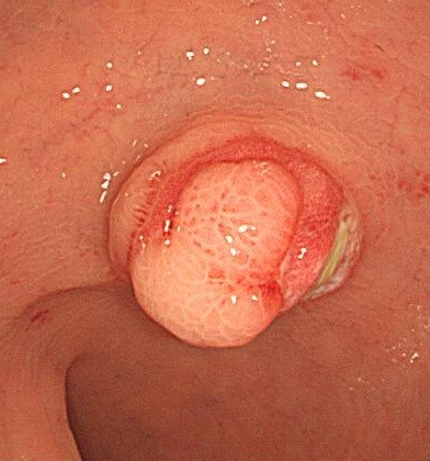
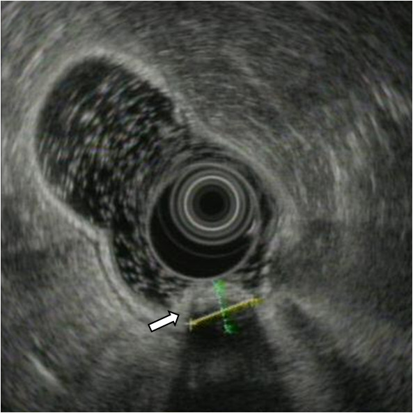
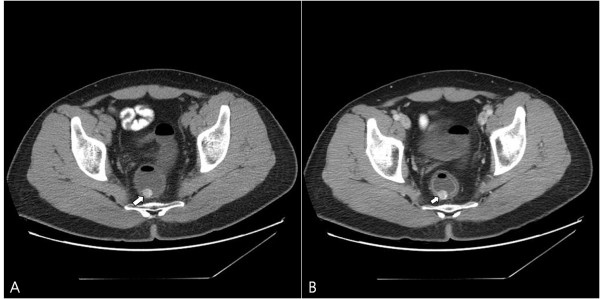
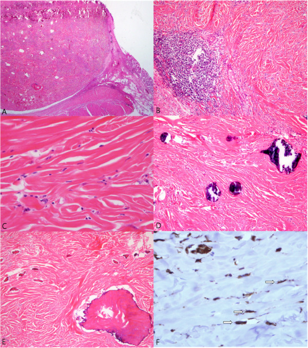
Calcifying fibrous tumor (CFT) is a recently recognized rare benign lesion characterized by dense hyalinized collagenous tissue with interspersed spindle cells and a lymphoplasmocytic infiltrate. Calcification is the hallmark of CFT and may present in the form of psammomatous bodies or dystrophic calcifications. CFT of the intestinal tract is uncommon and rectal CFT has never been reported. Recently, we experienced a case of CFT found in the rectum of a 36-year-old man. In this study, we described the characteristic histopathological findings with a review of the relevant literature. Although CFT of the intestinal tract as an intrinsic visceral lesion is unusual and clinically unexpected, CFT should be considered in the differential diagnosis of rectal submucosal tumor.


Figures

Figure 1
Figure 1
Colonoscopic finding shows an ovoid polypoid mass with normal overlying mucosa in the proximal rectum.
Figure 2
Figure 2
Endoscopic ultrasonographic finding shows a well-demarcated submucosal mass (arrow).
Figure 3
Figure 3
Plain (A) and enhanced (B) computed tomography of the abdomen reveals a prominent contrast-enhancing rectal wall mass with internal calcification (arrow).
Figure 4
Figure 4
Microscopic findings of the tumor. (A) Whole mount of the well-circumscribed tumor located in the submucosa (H&E x1.4). (B) Microscopic finding of the tumor shows dense fibrous tissue and lymphoid aggregates (H&E x100). (C) The spindle cells have bland nuclei and indistinct cytoplasm (H&E x400). (D)Psammomatous or dystrophic calcifications are seen (H&E x100). (E) Metaplastic ossifications are also noted (H&E x100). (F) Immunohistochemical staining for vimentin shows diffuse positive for tumor cells (arrow) (x400).

References

    1. Fetsch JF, Montgomery EA, Meis JM. Calcifying fibrous pseudotumor. Am J Surg Pathol. 1993;17:502–508. doi: 10.1097/00000478-199305000-00010. - DOI - PubMed
    1. Shibata K, Yuki D, Sakata K. Multiple calcifying fibrous pseudotumors disseminated in the pleura. Ann Thorac Surg. 2008;85:e3–e5. doi: 10.1016/j.athoracsur.2007.10.059. - DOI - PubMed
    1. Bajpai J, Rekhi B, Iyer K, Samar A, Mahajan A, Jambhekar NA. Multifocal calcifying fibrous tumor of the mesentery: an unusual case report with literature review and therapeutic implications. J Cancer Res Ther. 2011;7:500–502. doi: 10.4103/0973-1482.92022. - DOI - PubMed
    1. Soyer T, Ciftci AO, Gucer S, Orhan D, Senocak ME. Calcifying fibrous pseudotumor of lung: a previously unreported entity. J Pediatr Surg. 2004;39:1729–1730. doi: 10.1016/j.jpedsurg.2004.07.024. - DOI - PubMed
    1. Jeong HS, Lee GK, Sung R, Ahn JH, Song HG. Calcifying fibrous pseudotumor of mediastinum - a case report. J Korean Med Sci. 1997;12:58–62. - PMC - PubMed

Publication types

LinkOut - more resources