. 2014 Jan 30;156(3):522-36.
doi: 10.1016/j.cell.2013.12.040.

Control of stress-induced persistent anxiety by an extra-amygdala septohypothalamic circuit

Affiliations

Control of stress-induced persistent anxiety by an extra-amygdala septohypothalamic circuit

Todd E Anthony et al. Cell. .

Abstract

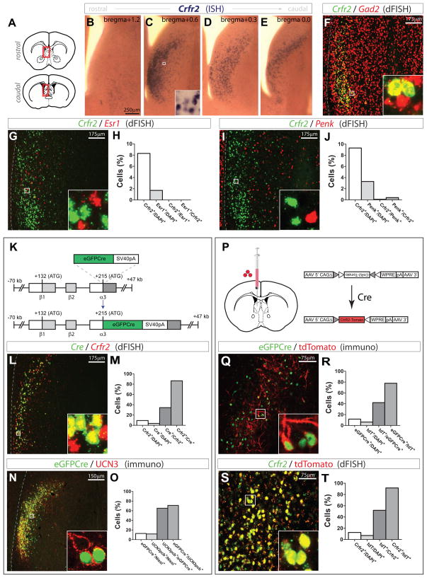
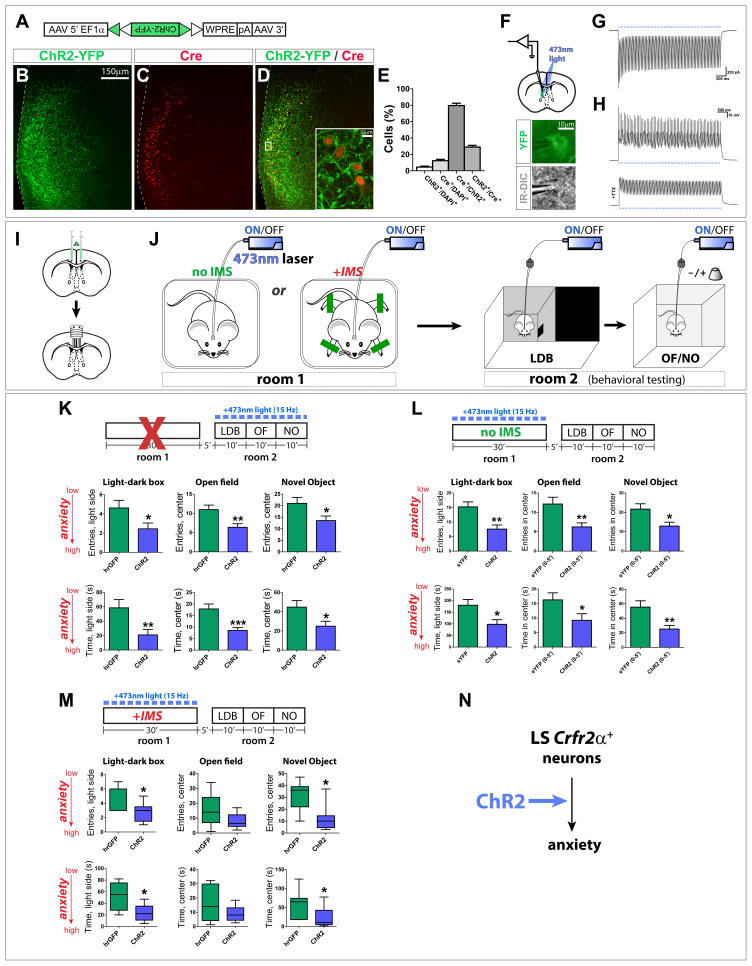
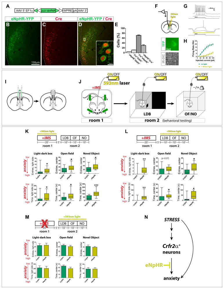
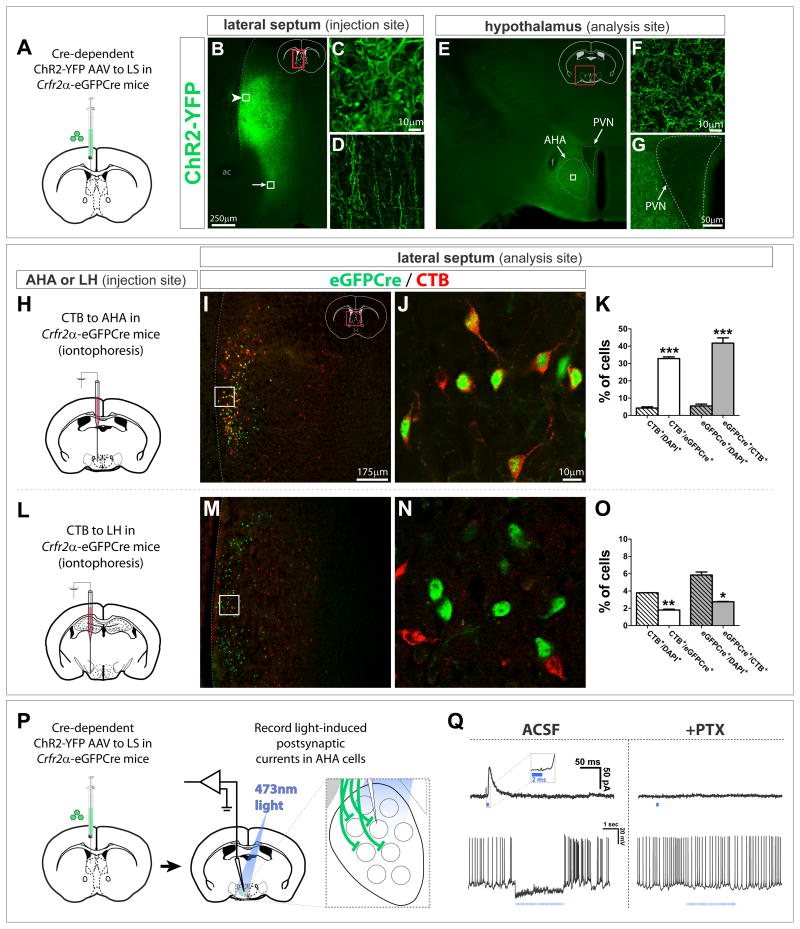
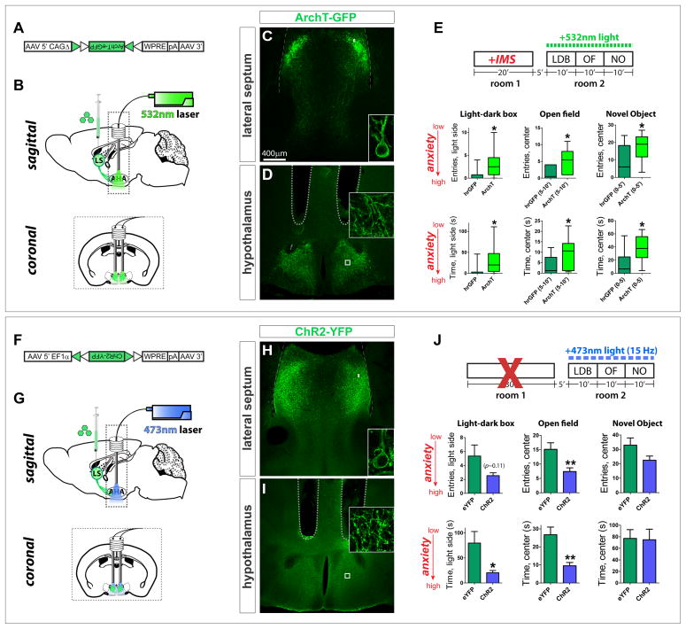
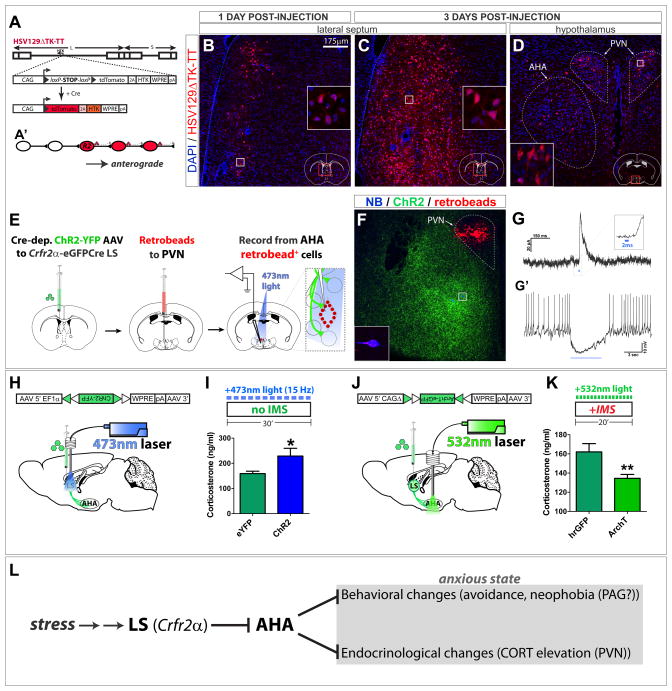
The extended amygdala has dominated research on the neural circuitry of fear and anxiety, but the septohippocampal axis also plays an important role. The lateral septum (LS) is thought to suppress fear and anxiety through its outputs to the hypothalamus. However, this structure has not yet been dissected using modern tools. The type 2 CRF receptor (Crfr2) marks a subset of LS neurons whose functional connectivity we have investigated using optogenetics. Crfr2(+) cells include GABAergic projection neurons that connect with the anterior hypothalamus. Surprisingly, we find that these LS outputs enhance stress-induced behavioral measures of anxiety. Furthermore, transient activation of Crfr2(+) neurons promotes, while inhibition suppresses, persistent anxious behaviors. LS Crfr2(+) outputs also positively regulate circulating corticosteroid levels. These data identify a subset of LS projection neurons that promote, rather than suppress, stress-induced behavioral and endocrinological dimensions of persistent anxiety states and provide a cellular point of entry to LS circuitry.


Figures

Figure 1
Figure 1. Circuit models for LS CRFR2+ neuronal regulation of anxiety
(A) Sagittal plane of LS anatomy showing simplified feedforward connections. (B) Coronal section at plane indicated by dashed line in (A). Modified from Allen Brain Atlas (www.brain-map.org). (C, D) Circuit models consistent with known anxiogenic effect of CRFR2 agonists in the LS. Depending on whether agonists excite (C) or inhibit (D) CRFR2+ (“R2”) neurons, increased R2 neuronal activity would be anxiogenic (C) or anxiolytic (D). GABAergic R2 neurons (Fig. 2F) may be local interneurons (“IN;” C1, D3) or projection neurons (“PN;” C2, D4). LS output may be anxiolytic (C1, D4) or anxiogenic (C2, D3).
Figure 2
Figure 2. Characterization and transgenic targeting of LS Crfr2α+ neurons
(A–E) Crfr2 mRNA ISH; coronal diagrams in (A) depict region analyzed. (F) dFISH for Crfr2 (green) and Gad2 (red). (G,H) dFISH for Crfr2 (green) vs Esr1 (red): Crfr2+/DAPI+ = 8.3%, Esr1+/DAPI+ = 1.7%, Crfr2+/Esr1+ = 0, Esr1+/Crfr2+ = 0; total n = 11,086 DAPI+ cells (I,J) dFISH for Crfr2 (green) vs Penk (red): Crfr2+/DAPI+ = 9.3%, Penk+/DAPI+ = 3.3%, Crfr2+/Penk+ = 0.1%, Penk+/Crfr2+ = 0.4%; total n = 8,274 DAPI+ cells (K) Crfr2α-eGFPCre BAC transgene design. (L,M) dFISH, Cre mRNA (green) vs. Crfr2 mRNA (red): Crfr2+/DAPI+ = 9.3%, Cre+/DAPI+ = 3.7%, Cre+/Crfr2+ = 34.4%, Crfr2+/Cre+ = 86.6%; total n = 17,537 DAPI+ cells (N,O) eGFPCre+ nuclei (green) vs. UCN3 pericellular baskets (pcbs; red); counts from areas with detectable transgene expression. UCN3+ pcbs colocalize with synaptophysin (data not shown). eGFPCre+/DAPI+ = 12.8%, UCN3pcb+/DAPI+ = 11.8%, UCN3pcb+/eGFPCre+ = 65.2%, eGFPCre+/UCN3pcb+ = 71.0%; total n = 2,665 Nissl+ cells (P) Injection site (left) and Cre-dependent AAV (right). (Q,R) Transgenic eGFPCre+ nuclei (green) vs. recombined, viral tdT (red); counts from areas containing tdT+ cells. eGFPCre+/DAPI+ = 11.7%, tdT+/DAPI+ = 6.3%, tdT+/eGFPCre+ = 42.1%, eGFPCre+/tdT+ = 77.9%; total n = 3,586 DAPI+ cells (S,T) dFISH for Crfr2 mRNA (green) vs. tdT mRNA (red); counts from areas containing tdT+ cells. Crfr2+/DAPI+ = 12.5%, tdT+/DAPI+ = 7.1%, tdT+/Crfr2+ = 51.9%, Crfr2+/tdT+ = 91.6%; total n = 5,829 DAPI+ cells In this and subsequent figures, dashed lines at left indicate lateral ventricle (LV), boxed regions shown at higher magnification in insets. See also Figures S1 and S2.
Figure 3
Figure 3. Optogenetic stimulation of LS Crfr2α+ neurons induces a persistent, anxious state
(A) Cre-dependent ChR2-YFP AAV. (B–D) Injection of (A) into Crhr2α-eGFPCre LS yielded ChR2-YFP (green) in Cre+ cells (red). (E) Recombination specificity and sensitivity; counts done in areas containing ChR2-YFP+ cells: ChR2+/DAPI+ = 4.6±1.1%, Cre+/DAPI+ = 12.4±1.7%, Cre+/ChR2+=79.7±2.9%, ChR2+/Cre+=29.0±2.1% (mean±s.e.m.). Total n = 9,673 DAPI+ cells from 2 mice. (F) top, ChR2 activity in Crfr2α+ cells assessed in acute slices; bottom, patched YFP+ neuron under infrared differential interference contrast (IR-DIC) microscopy. (G,H) Robust photocurrents were observed (G) that were sufficient to drive repetitive, tetrodotoxin(TTX)-sensitive spiking in ChR2-YFP+ neurons (H). (I,J) Surgical manipulations and behavioral testing. (K) Photostimulation of Crfr2α+ neurons during behavioral testing increased anxiety. hrGFP(n=39) vs. ChR2(n=38): LDB, entries in light side (4.6±0.8 vs. 2.5±0.6, P<0.05), time in light side (58.6±11.4 vs. 21.0±7.6, P<0.01); OF, entries in center (11.0±1.2 vs 6.4±0.9, P<0.01), time in center (17.9±2.1 vs. 8.5±1.2, P<0.001); NO, entries in center (21.0±2.6 vs. 13.6±2.0, P<0.05), time in center (44.7±6.8 vs. 24.9±5.1, P<0.05). P values represent two-tailed unpaired t-tests. Values indicate mean±s.e.m. (L) Photostimulation of Crfr2α+ neurons increased anxiety during subsequent testing. eYFP(n=13) vs. ChR2(n=12): LDB, entries in light side (15.2±1.7 vs. 7.6±1.4, P<0.01), time in light side (180.2±24.8 vs. 98.0±20.1, P<0.05); OF(0–5′), entries in center (12.2±1.8 vs. 6.3±1.0, P<0.01), time in center (16.3±2.4 vs. 9.2±2.3, P<0.01); NO(0–5′), entries in center (21.7±2.8 vs. 13.0±2.0, P<0.05), time in center (55.4±8.7 vs. 25.3±5.0, P<0.01). (M) Stimulating Crfr2α+ neurons exacerbates the anxiogenic effects of IMS (mice were habituated to minimize ceiling effect confound, see methods). hrGFP (n=7) vs. ChR2 (n=8), data presented as Mann-Whitney U value, P value: LDB, entries in light side (U = 7.5, P<0.05), time in light side (U = 10.5, P<0.05); OF, entries in center (U = 15.5, P>0.15), time in center (U = 22.5, P>0.55); NO, entries in center (U = 7.0, P<0.05), time in center (U = 10.0, P<0.05). (N) Model of LS Crfr2α+ neuronal valence based on optogenetic stimulation. See also Figure S3.
Figure 4
Figure 4. LS Crfr2α+ neurons are necessary for the induction and expression of a stress-induced, anxiogenic state
(A) Cre-dependent eNpHR(2.0)-YFP AAV. (B–D) Injection of (A) into Crhr2α-eGFPCre LS yielded eNpHR-YFP (green) in Cre+ cells (red). (E) Recombination specificity and sensitivity; counts from areas containing eNpHR-YFP+ cells: eNpHR+/DAPI+ = 4.7±0.2%, Cre+/DAPI+ = 11.4±0.4%, Cre+/eNpHR+=71.2±1.6%, eNpHR+/Cre+=29.8±3.0% (mean±s.e.m.). Total n = 8,216 DAPI+ cells from 2 mice. (F–H) Recordings from eNpHR-YFP+Crfr2α+ neurons. (G) Cell in response to the same current injections in absence (top) or presence (bottom) of 593nm light. (H) 593nm light significantly suppressed firing of eNpHR-YFP+ neurons (F1,108 = 28.69, P<0.001). (I,J) Surgical manipulations and behavioral testing. (K) Photoinhibition of Crfr2α+ neurons only during IMS reduced anxiety in subsequent testing. hrGFP (n=15) vs. eNpHR (n=17), data presented Mann-Whitney U value, P value: LDB, entries in light side (U = 64.0, P<0.05), time in light side (U = 62.5, P<0.05); OF, entries in center (U = 69.0, P<0.05), time in center (U = 64.5, P<0.05); NO, entries in center (U = 73.5, P<0.05), time in center (U = 34.0, P<0.001). (L) IMS mice that received photoinhibition of Crfr2α+ neurons only during behavioral testing showed reduced anxiety relative to controls. hrGFP (n=11) vs. eNpHR (n=11): LDB, entries in light side (U = 21.5, P<0.01), time in light side (U = 20.0, P<0.01); OF, entries in center (U = 25.5, P=0.07), time in center (U = 23.0, P<0.05); NO, entries in center (U = 22.0, P<0.05), time in center (U = 21.0, P<0.05). (M) Inhibiting Crfr2α+ neurons in the absence of IMS does not alter anxiety. hrGFP(n=11) vs. eNpHR (n=11): LDB, entries in light side (11.5±1.9 vs. 11.7±2.3, P>0.9), time in light side (218.7±44.6 vs. 206.4±45.2, P>0.8); OF, entries in center (23.1±2.6 vs. 20.4±2.8, P>0.45), time in center (34.4±3.2 vs. 26.7±4.4, P>0.15); NO, entries in center (43.0±6.3 vs. 39.0±3.9, P>0.6), time in center (97.0±15.8 vs. 97.5±14.9, P>0.95). (N) Model for LS Crfr2α+ neuronal valence based on optogenetic inhibition. See also Figure S4.
Figure 5
Figure 5. LS Crfr2α+ neurons make GABAergic synapses in the AHA
(A–G) Cre-dependent ChR2-YFP AAV injected into Crfr2α-eGFPCre LS labeled cell bodies (arrowhead in B, enlarged in C) and axons (arrow in B, enlarged in D) Terminals were densest in AHA (E). All AHA labeling was axonal (F, enlarged from boxed area in E). YFP+ axons appeared to be excluded from the PVN (G). ac, anterior commissure; f, fornix; PVN, paraventricular hypothalamic nucleus. (H–O) CTB retrograde tracing. eGFPCre (green) and CTB (red) in the LS of Crfr2α-eGFPCre mice that received iontophoretic injection of CTB into the AHA (I,J) or LH (M,N). (K) A significant fraction of Crfr2α+ neurons project to AHA (CTB+/DAPI+ = 4.3±0.7% vs. CTB+/eGFPCre+ = 33.0±1.0%, P<0.001), and Crfr2α+ neurons comprise a large fraction of the neurons that project to AHA from middle levels of LS (GFPCre+/DAPI+ = 5.5±1.1% vs. eGFPCre+/CTB+ = 42.0±3.1%, P<0.001). (O) Few Crfr2α+ neurons innervate the LH (CTB+/DAPI+ = 3.8±0.01% vs. CTB+/eGFPCre+ = 1.8±0.1%, P<0.01), with most projections to this region coming from Crfr2α cells (GFPCre+/DAPI+ = 5.9±0.4% vs. eGFPCre+/CTB+ = 2.8±0.1%, P<0.05). Counts done at bregma+0.6 in LS regions that contained CTB+ cells. Total n = 7,737 DAPI+ cells (AHA) and n = 5,767 DAPI+ cells (LH). Values indicate mean±s.e.m. (P,Q) CRACM in standard ACSF or plus 100μM picrotoxin (PTX). (Q) Single 2ms pulses of 473nm light evoked IPSCs blocked by PTX (top). Inset, higher temporal resolution to illustrate response latency. (bottom), picrotoxin-sensitive inhibition of spontaneous firing observed by presynaptic terminal photostimulation (15Hz, 2ms pulses). See also Figure S5.
Figure 6
Figure 6. LS Crfr2α+ projections to AHA are necessary for stress-induced anxiety
(A) Cre-dependent ArchT-GFP AAV. (B–E) Axon terminal inhibition. (C,D) ArchT-GFP in LS Crfr2α+ (C) cell bodies and (D) and axons in AHA. (E) Photoinhibition of Crfr2α+ axon terminals in AHA during behavioral testing in IMS-stressed mice reduces anxiety. hrGFP (n=10) vs. ArchT (n=12), data presented Mann-Whitney U value, P value: LDB, entries in light side (U = 25.0, P<0.05), time in light side (U = 24.0, P<0.05); OF(5–10′), entries in center (U = 21.5, P<0.05), time in center (U = 26.0, P<0.05); NO(0–5′), entries in center (U = 29.5, P<0.05), time in center (U = 19.0, P<0.05). (F) Cre-dependent ChR2-YFP AAV. (G–J) Axon terminal stimulation. (H,I) ChR2-YFP in LS Crfr2α+ (H) cell bodies and (I) axons. (J) Stimulation of Crfr2α+ axon terminals in AHA during testing increased anxiety. eYFP(n=17) vs. ChR2(n=15): LDB, entries in light side (5.2±1.6 vs. 2.5±0.4, P>0.1), time in light side (79.9±23.2 vs. 20.3±4.6, P<0.05); OF, entries in center (15.1±2.3 vs. 7.4±1.3, P<0.01), time in center (26.8±4.2 vs. 9.5±1.8, P<0.01); NO, entries in center (32.9±5.0 vs. 22.5±2.9, P=0.08), time in center (77.2±15.2 vs. 74.9±18.3, P>0.1); values indicate mean±s.e.m. P values represent two-tailed unpaired t-tests. Dashed lines in (D,I) indicate guide tips. See also Figure S6.
Figure 7
Figure 7. LS Crfr2α+ projections to AHA are polysynaptically upstream of PVN and positively regulate corticosterone levels
(A,A′) HSV129ΔTK-TT. (B–D) tdT (red) in HSV129ΔTK-TT-injected Crfr2α-eGFPCre mice counterstained with DAPI (blue) at 1 (B) or 3 (C,D) days post-injection (dpi) in LS (B,C) or medial hypothalamus (D). Dashed lines in (D) demarcate PVN (upper right) and AHA (lower left). (E–G) Combined CRACM and retrograde tracing. (F) A slice used for recording; Crfr2α+ChR2-YFP+ axons originating from LS (green), retrobeads (red), and two neurobiotin-filled neurons (blue). The cell that yielded the traces in (G) is shown at higher magnification in the inset. (G) A 2ms pulse of 473nm light induced monosynaptic IPSCs in 4/9 retrobead+ AHA neurons that were sufficient to inhibit firing (G′). (H,I) Optogenetic stimulation of LS Crfr2α+ neurons increased CORT. eYFP(n=9) vs. ChR2(n=9): CORT, ng/ml (159.2±9.3 vs. 228.8±30.9, P<0.05). (J,K) Optogenetic inhibition of LS Crfr2α+ projections to AHA decreased CORT. hrGFP(n=15) vs. ArchT-GFP(n=17): CORT, ng/ml (162.1±8.6 vs. 134.5±4.1, P<0.01). Values indicate mean±s.e.m. (L) Proposed model for LS Crfr2α+ neuronal control of stress-induced anxiety. See also Figure S7.

References

    1. Adamec R, Head D, Blundell J, Burton P, Berton O. Lasting anxiogenic effects of feline predator stress in mice: sex differences in vulnerability to stress and predicting severity of anxiogenic response from the stress experience. Physiology & behavior. 2006;88:12–29. - PubMed
    1. Albert DJ, Richmond SE. Hyperreactivity and aggressiveness following infusion of local anesthetic into the lateral septum or surrounding structures. Behavioral biology. 1976;18:211–226. - PubMed
    1. Albert DJ, Wong RC. Hyperreactivity, muricide, and intraspecific aggression in the rat produced by infusion of local anesthetic into the lateral septum or surrounding areas. J Comp Physiol Psychol. 1978;92:1062–1073. - PubMed
    1. Almeida-Santos AF, Gobira PH, Rosa LC, Guimaraes FS, Moreira FA, Aguiar DC. Modulation of anxiety-like behavior by the endocannabinoid 2-arachidonoylglycerol (2-AG) in the dorsolateral periaqueductal gray. Behav Brain Res. 2013;252:10–17. - PubMed
    1. Atasoy D, Aponte Y, Su HH, Sternson SM. A FLEX switch targets Channelrhodopsin-2 to multiple cell types for imaging and long-range circuit mapping. J Neurosci. 2008;28:7025–7030. - PMC - PubMed

Publication types

Substances