. 2014 Feb 13;6(3):553-64.
doi: 10.1016/j.celrep.2014.01.009. Epub 2014 Jan 30.

Breast cancer proteins PALB2 and BRCA2 stimulate polymerase η in recombination-associated DNA synthesis at blocked replication forks

Affiliations

Breast cancer proteins PALB2 and BRCA2 stimulate polymerase η in recombination-associated DNA synthesis at blocked replication forks

Rémi Buisson et al. Cell Rep. .

Abstract

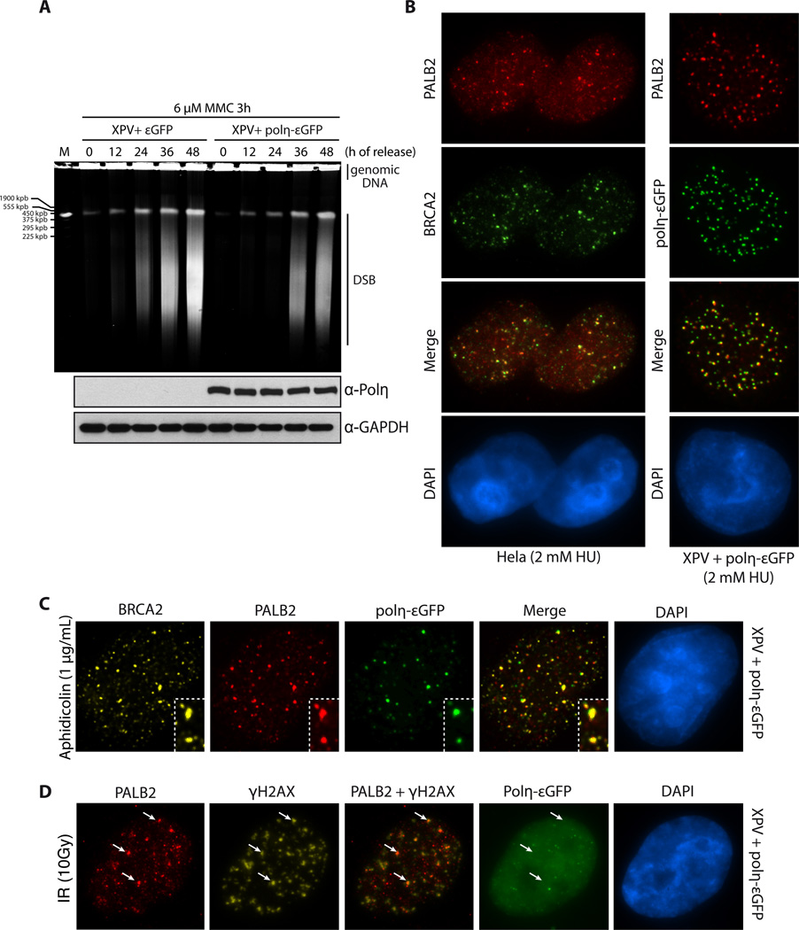
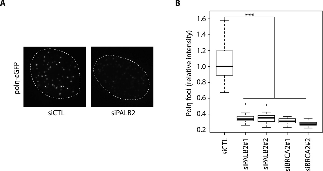
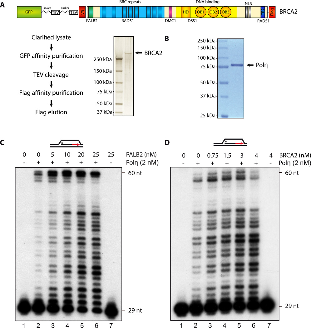
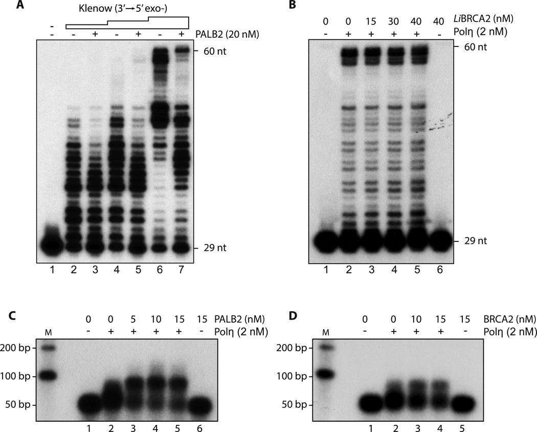
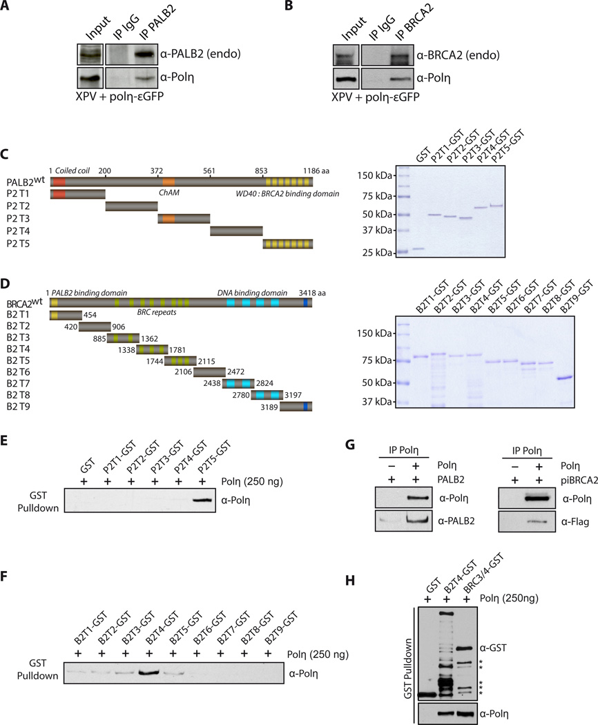
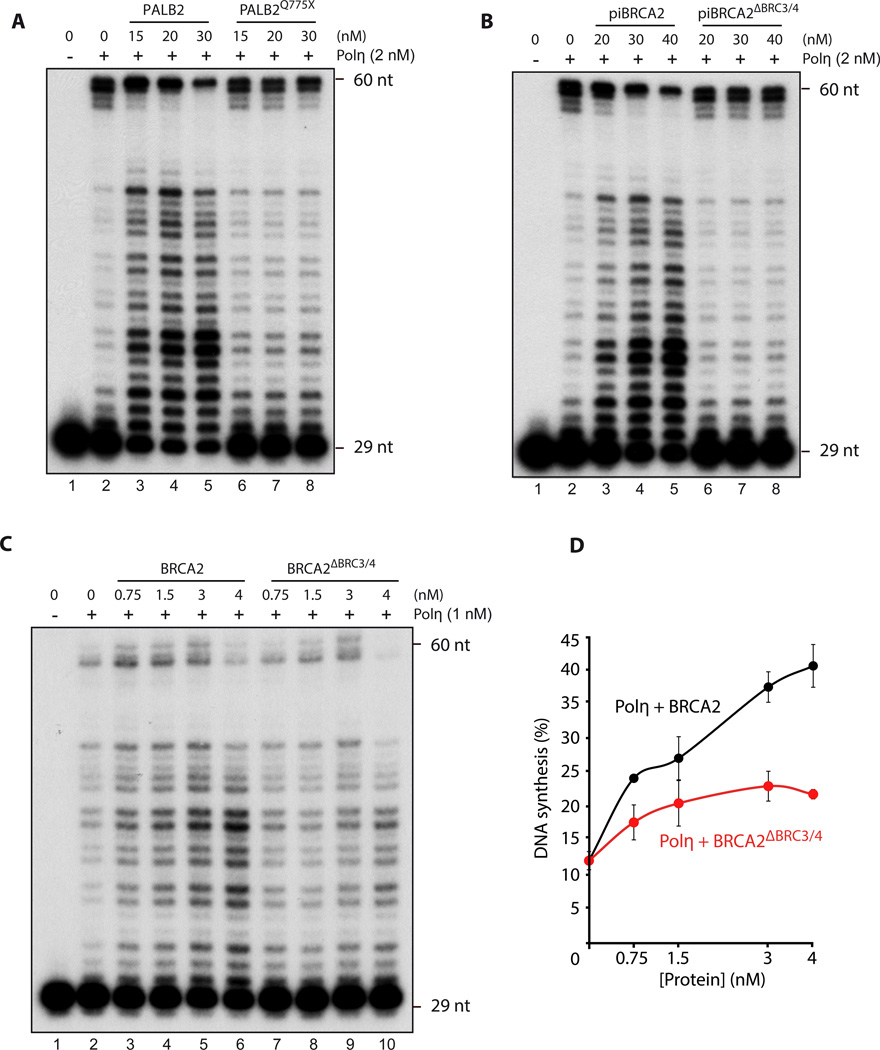
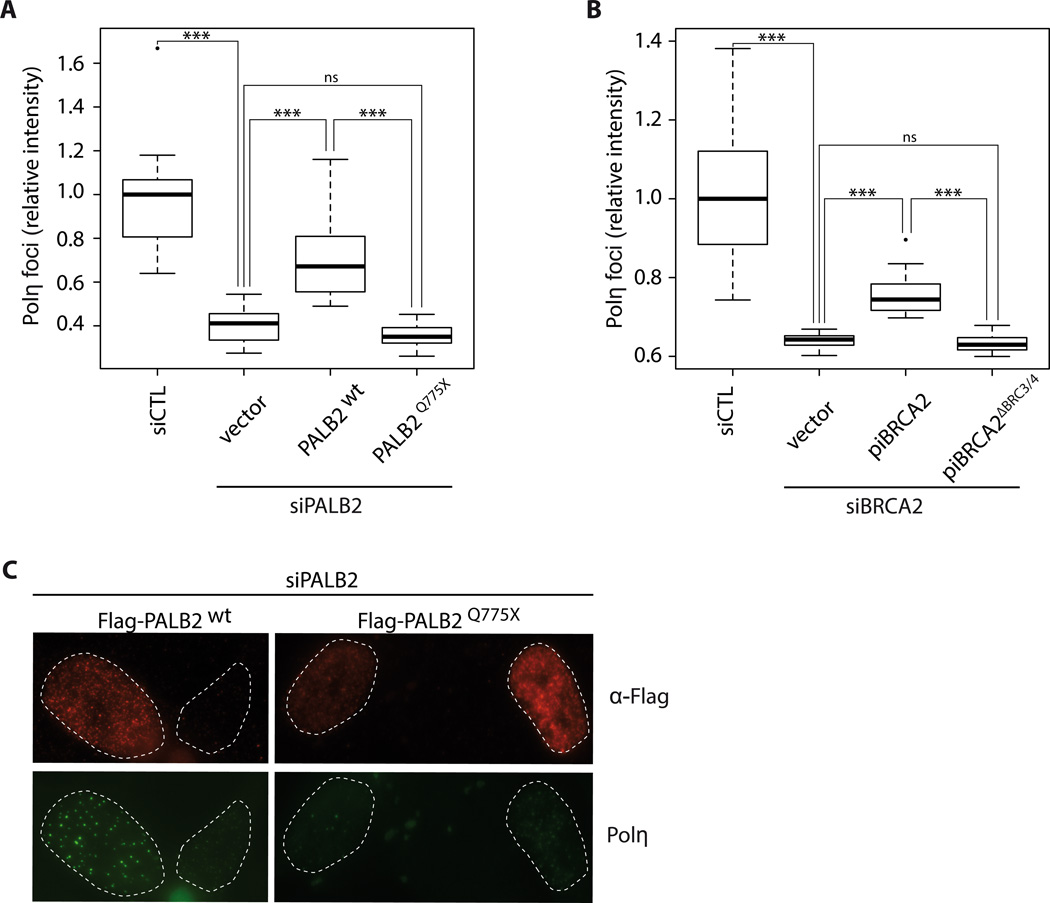
One envisioned function of homologous recombination (HR) is to find a template for DNA synthesis from the resected 3'-OH molecules that occur during double-strand break (DSB) repair at collapsed replication forks. However, the interplay between DNA synthesis and HR remains poorly understood in higher eukaryotic cells. Here, we reveal functions for the breast cancer proteins BRCA2 and PALB2 at blocked replication forks and show a role for these proteins in stimulating polymerase η (Polη) to initiate DNA synthesis. PALB2, BRCA2, and Polη colocalize at stalled or collapsed replication forks after hydroxyurea treatment. Moreover, PALB2 and BRCA2 interact with Polη and are required to sustain the recruitment of Polη at blocked replication forks. PALB2 and BRCA2 stimulate Polη-dependent DNA synthesis on D loop substrates. We conclude that PALB2 and BRCA2, in addition to their functions in D loop formation, play crucial roles in the initiation of recombination-associated DNA synthesis by Polη-mediated DNA repair.


Figures

Figure 1
Figure 1. Polη, PALB2 and BRCA2 recruitment at replication dependent DNA double-strand breaks
(A) Pulsed-field gel electrophoresis was used to visualize double-strand break (DSB) formation in XPV cells complemented with εGFP or Polη-εGFP, after treatment with 6 µM MMC for 3h and release for the times indicated. (B) Co-localization of PALB2 and BRCA2 or PALB2 and Polη-εGFP at DSBs induced by HU. DNA was counterstained with DAPI. (C–D) Immunofluorescence staining of the indicated DNA repair proteins at DSBs induced by aphidicolin or IR treatment in XPV cells complemented with Polη-εGFP. DNA was counterstained with DAPI. The white arrows indicate co-localization of PALB2 and γ-H2AX.
Figure 2
Figure 2. Knockdown of PALB2 or BRCA2 decreases Polη foci formation
(A) A representative example of Polη-εGFP foci intensity in cells transfected with a control siRNA or PALB2 siRNA. (B) Quantification of Polη-εGFP foci intensity induced by HU treatment in control, PALB2 or BRCA2 knockdowns. *** P < 0.001. The median is represented by a black line.
Figure 3
Figure 3. PALB2 and BRCA2 stimulate D-loop extension by Polymerase η
SDS-PAGE of purified human BRCA2 (A), or Polη ((B), 1 µg) stained by silver stain or Coomassie blue, respectively. (C–D) 32P-labeled D-loop was incubated with Polη (2 nM) with the indicated amounts of purified PALB2 or BRCA2. DNA synthesis products were analyzed by denaturing PAGE.
Figure 4
Figure 4
(A) Klenow DNA polymerase activity is not enhanced by PALB2 D-loop substrate was incubated with PALB2 (20 nM) followed by the addition of Klenow polymerase (0.25, 0.5 and 2.5 µU). DNA synthesis products were analyzed by denaturing PAGE. (B) Leishmania infantum BRCA2 does not stimulate Polη 32P-labeled D-loop was incubated with the indicated amounts of LiBRCA2 prior to the addition of Polη (2 nM). DNA synthesis products were analyzed by denaturing PAGE. (C–D) PALB2 and BRCA2 stimulate D-loop extension by Polη Purified D-loop substrate was incubated with increasing concentrations of PALB2 or BRCA2 following by addition of Polη. DNA synthesis products were analyzed by alkaline gel electrophoresis.
Figure 5
Figure 5. PALB2 and BRCA2 interact directly with Polη
(A) Cell extracts from complemented XPV cells were subjected to immunoprecipitation with anti-PALB2 or (B) anti-BRCA2 antibodies. Immunoprecipitated proteins were detected by Western Blotting with the indicated antibodies. (C) Left: Scheme of the PALB2 or (D) BRCA2 deletion variants fused to GST. Right: SDS-PAGE of the corresponding purified proteins. (E) GST alone or GST-PALB2 truncation (P2T1 to P2T5) were incubated with Polη followed by GST pulldown and detection of Polη by Western blotting. (F) GST alone or GST-BRCA2 truncations (B2T1 to B2T9) were incubated with Polη, followed by GST pulldown. The beads were washed and bound proteins were eluted with Laemmli buffer, and revealed by western blotting with the antibodies indicated. The input for PALB2 or BRCA2 truncations are shown in (C) and (D). (G-H) Co-immunoprecipitation of purified PALB2, piBRCA2, B2T4 or BRC3/4 and Polη. Asterisk: degradation products.
Figure 6
Figure 6. Polη interactions domains within PALB2 or piBRCA2 are essential for Polη stimulation
(A–B–C) 32P-labeled D-loop oligonucleotides was first incubated with indicated concentrations of PALB2, piBRCA2 with/without the Polη interacting domains following by the addition of Polη. DNA synthesis products were analyzed by denaturing PAGE. (D) quantification of the results shown in (C).
Figure 7
Figure 7. Deletion of Polη interaction domains in PALB2 and piBRCA2 affect polymerase η foci formation
(A) quantification of Polη-εGFP foci intensity induced by HU treatment in control, and siPALB2 knockdowns complemented with vector alone, wild-type PALB2, or PALB2Q775X. (B) Quantification of Polη-εGFP foci intensity induced by HU treatment in control, and siBRCA2 knockdowns complemented with vector alone, wild-type piBRCA2, or piBRCA2ΔBRC3/4. *** P < 0.001, ns not significant between the identified groups. The median is represented by a black line. (C) Representative images of Figure 7A. XPV complemented cells knockdown for PALB2, and complemented with Flag-PALB2 wild-type, and Flag-PALB2 Q775X were subjected for immunofluorescence against Polη (green) or anti-Flag antibodies to discriminate transfected cells. siRNA PALB2 resistant constructs were used.

References

    1. Arad G, Hendel A, Urbanke C, Curth U, Livneh Z. Single-stranded DNA-binding protein recruits DNA polymerase V to primer termini on RecA-coated DNA. J Biol Chem. 2008;283:8274–8282. - PubMed
    1. Bergoglio V, Boyer AS, Walsh E, Naim V, Legube G, Lee MY, Rey L, Rosselli F, Cazaux C, Eckert KA, et al. DNA synthesis by Pol eta promotes fragile site stability by preventing under-replicated DNA in mitosis. The Journal of cell biology. 2013;201:395–408. - PMC - PubMed
    1. Bleuyard JY, Buisson R, Masson JY, Esashi F. ChAM, a novel motif that mediates PALB2 intrinsic chromatin binding and facilitates DNA repair. EMBO Rep. 2012;13:135–141. - PMC - PubMed
    1. Bryant HE, Schultz N, Thomas HD, Parker KM, Flower D, Lopez E, Kyle S, Meuth M, Curtin NJ, Helleday T. Specific killing of BRCA2-deficient tumours with inhibitors of poly(ADP-ribose) polymerase. Nature. 2005;434:913–917. - PubMed
    1. Buisson R, Dion-Cote AM, Coulombe Y, Launay H, Cai H, Stasiak AZ, Stasiak A, Xia B, Masson JY. Cooperation of breast cancer proteins PALB2 and piccolo BRCA2 in stimulating homologous recombination. Nat Struct Mol Biol. 2010;17:1247–1254. - PMC - PubMed

Publication types