. 2014 Apr;35(12):3786-93.
doi: 10.1016/j.biomaterials.2014.01.037. Epub 2014 Jan 30.

The influence of a spatiotemporal 3D environment on endothelial cell differentiation of human induced pluripotent stem cells

Affiliations

The influence of a spatiotemporal 3D environment on endothelial cell differentiation of human induced pluripotent stem cells

Sophia Zhang et al. Biomaterials. 2014 Apr.

Abstract

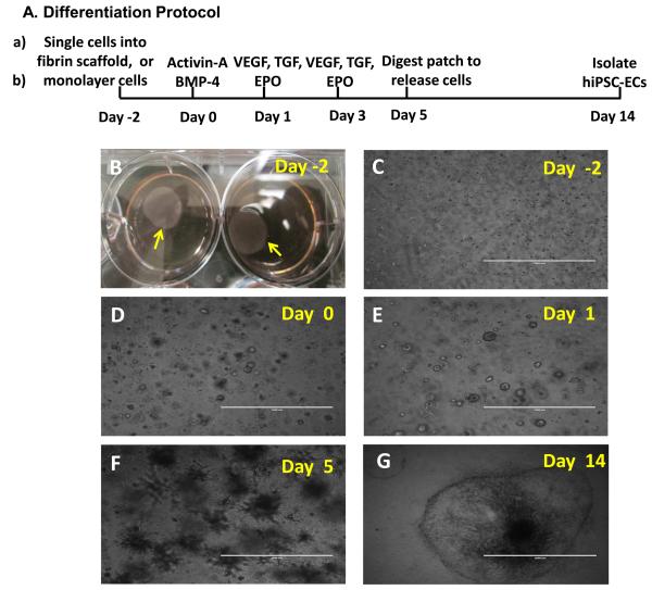
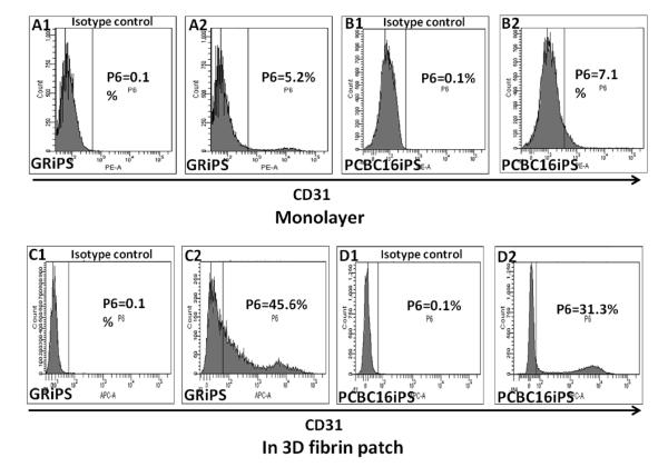
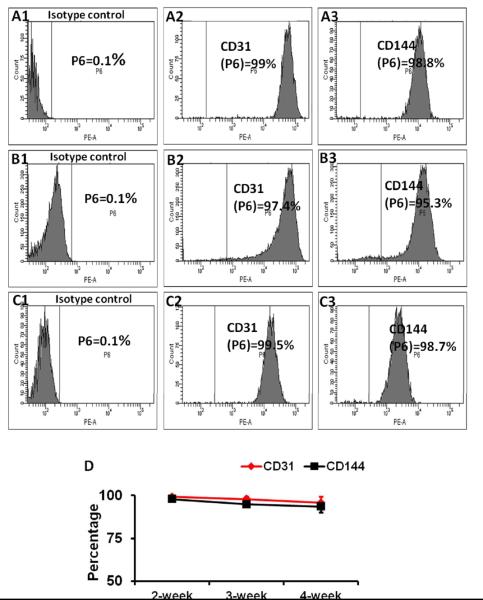
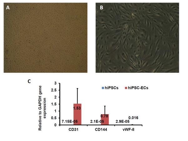
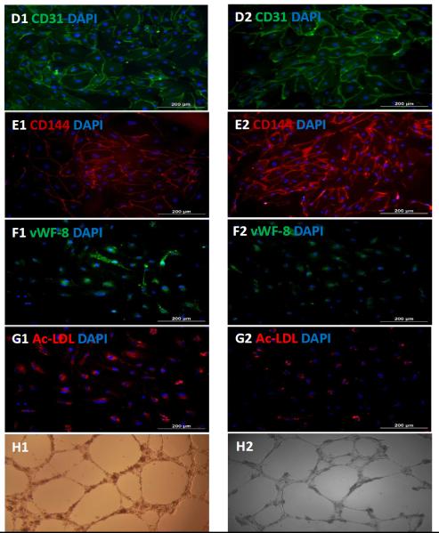
Current EC differentiation protocols are inefficient, and the phenotypes of the differentiated ECs are only briefly stable, which significantly inhibits their utility for basic science research. Here, a remarkably more efficient hiPSC-EC differentiation protocol that incorporates a three-dimensional (3D) fibrin scaffold is presented. With this protocol, up to 45% of the differentiated hiPSCs assumed an EC phenotype, and after purification, greater than 95% of the cells displayed the EC phenotype (based on CD31 expression). The hiPSC-ECs continued to display EC characteristics for 4 weeks in vitro. Gene and protein expression levels of CD31, CD144 and von Willebrand factor-8 (vWF-8) were significantly up-regulated in differentiated hiPSC-ECs. hiPSC-ECs also have biological function to up-take Dil-conjugated acetylated LDL (Dil-ac-LDL) and form tubular structures on Matrigel. Collectively, these data demonstrate that a 3D differentiation protocol can efficiently generate ECs from hiPSCs and, furthermore, the differentiated hiPSC-ECs are functional and can maintain EC fate up to 4 weeks in vitro.

Keywords: Cell differentiation; Eddothelial cells; Human induced pluripotent stem cells; Scaffold.


Figures

Figure 1
Figure 1. 3D scaffold-mediated differentiation of hiPSCs into hiPSC-ECs
(A) A schematic diagram of the hiPSC-EC differentiation protocol is displayed. (B) Two days before the differentiation protocol was initiated (i.e., on Day -2), fibrinogen solution was loaded with hiPSCs and mixed with thrombin to form a hiPSC-containing scaffold (yellow arrow). (C) The cells self-assembled into small clusters within the patch and grew to form sphere-shaped structures on (D) Day 0, when stage 1 of differentiation was initiated by culturing the cells with activin A and BMP-4. (E) On Day 1 (i.e., 24 hours later), stage 2 of differentiation was initiated by replacing the activin A/BMP-4 medium with medium containing VEGF, EPO, and TGFβ1, and the cells were cultured for 96 hours (i.e., until Day 5). (F) After 5 days of differentiation, the spheres had grown into spike-like structures, which were released from the patch via collagenase IV digestion. (G) By Day 14, the differentiated hiPSCs (visible as a sphere attached to the cell culture surface and formed a single layer of cells. (C-F: Bar=1 mm; G: Bar=2 mm).
Figure 2
Figure 2. The efficiency of hiPSC-EC differentiation increases when the cells are encapsulated in a fibrin patch
hiPSCs were differentiated (A1-B2) in monolayers or (C1-D2) after suspension in a fibrin patch. Differentiation efficiency was evaluated by comparing (A2, B2, C2, D2) CD31 expression with isotype controls (A1, B1, C1, D1) via flow cytometry. The maximum efficiencies obtained with each differentiation protocol are shown for both GRiPS- and PCBC16iPS-lineage cells.
Figure 3
Figure 3. Differentiated hiPSC-ECs maintained EC characteristics for at least 4 weeks in vitro
Differentiated hiPSC-ECs were purified to >95% CD31+ cells via flow cytometry, and maintained in medium supplemented with VEGF and SB431542 for 14 days (A1-A3), 21 days (B1-B3), and 28 days (C1-C3). The proportion of cells that expressed CD31 (A2, B2, C2) or CD144 (A3, B3, C3) were compared with isotype controls (A1, B1, and C1) and (D) presented as a function of time after purification.
Figure 4
Figure 4. Characterization of differentiated hiPSC-ECs
The morphology of the differentiated hiPSC-ECs was evaluated via images obtained at (A) 25X or (B) 100X magnification. (C) The gene expression levels of CD31, CD144, and vWF-8 mRNA were normalized to GAPDH, and presented as fold changes. Protein expression of CD31, CD144, and vWF-8 at week-1 (D1, E1, and F1) and week-4 (D2, E2, and F2) after isolation were evaluated via immunofluorescence; nuclei were counterstained with DAPI. The biological function of hiPSC-ECs was evaluated via Dil-ac-LDL uptake and the formation of tube-like structures on Matrigel at week-1 (G1 and H1) and week-4 (G2 and H2) after isolation; (Bar=200 μm, Panel E: magnification=200x).

Similar articles

Cited by

References

    1. Bolognese L, Neskovic AN, Parodi G, Cerisano G, Buonamici P, Santoro GM, et al. Left ventricular remodeling after primary coronary angioplasty: patterns of left ventricular dilation and long-term prognostic implications. Circulation. 2002;106:2351–7. - PubMed
    1. Hu Q, Wang X, Lee J, Mansoor A, Liu J, Zeng L, et al. Profound bioenergetic abnormalities in peri-infarct myocardial regions. Am J Physiol Heart Circ Physiol. 2006;291:H648–57. - PubMed
    1. Hatzistergos KE, Quevedo H, Oskouei BN, Hu Q, Feigenbaum GS, Margitich IS, et al. Bone marrow mesenchymal stem cells stimulate cardiac stem cell proliferation and differentiation. Circ Res. 2010;107:913–22. - PMC - PubMed
    1. Leri A, Kajstura J, Anversa P. Role of cardiac stem cells in cardiac pathophysiology: a paradigm shift in human myocardial biology. Circ Res. 2011;109:941–61. - PMC - PubMed
    1. Wang X, Hu Q, Nakamura Y, Lee J, Zhang G, From AH, et al. The role of the sca-1+/CD31- cardiac progenitor cell population in postinfarction left ventricular remodeling. Stem Cells. 2006;24:1779–88. - PubMed

Publication types