. 2014 Feb 20;53(4):606-16.
doi: 10.1016/j.molcel.2014.01.003. Epub 2014 Jan 30.

A phosphate-binding pocket within the platform-PAZ-connector helix cassette of human Dicer

Affiliations

A phosphate-binding pocket within the platform-PAZ-connector helix cassette of human Dicer

Yuan Tian et al. Mol Cell. .

Abstract

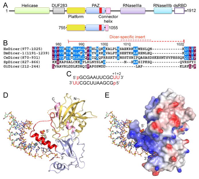
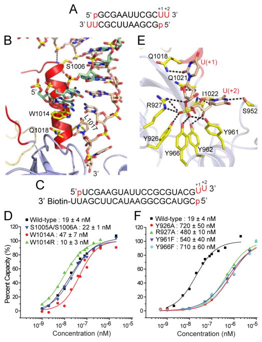
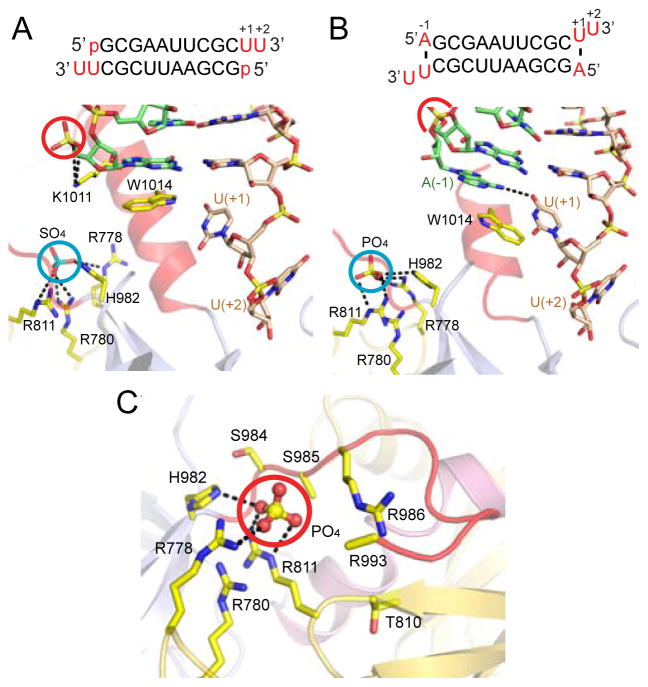
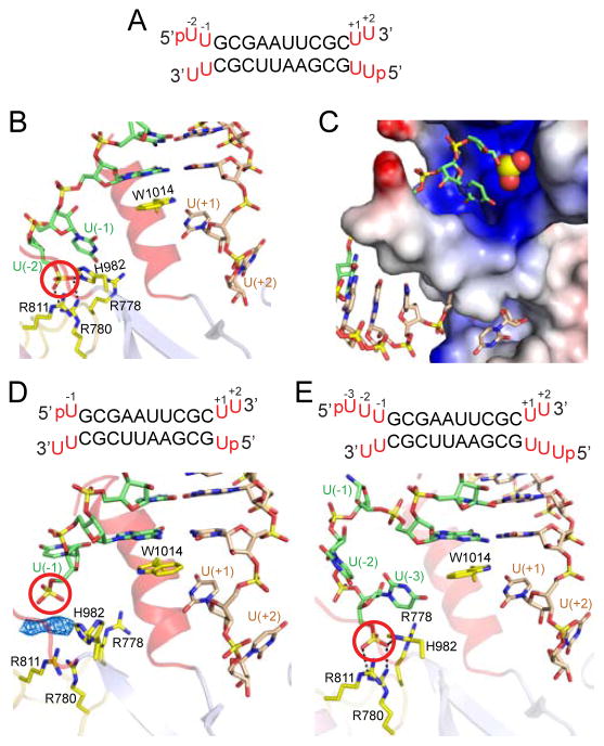
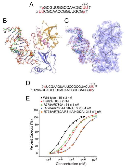
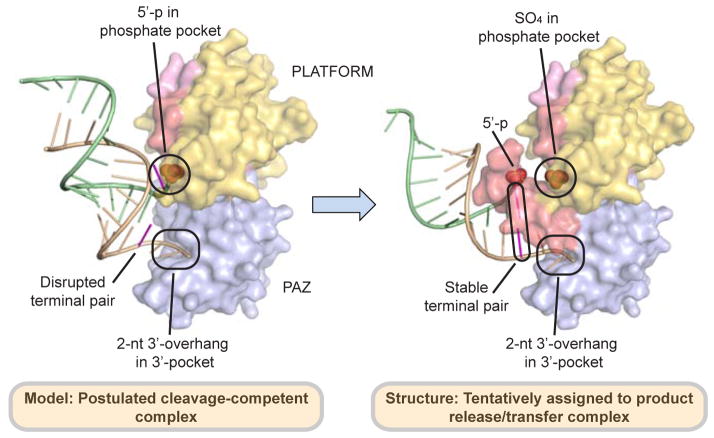
We have solved two families of crystal structures of the human Dicer "platform-PAZ-connector helix" cassette in complex with small interfering RNAs (siRNAs). The structures possess two adjacently positioned pockets: a 2 nt 3'-overhang-binding pocket within the PAZ domain (3' pocket) and a phosphate-binding pocket within the platform domain (phosphate pocket). One family of complexes contains a knob-like α-helical protrusion, designated "hDicer-specific helix," that separates the two pockets and orients the bound siRNA away from the surface of Dicer, which could be indicative of a product release/transfer state. In the second complex, the helical protrusion is melted/disordered and the bound siRNA is aligned toward the surface of Dicer, suggestive of a cleavage-competent state. These structures allow us to propose that the transition from the cleavage-competent to the postulated product release/transfer state may involve release of the 5'-phosphate from the phosphate pocket while retaining the 3' overhang in the 3' pocket.


Figures

Figure 1
Figure 1. Dicer Domain Architecture, Species-Specific Sequences of ‘Dicer PAZ Insertion Element’ and Structure of hDicer PAZ Cassette Bound to a 12-mer siRNA
(A) Domain architecture of hDicer (top) and hDicer ‘platform-PAZ-connector helix’ cassette (below), with the latter used for structural studies of complexes with bound siRNAs and dsRNA. The Dicer-specific insert is shown in red. (B) Comparison of sequences (human, D. melanogaster-1, C. elegans, S. pombe and G. intestinalis) of the ‘Dicer PAZ insert’ element. (C) Sequence of the self-complementary 12-mer siRNA containing 5′-p and UU-overhangs at 3′-ends. (D) Structure of hDicer PAZ cassette bound to a 12-mer siRNA. The platform, PAZ and connector helix are colored in yellow, blue and pink, respectively. The ‘Dicer PAZ-insertion element’ is colored in red and composed of a disordered segment followed by the ‘hDicer-specific helix’. The 5′-phosphate is shown in a space-filling representation. (E) A view of the complex in the same orientation as in panel D, with the protein in an electrostatic surface representation. See also Figure S1, S2 and S6, and Table S2.
Figure 2
Figure 2. Structural Details of hDicer PAZ Cassette Bound to 12-mer siRNA and Impact of Mutations of Residues in the hDicer-Specific Helix and 3′-pocket
(A) Sequence of the self-complementary 12-mer siRNA containing 5′-p and UU-overhangs at 3′-ends. (B) Intermolecular hydrogen-bonding contacts involving either end of the ‘hDicer-specific helix’, with Trp1014 stacking over the terminal base pair. (C) Sequence of immobilized siRNA used for SPR measurements containing 5′-p, UU 3′-overhang and stable terminal G-C pair at one end and biotin attached to the 3′-overhang at the other end. (D) Measurement of SPR-based siRNA (sequence in panel C)-binding affinities for hDicer PAZ cassette mutants located within the ‘hDicer-specific helix’. (E) Intermolecular hydrogen bonds involving the backbone phosphate and 2′-OH groups of the 2-nt overhang at the 3′-end of the 12-mer siRNA and residues lining the 3′-pocket in the complex. (F) Measurement of SPR-based siRNA (sequence in panel C)-binding affinities for hDicer PAZ cassette mutants lining the 3′-pocket. See also Figures S3, S4, S5 and S6 and Table S2.
Figure 3
Figure 3. Dicer rescue assay in Dicer knockout embryonic stem cells using ‘hDicer-specific helix’ mutants
(A) Experimental scheme of the Dicer rescue assay. (B) Sequences of ‘hDicer-specific helix’ mutants. Residues comprising the ‘hDicer-specific helix’ are colored in blue. Mutant residues are in red. (C) Results of Taqman miRNA qPCR using miR-16 and miR-21 probes (left panel). The plasmids were transfected in two different concentrations (20 μg and 30μg). Western blot showed comparable expression of Dicer (right panel).
Figure 4
Figure 4. Structural Details of hDicer PAZ Cassette Bound to 12-mer and 13-mer siRNAs that Identifed a Phosphate-binding Pocket Within the Platform Domain
(A) Details of hDicer PAZ cassette bound to 12-mer siRNA (sequence in insert), containing a 5′-phosphate. Note the positioning and hydrogen-bonding of a bound sulfate (cyan circle) in a basic pocket, designated the ‘phosphate pocket’. (B) Details of hDicer PAZ cassette bound to 13-mer siRNA (sequence in insert), containing an adenine, labeled A(−1), which base pairs with U(+1), with the purine ring of A partially inserting between the terminal base pair and Trp1014. Note the positioning and hydrogen-bonding of a bound phosphate (cyan circle) in the basic phosphate pocket. (C) Basic patches on the surface of the platform domain in the structure of the hDicer PAZ cassette bound to 13-mer siRNA, with one basic patch bound to phosphate originating from the crystallization (Na/K phosphate) buffer. The phosphate is anchored in a pocket composed of Arg778, Arg780, Arg811 and His982. A second basic patch is composed of Arg986, Arg993 and His994 (disordered in the structure) and is positioned adjacent to the first patch. The side chain atoms of Ser985 and Arg993 are disordered in this structure. See also Table S3.
Figure 5
Figure 5. Structural Details of hDicer PAZ Cassette Bound to RNAs containing pUU (14-mer), pU (13-mer) and pUUU (15-mer) Overhangs at 5′-ends and UU Overhangs at 3′-ends
(A) Sequence of the self-complementary 14-mer RNA containing pUU and UU-overhangs at 5′ and 3′-ends, respectively. (B) A view of the positioning of the p-U(−1)-U(−2) at the 5′-end (green) and U(+1)-U(+2) at the 3′-end (wheat) on either side of the ‘hDicer-specific helix’ in the complex. Note that the 5′-phosphate (red circle) is positioned in the phosphate pocket and forms hydrogen bonds with basic residues. The base at position U(−2) is disordered. (C) An expanded view of the phosphate and 3′-pockets in the 14-mer RNA complex, with the protein represented in an electrostatic surface representation. Note that the ‘hDicer-specific helix’ separates the basic phosphate pocket (above) from the hydrophobic 3′-overhang-binding pocket (below). The base at position U(−2) is disordered. (D) Sequence of the 13-mer RNA duplex containing pU overhangs at 5′-ends and UU-overhangs at 3′-ends. A view of the positioning of the p-U(−1) at 5′-end (green) and U(+1)-U(+2) at 3′-end (wheat) on either side of the ‘hDicer-specific helix’ in the complex. The 5′-phosphate does not reach into the phosphate pocket that is represented by cluster of His and Arg residues. Unaccounted electron density is shown in a blue mesh representation, which could have originated from an additive used to facilitate crystallization. (E) Sequence of the 15-mer RNA duplex containing pUUU overhangs at 5′-ends and UU-overhangs at 3′-ends. A view of the positioning of the p-U(−1)-U(−2)-U(−3) at 5′-end (green) and U(+1)-U(+2) at 3′-end (wheat) on either side of the ‘hDicer-specific helix’ in the complex. The base at position U(−1) is disordered. Intermolecular hydrogen-bonds are observed between the 5′-phosphate and the side chains of His982 and Arg780, with the side chains of Arg788 and Arg911 contributing to the basic nature of the phosphate-binding pocket in the complex. See also Figure S7 and Table S2 and S3.
Figure 6
Figure 6. Structural Details of hDicer PAZ Cassette Bound to 16-mer siRNA and Impact of Phosphate-Pocket Mutants in the hDicer PAZ Cassette on Binding Affinity to a siRNA duplex
(A) Sequence of the self-complementary 16-mer siRNA containing 5′-p and UU-overhangs at 3′-ends. (B) Structure of hDicer PAZ cassette bound to a 16-mer siRNA. The platform, PAZ and connector helix are colored in yellow, blue and pink, respectively. The ‘Dicer PAZ-insertion element’ is disordered in this complex. The 5′-phosphate is highlighted in a space-filling representation. (C) Omit map (1σ) of the protein and RNA in the hDicer PAZ cassette-16-mer siRNA complex. (D) Sequence of immobilized RNA used for SPR measurements containing accessible 5′-p, UU 3′-overhang and terminal U•U mismatch at one end and biotin attached to the 3′-overhang at the other end. (E) Measurement of SPR-based RNA-binding affinities for hDicer PAZ cassette mutants located within the phosphate pocket. See also Table S1.
Figure 7
Figure 7. Proposed Alignments of hDicer PAZ Cassette and siRNA in Postulated Model of Cleavage-Competent and in the Structure Tentatively Assigned to the Product Release/Transfer Complexes
(Left panel) The proposed features of the postulated model of the cleavage-competent complex are as follows: The Dicer-specific helix is disrupted, with the RNA duplex directed towards the protein. The 2-nt 3′-overhang is positioned in the 3′-pocket, with disruption of the terminal mismatch pair most likely facilitates positioning of the 5′-phosphate in the phosphate pocket. (Right panel) The unique features of the structure tentatively assigned to the product release/transfer complex are as follows: The Dicer-specific helix adopts a knob-like structure, thereby directing the RNA duplex away from the protein. The 2-nt 3′-overhang remains positioned in the 3′-pocket, while the 5′-phosphate is released from the phosphate pocket, which is now occupied by a sulfate/phosphate from the buffer.

References

    1. Adams PD, Grosse-Kunstleve RW, Hung LW, Ioerger TR, McCoy AJ, Moriarty NW, Read RJ, Sacchettini JC, Sauter NK, Terwilliger TC. PHENIX: building new software for automated crystallographic structure determination. Acta Crystallogr D Biol Crystallogr. 2002;58:1948–1954. - PubMed
    1. Afonine PV, Grosse-Kunstleve RW, Adams PD. CCP4 Newsl 2005
    1. Bernstein E, Caudy AA, Hammond SM, Hannon GJ. Role for a bidentate ribonuclease in the initiation step of RNA interference. Nature. 2001;409:363–366. - PubMed
    1. Carmell MA, Hannon GJ. RNase III enzymes and the initiation of gene silencing. Nature structural & molecular biology. 2004;11:214–218. - PubMed
    1. Chendrimada TP, Gregory RI, Kumaraswamy E, Norman J, Cooch N, Nishikura K, Shiekhattar R. TRBP recruits the Dicer complex to Ago2 for microRNA processing and gene silencing. Nature. 2005;436:740–744. - PMC - PubMed

Publication types

MeSH terms

LinkOut - more resources