. 2014 Feb 6;53(3):420-32.
doi: 10.1016/j.molcel.2013.12.027. Epub 2014 Jan 30.

Rad54 functions as a heteroduplex DNA pump modulated by its DNA substrates and Rad51 during D loop formation

Affiliations

Rad54 functions as a heteroduplex DNA pump modulated by its DNA substrates and Rad51 during D loop formation

William Douglass Wright et al. Mol Cell. .

Abstract

The displacement loop (D loop) is the product of homology search and DNA strand invasion, constituting a central intermediate in homologous recombination (HR). In eukaryotes, the Rad51 DNA strand exchange protein is assisted in D loop formation by the Rad54 motor protein. Curiously, Rad54 also disrupts D loops. How these opposing activities are coordinated toward productive recombination is unknown. Moreover, a seemingly disparate function of Rad54 is removal of Rad51 from heteroduplex DNA (hDNA) to allow HR-associated DNA synthesis. Here, we uncover features of D loop formation/dissociation dynamics, employing Rad51 filaments formed on ssDNAs that mimic the physiological length and structure of in vivo substrates. The Rad54 motor is activated by Rad51 bound to synapsed DNAs and guided by a ssDNA-binding domain. We present a unified model wherein Rad54 acts as an hDNA pump that drives D loop formation while simultaneously removing Rad51 from hDNA, consolidating both ATP-dependent activities of Rad54 into a single mechanistic step.


Figures

Figure 1
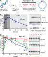
Figure 1. The D-loop cycle catalyzed by Rad51 and Rad54
A, Reaction scheme. B, Quantitation of D-loop reaction of a 100-mer paired with supercoiled plasmid DNA (denoted 100::sc), with interpretation of net formation and dissociation phases of the reaction. A representative time course is shown. Here, and in all subsequent figures, unless stated otherwise: Reactions are at 30 °C; homologous ssDNA is present at 0.61 μM nt, donor dsDNA at 21 μM bp (3 kb, 7 nM molecules); Rad51 is saturating with respect to the invading ssDNA at 1 Rad51 to 3 nts ssDNA; RPA is at 1 heterotrimer to 25 nt ssDNA; Rad54 is at 84 nM monomers; Rad51 is added to ssDNA for 10 min, then RPA for 10 min, then Rad54 is added with the dsDNA. C, Reaction time courses of 197::sc reactions. Rad51 filaments were allowed to form at 30 °C, then reactions were completed after dsDNA/Rad54 addition at 23, 30 or 37 °C (time ‘0’ onward). Shown are means ± standard deviations of three or more reactions.
Figure 2
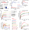
Figure 2. Physiological-length, tailed substrates form high levels of D-loops which are stable to dissociation
A, Scheme and nomenclature of substrates. B, Time courses of D-loop reactions with substrates shown in A paired with supercoiled plasmid DNA (0, 6, and 40 min time points). C, Quantitation of full D-loop time courses. Total D-loops are quantified, including single and MI species, if applicable. Fully single-stranded DNAs are plotted on the left and 5′-dsDNA-tailed DNA on the right. D, Time course gels (0, 1, 3, 6, 10, 15, 20, 40, and 60 min) showing the turnover of MI species. E, Quantitation of reactions shown in C. Shown are means ± standard deviations of three or more reactions.
Figure 3
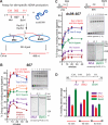
Figure 3. D-loops with short homologous ssDNAs are stabilized by heterologies flanking either side of the homology
D-loop reaction time courses of ssDNAs with 65 nt homology and the indicated heterologous double or single-stranded regions. The 65 nt homology substrates plotted on the left graph (A) have heterology on neither or one side of the homology, while those plotted on the right have heterologies on both sides (B). Shown are means ± standard deviations of three or more reactions.
Figure 4
Figure 4. Rad51 and Rad54, unlike RecA, form D-loops with linear donor dsDNA
A, Schematic of the homology of the 607 nt invading ssDNA within the Bsa1-linearized dsDNA substrate. B, RecA protein D-loops, formed with supercoiled or linearized dsDNA (21 μM bps). C, Rad51/Rad54 197 and ds98-197 D-loops formed with linearized (left) or supercoiled (inset; same data as Figure 2B). D, Comparison of 607 nt homology substrates with 5′ or 3′ double or single-stranded heterologies. Linear donor reactions are plotted on the left (above, total D-loops; below, multiple invasions) and supercoiled reactions are plotted on the right. Gel inset: 15 min time point of linear dsDNA reactions. The last lane is a control/migration marker ds98-607::sc reaction. Shown are means ± standard deviations of three or more reactions.
Figure 5
Figure 5. hDNA forms preferentially, but not obligatorily, at DNA ends with a modest preference for 3′ ssDNA ends
A, Scheme of the site-specific hDNA assay. B, 607::sc and C, 98ds-607::sc D-loop reaction quantitations, with representative gels shown beside. The invading substrates used in C are shown. D, hDNA analysis of the indicated 607 homology substrates including an internal BtsCI site. Dotted lines indicate the optional dsDNA heterologies.
Figure 6
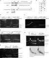
Figure 6. N-terminal fragments of yeast and human Rad54 preferentially bind ssDNA
A, Rad54 fragments cloned from yeast (y54-N) and human (h54-N) cDNAs contain the majority of the N-terminal domain. B, SDS-PAGE of purified GST fusions (2 μg) stained with coomassie blue. C, y54-N titration (0.6. 1.2, 2.3, 4.6, and 9.2 μM) on ssDNA (7.6 μM nt; ~ 3 kb, circular), dsDNA (7.6 μM bp; ~ 3 kb, linearized), and with ssDNA and dsDNA present (7.6 μM nt/bp each). The gel is stained with SYBR Gold. * and ^ mark minor dsDNA and ssDNA contaminants, respectively, in the ssDNA preparation. D, Pre-bound dsDNA or ssDNA y54-N complexes (7.6 μM nt/bp + 3 μM y54-N) were challenged by titrating in the other DNA species (1, 2, 3, 4, 6, 8, 10, 15 μM bp or nt). E, Comparison of the ability of protein-free ssDNA versus yRPA-ssDNA to disrupt pre-formed dsDNA:y54-N complexes (7.6 μM nt/bp plus 3 μM y54-N). ssDNA concentrations are 2, 4, 6, 10 and 15 μM nt, and when present yRPA is at 1 heterotrimer to 25 nts (saturating). F, h54-N was titrated on the indicated DNA species as in C. The lanes marked “GST” contain 11 μM purified GST. G, Challenge of pre-formed dsDNA:h54-N complexes with ssDNA or hRPA:ssDNA as in E. The gel was stained with CYBR Gold (upper), and then transferred to PVDF membrane and probed with anti-hRAD54 antibody (lower).
Figure 7
Figure 7. Heteroduplex DNA pump model for Rad54 function during HR. A, D-loop formation
The initial Rad51-mediated homologous joint DNA molecule is a three-stranded structure previously called a paranemic joint (Bianchi et al., 1983). Homologous base pairing occurs between the strands in some poorly understood fashion, but the invading strand is not yet in hDNA and the donor is no longer B-form dsDNA. To translocate, Rad54 requires dsDNA. A filament-terminal Rad51 monomer hands off the correct two DNA strands to Rad54, which begins to translocate on them, effectively pushing the two heteroduplex strands together ahead of itself to form the hDNA which it translocates upon. Rad51 filaments stimulate Rad54 translocation giving it directionality, resulting in the removal of Rad51 from the hDNA simultaneous with its creation. The displaced strand also stimulates Rad54 activity by binding the Rad54 N-terminal region, activating the Rad54 ATPase. B, D-loop dissociation. Rad54, translocating on the two nascent hDNA strands, can only incorporate the invading strand into hDNA until it reaches the end of that strand. Single-molecule studies of Rad54 dsDNA translocation have shown complex behavior, such as pausing, reversing direction, and restarting at new translocation velocities, suggesting a change in the molecular species responsible for translocation (Amitani et al., 2006). At the D-loop, such a conformational change may involve switching the strands occupied by the core dsDNA binding (motor) domain and the N-terminal ssDNA binding domain (case I). Binding of the invading strand by the N-terminal domain orients the motor to translocate on two original dsDNA strands and displaces the original invading strand. The result is D-loop disruption by a similar mechanism as formation, but with different roles taken by the individual strands. We suggest that the proposed reversal in hDNA pumping, like hDNA formation, preferentially initiates from ssDNA ends, which are absent when branchpoints flank the hDNA region (case II). The greatest effect in stabilizing D-loops is imparted by terminal dsDNA (Fig. 3), which would not be bound productively by the N-terminal regulatory domain of Rad54 to promote D-loop disruption. Protein and DNA elements are not meant to represent actual structure or scale.

References

    1. Alexeev A, Mazin A, Kowalczykowski SC. Rad54 protein possesses chromatin-remodeling activity stimulated by a Rad51-ssDNA nucleoprotein filament. Nature Struct Biol. 2003;10:182–186. - PubMed
    1. Alexiadis V, Lusser A, Kadonaga JT. A conserved N-terminal motif in Rad54 is important for chromatin remodeling and homologous strand pairing. J Biol Chem. 2004;279:27824–27829. - PubMed
    1. Amitani I, Baskin RJ, Kowalczykowski SC. Visualization of Rad54, a chromatin remodeling protein, translocating on single DNA molecules. Mol Cell. 2006;23:143–148. - PubMed
    1. Bianchi M, DasGupta C, Radding CM. Synapsis and the formation of paranemic joints by E. coli RecA protein. Cell. 1983;34:931–939. - PubMed
    1. Bigot S, Saleh OA, Lesterlin C, Pages C, El Karoui M, Dennis C, Grigoriev M, Allemand JF, Barre FX, Cornet F. KOPS: DNA motifs that control E. coli chromosome segregation by orienting the FtsK translocase. EMBO J. 2005;24:3770–3780. - PMC - PubMed

Publication types

MeSH terms