Review
. 2014 Apr 17;588(8):1221-9.
doi: 10.1016/j.febslet.2014.01.031. Epub 2014 Jan 30.

Degradation of connexins and gap junctions

Affiliations
Review

Degradation of connexins and gap junctions

Matthias M Falk et al. FEBS Lett. .

Abstract

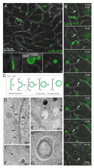
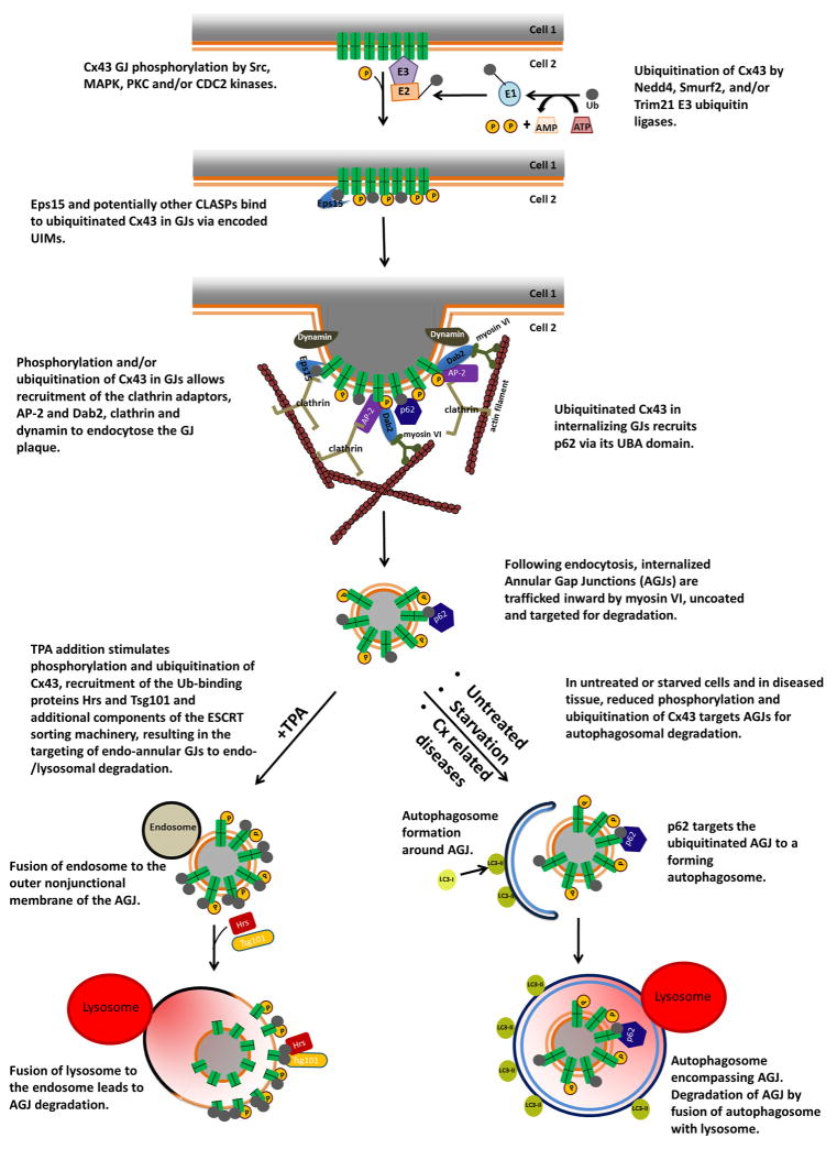
Connexin proteins are short-lived within the cell, whether present in the secretory pathway or in gap junction plaques. Their levels can be modulated by their rate of degradation. Connexins, at different stages of assembly, are degraded through the proteasomal, endo-/lysosomal, and phago-/lysosomal pathways. In this review, we summarize the current knowledge about connexin and gap junction degradation including the signals and protein-protein interactions that participate in their targeting for degradation.

Keywords: Autophagosome; Connexin; Gap junction; Lysosome; Proteasome; Ubiquitin.


Figures

Figure 1
Figure 1
Internalization of gap junction plaques. (A) Combined phase-contrast and fluorescence images of HeLa cells transfected with Cx43-GFP reveal efficient expression and assembly of gap junctions at appositional membranes of transfected cells (visible as green fluorescent lines and puncta such as the one shown in insert (1). Over time, gap junction plaques invaginate into the cytoplasm of one of the two cells (insert 2), detach from the plasma membrane and form endocytosed cytoplasmic annular gap junctions or connexosomes (insert 3). (B) Selected still-images of a time-lapse recording of stably transfected Cx43-YFP expressing HeLa cells showing the formation of a gap junction, its internalization into the cytoplasm of one of the adjacent cells, and subsequent degradation of the generated annular gap junction, indicated by the loss of its fluorescence (marked with arrows). (C) Schematic representation depicting the progressive stages of gap junction internalization shown in A and B. (D) Electron micrograph showing GJs (arrows) at the appositional membranes of mouse embryonic fibroblasts. (E) Electron micrograph showing an internalized GJ structure within a membrane-surrounded compartment, which probably corresponds to a secondary lysosome or autolysosome. Note that the pentalaminar pattern characteristic of GJs is being lost (arrow) in the internalized structure, probably because it is undergoing degradation. (F) Electron micrograph obtained from mouse embryonic fibroblasts starved by incubation in Hank’s balanced salt solution for 1 hour showing a double membrane structure, probably an autophagosome (arrow) that encloses an internalized GJ in close proximity to rough endoplasmic reticulum (short arrow). (G) Electron micrograph showing the internalized GJ enclosed within the autophagosome at higher magnification; the rough endoplasmic reticulum membrane is indicated by the short arrow. Nu: nucleus (A–C reproduced with permission from Landes Bioscience from ref. [91], D–G reproduced with permission from The Company of Biologists from ref. [93]).
Figure 2
Figure 2
Schematic representation of the signals participating in the proposed steps that lead to gap junction internalization, formation of annular gap junctions in the cytoplasm of the acceptor cell, and annular gap junction degradation through the phago-/lysosomal or the endo-/lysosomal pathway based on studies published in the literature. (Abbreviations are: AGJ, annular gap junction; CLASPs, clathrin-coat-associated proteins; ESCRT, endosomal sorting complexes required for transport; GJ, gap junction; UBA, ubiquitin-associated domain; UIMs, ubiquitin-interacting motifs.)

Similar articles

Cited by

References

    1. Beardslee MA, Laing JG, Beyer EC, Saffitz JE. Rapid turnover of connexin43 in the adult rat heart. Circ Res. 1998;83:629–35. - PubMed
    1. Berthoud VM, Minogue PJ, Laing JG, Beyer EC. Pathways for degradation of connexins and gap junctions. Cardiovasc Res. 2004;62:256–67. - PubMed
    1. Falk MM, Baker SM, Gumpert AM, Segretain D, Buckheit RW., 3rd Gap junction turnover is achieved by the internalization of small endocytic double-membrane vesicles. Mol Biol Cell. 2009;20:3342–52. - PMC - PubMed
    1. Fallon RF, Goodenough DA. Five-hour half-life of mouse liver gap-junction protein. J Cell Biol. 1981;90:521–6. - PMC - PubMed
    1. Gaietta G, et al. Multicolor and electron microscopic imaging of connexin trafficking. Science. 2002;296:503–7. - PubMed

Publication types