

Composite bone models in orthopaedic surgery research and education
- PMID: 24486757
- PMCID: PMC4251767
- DOI: 10.5435/JAAOS-22-02-111
Composite bone models in orthopaedic surgery research and education
Abstract
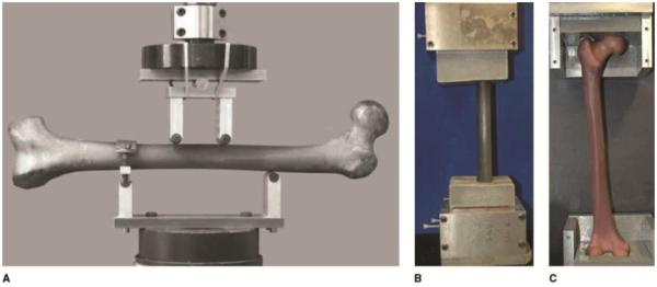
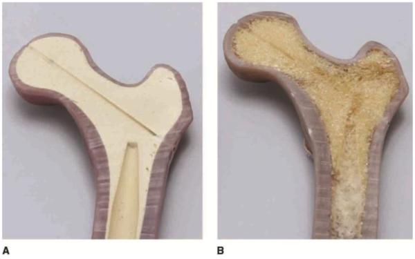
Composite bone models are increasingly used in orthopaedic biomechanics research and surgical education-applications that traditionally relied on cadavers. Cadaver bones are suboptimal for many reasons, including issues of cost, availability, preservation, and inconsistency between specimens. Further, cadaver samples disproportionately represent the elderly, whose bone quality may not be representative of the greater orthopaedic population. The current fourth-generation composite bone models provide an accurate reproduction of the biomechanical properties of human bone when placed under bending, axial, and torsional loads. The combination of glass fiber and epoxy resin components into a single phase has enabled manufacturing by injection molding. The high level of anatomic fidelity of the cadaver-based molds and negligible shrinkage properties of the epoxy resin results in a process that allows for excellent definition of anatomic detail in the cortical wall and optimized consistency of features between models. Recent biomechanical studies of composites have validated their use as a suitable substitute for cadaver specimens.
Figures
References
-
- Ferrari G. Public anatomy lessons and the carnival: The anatomy theatre of Bologna. Past Present. 1987;(117):50–106. Medline. - PubMed
-
- Sawbones . Product Catalogue. Pacific Research Laboratories; Vashon, WA: 2012. 2012. Available at: http://www.sawbones.com/products/productlist.aspx?111. Accessed December 6, 2013.
-
- Holland JP, Waugh L, Horgan A, Paleri V, Deehan DJ. Cadaveric hands-on training for surgical specialties: Is this back to the future for surgical skills development? J Surg Educ. 2011;68(2):110–116. Medline. - PubMed
-
- Kerckaert I, Van Hoof T, Pattyn P, D’Herde K. Endogent: Centre for Anatomy and Invasive Techniques. International Journal of Experimental and Clinical Anatomy. 2008;2:28–33.
-
- Thiel W. The preservation of the whole corpse with natural color [German] Ann Anat. 1992;174(3):185–195. Medline. - PubMed
Publication types
MeSH terms
Substances
Grants and funding
LinkOut - more resources
Full Text Sources
Other Literature Sources
