. 2014 Apr 1;306(7):L604-19.
doi: 10.1152/ajplung.00272.2013. Epub 2014 Jan 31.

MKK3 regulates mitochondrial biogenesis and mitophagy in sepsis-induced lung injury

Affiliations

MKK3 regulates mitochondrial biogenesis and mitophagy in sepsis-induced lung injury

Praveen Mannam et al. Am J Physiol Lung Cell Mol Physiol. .

Abstract

Sepsis is a systemic inflammatory response to infection and a major cause of death worldwide. Because specific therapies to treat sepsis are limited, and underlying pathogenesis is unclear, current medical care remains purely supportive. Therefore targeted therapies to treat sepsis need to be developed. Although an important mediator of sepsis is thought to be mitochondrial dysfunction, the underlying molecular mechanism is unclear. Modulation of mitochondrial processes may be an effective therapeutic strategy in sepsis. Here, we investigated the role of the kinase MKK3 in regulation of mitochondrial function in sepsis. Using clinically relevant animal models, we examined mitochondrial function in primary mouse lung endothelial cells exposed to LPS. MKK3 deficiency reduces lethality of sepsis in mice and by lowering levels of lung and mitochondrial injury as well as reactive oxygen species. Furthermore, MKK3 deficiency appeared to simultaneously increase mitochondrial biogenesis and mitophagy through the actions of Sirt1, Pink1, and Parkin. This led to a more robust mitochondrial network, which we propose provides protection against sepsis. We also detected higher MKK3 activation in isolated peripheral blood mononuclear cells from septic patients compared with nonseptic controls. Our findings demonstrate a critical role for mitochondria in the pathogenesis of sepsis that involves a previously unrecognized function of MKK3 in mitochondrial quality control. This mitochondrial pathway may help reveal new diagnostic markers and therapeutic targets against sepsis.

Keywords: biogenesis; lung injury; mitochondria; mitogen-activated protein kinases; mitophagy; sepsis.


Figures

Fig. 1.
Fig. 1.
MKK3−/− mice are resistant to lethal LPS. A: a lethal dose of intraperitoneal LPS (40 mg/kg) was given to wild-type (WT) mice and MKK3−/− mice and survival was assessed (n = 15, *P < 0.001). B: a lethal dose of intraperitoneal live Escherichia coli (1×10⁷ CFU/mouse) was given to WT mice and MKK3−/− mice and survival was assessed (n = 10, *P < 0.05). C: WT and MKK3−/− mice underwent either cecal ligation and puncture (n = 10 per group) or sham surgery (n = 4 per group) and survival was assessed (*P < 0.05). D: a sublethal dose of intraperitoneal LPS (5 mg/kg, 6 h) was administered to WT and MKK3−/− mice, and temperature and blood pressure were measured (n = 9, horizontal lines mark means, *P < 0.001). All results are representative of at least 3 independent experiments.
Fig. 2.
Fig. 2.
MKK3−/− mice were protected against lung injury after LPS. A: representative electron microscopy images of lungs of mice after intraperitoneal LPS (40 mg/kg, 12 h). Arrows point to inflammatory cells. Bar = 10 μM. B: representative electron microscopy images of lungs of mice after intraperitoneal LPS (40 mg/kg, 12 h). WT lungs show more mitochondrial damage characterized by pale swollen mitochondria with loss of normal architecture and distortion of cristae. Arrows indicate mitochondria. Bar = 1 μM. C: representative electron microscopy images of lungs of mice after intraperitoneal LPS (40 mg/kg, 12 h). Endothelial damage is shown by increased swelling, vacuolization and disruption of the inter-endothelial gap junctions in WT (arrows) compared with MKK3−/− mice (arrowheads). Top, bar = 1 μM; bottom, bar = 200 nm. D: bar graph of absolute number of neutrophils and macrophages were calculated as a fraction of the total number of cells in lung digests. WT and MKK3−/− mice were given intraperitoneal LPS (40 mg/kg, 6 h), the lungs were digested and analyzed by flow cytometry for neutrophils (Ly6G+, GR-1+) and macrophages (F4/80+, CD45+) (mean ± SE, n = 3, *P < 0.05). All results are representative of at least 3 independent experiments.
Fig. 3.
Fig. 3.
Mitochondrial injury and reactive oxygen species (ROS) production were lower in MKK3−/− mice and endothelial cells after LPS. A: bar graphs of mitochondrial DNA in serum of septic mice. WT and MKK3−/− mice given intraperitoneal LPS (40 mg/kg, 6 h) and serum was checked for the presence of mitochondrial DNA by quantitative PCR (qPCR). Mitochondrial genes detected were cytochrome b (CYTB), NADH dehydrogenase subunit 1 (ND1), ATPase 6 (AP6) and cytochrome c oxidase subunit I (COX1) (*P < 0.05). B: representative fluorescence quantification of LPS-exposed cells. WT and MKK3−/− lung endothelial cells were exposed to LPS (1 μg/ml, 6 h). C, control. Cells were stained with Mitosox red, which detects mitochondrial ROS, and levels were measured by flow cytometry. Values are expressed as mean fluorescent intensity ± SE (P < 0.05). All results are representative of at least 3 independent experiments.
Fig. 4.
Fig. 4.
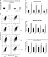
MKK3−/− endothelial cells have higher mitochondrial mass and potential compared with WT cells. A: bar graphs of relative mitochondrial DNA. Mitochondrial mass was measured in lungs and endothelial cells as relative copy number of mitochondrial gene (cytochrome b) compared with nuclear gene (18s RNA gene) (*P < 0.05). B: flow cytometry of WT and MKK3−/− endothelial cells left untreated or stimulated with LPS (1 μg/ml, 6 h) or carbonyl cyanide m-chlorophenyl hydrazine (CCCP, 50 μM) or ATP (5 mM) for 15 min. Cells were stained with Mitotracker Deep Red and Mitotracker Green before analysis. Data are representative of 3 experiments. C: quantitative representation of mean fluorescent intensity of Mitotracker staining and % of cells positive for both Mitotracker Deep Red and Green. Data are representative of 3 experiments (*P < 0.05 compared with WT cells, #P < 0.05 compared with untreated controls). All results are representative of at least 3 independent experiments.
Fig. 5.
Fig. 5.
Sirt1 expression and activity are increased in MKK3−/− lungs and endothelial cells. A: levels of Sirt1 checked by Western blots of lung lysates and endothelial cells after LPS exposure (40 mg/kg, 6 h and 1 μg/ml, 6 h respectively). Densitometric quantification of Sirt1 is shown in the graph. (*P < 0.05 compared with WT). B: Sirt1 levels were measured in WT and MKK3−/− lungs by qPCR (*P < 0.05). C: levels of PGC-1α checked by Western blots for lung lysates in WT and MKK3−/− mice after LPS exposure (40 mg/kg, 6 h). Densitometric quantification of PGC-1α is shown in the graph. (*P < 0.05 compared with WT). D: PGC-1α levels were measured in WT and MKK3−/− lungs by qPCR (*P < 0.05). E: lung lysates from control and LPS-exposed mice (40 mg/kg, 6 h) were immunoprecipitated (IP) first with a PGC-1α-specific antibody followed by Western immunoblotting (IB) with a acetylated lysine antibody. − control, negative control in which lung lysates were processed without the immunoprecipitation antibody. Densitometric quantification of acetylated lysine is shown in the graph. (*P < 0.05 compared with WT). F: nuclear extracts from lungs of control and LPS-exposed mice (40 mg/kg, 6 h) were checked by Western blots for levels of transcription factor Nrf1. Lamin B was used as a nuclear loading control. Densitometric quantification of Nrf1 is shown in the graph. (*P < 0.05 compared with WT). G: Nrf1 levels were measured in WT and MKK3−/− lungs by qPCR (*P < 0.05). All results are representative of at least 3 independent experiments.
Fig. 6.
Fig. 6.
MKK3−/− endothelial cells have higher levels of mitophagy than WT endothelial cells. A: Western blots of endothelial cells. LC3B, a marker of mitophagy, was measured in control and LPS-exposed (1 μg/ml, 6 h) endothelial cells. Actin is the loading control. B: fluorescent microscopy of control and LPS exposed (1 μg/ml, 6 h) endothelial cells to show localization of LC3B with the mitochondria. LC3B is stained red and mitochondria are stained with antibodies against Hsp60 (green). Magnification ×100. C: quantification of colocalization is represented by the colocalization coefficient, the fraction of LC3B that colocalizes with Hsp60 (*P < 0.05 compared with WT cells, #P < 0.05 compared with untreated control). D: Western blots of MKK3−/− endothelial cells. Autophagy flux was checked in MKK3−/− endothelial cells by measuring LC3B levels after treatment with leupeptin (250 μM for 14 h). All results are representative of at least 3 independent experiments.
Fig. 7.
Fig. 7.
MKK3−/− mice have higher levels of Pink1 and Parkin than WT. Pink1 and Parkin (markers of mitophagy) were measured by Western blot analysis in WT and MKK3−/− lungs and endothelial cells. Densitometric quantifications of Pink1 and Parkin are shown in the graphs. (*P < 0.05 compared with WT). All results are representative of at least 3 independent experiments.
Fig. 8.
Fig. 8.
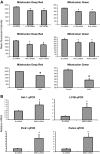
Inhibition of Sirt1 and mitophagy reverses the mitochondrial integrity of MKK3−/− endothelial cells. A: quantitative representation of mean fluorescent intensity of Mitotracker staining. Sirt-1 was inhibited by using siRNA and mitophagy was inhibited in MKK3−/− endothelial cells with LC3B, Pink1, and Parkin siRNA and 3-methylademine (3-MA, 5 mM for 4 h). Cells were stained with Mitotracker Deep Red and Mitotracker Green before analysis (*P < 0.05). B: qPCR of Sirt-1, LC3B, Pink1, and Parkin mRNA were measured after MKK3 silencing by using siRNA in WT endothelial cells with siRNA (*P < 0.05). All results are representative of at least 3 independent experiments.
Fig. 9.
Fig. 9.
Inhibition of mitophagy in mice increased susceptibility to sepsis and lung injury. A: a lethal dose of intraperitoneal LPS (40 mg/kg) was given to WT mice and heterozygous MKK3+/−, Pink1+/−, and Pink+/−, MKK3+/− mice and survival assessed (n = 7, *P < 0.05 compared with WT and Pink+/− mice). B: Pink1 mRNA levels were checked by qPCR in lungs of WT mice and heterozygous MKK3+/−, Pink1+/− and Pink+/−, MKK3+/− mice (*P < 0.05).
Fig. 10.
Fig. 10.
MKK3 and Pink1 levels were higher in septic patients. A: p-MKK3/6 levels were measured in peripheral blood mononuclear cells (PBMCs) of septic and nonseptic intensive care unit patients by AlphaScreen SureFire assay. Relative expression is normalized to HeLa negative controls (*P < 0.05). B: MKK3, p-MKK3, and Pink1 levels were measured in PBMCs of septic and nonseptic patients by Western blots (black arrow). Relative expression was normalized to HeLa negative controls and β-actin levels. Gray arrows in bottom panels indicate the relative expression of MKK3, p-MKK3, and Pink1 levels in a severely ill patient with gram-negative rod bacteremia and shock (*P < 0.05).
Fig. 11.
Fig. 11.
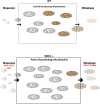
Summary of MKK3 effects on mitochondrial health. We postulate that MKK3 deficiency increases both biogenesis (through action of Sirt1, PGC-1α, and Nrf1) and mitophagy (through the actions of Pink1 and Parkin), leading to a larger pool of healthy mitochondria (gray) and less ROS-producing defective mitochondria (brown). The net effect is protection against lethal sepsis.

References

    1. Abramoff MD, Magalhães PJ, Ram SJ. Image processing with ImageJ. Biophotonics Int 11: 36–42, 2004
    1. Adrie C, Bachelet M, Vayssier-Taussat M, Russo-Marie F, Bouchaert I, Adib-Conquy M, Cavaillon JM, Pinsky MR, Dhainaut JF, Polla BS. Mitochondrial membrane potential and apoptosis peripheral blood monocytes in severe human sepsis. Am J Respir Crit Care Med 164: 389–395, 2001 - PubMed
    1. Bone RC, Balk RA, Cerra FB, Dellinger RP, Fein AM, Knaus WA, Schein RM, Sibbald WJ; ACCP/SCCM Consensus Conference Committee. Definitions for sepsis and organ failure and guidelines for the use of innovative therapies in sepsis. The ACCP/SCCM Consensus Conference Committee American College of Chest Physicians/Society of Critical Care Medicine. 1992. Chest 136: e28, 2009 - PubMed
    1. Brealey D, Brand M, Hargreaves I, Heales S, Land J, Smolenski R, Davies NA, Cooper CE, Singer M. Association between mitochondrial dysfunction and severity and outcome of septic shock. Lancet 360: 219–223, 2002 - PubMed
    1. Carchman EH, Whelan S, Loughran P, Mollen K, Stratamirovic S, Shiva S, Rosengart MR, Zuckerbraun BS. Experimental sepsis-induced mitochondrial biogenesis is dependent on autophagy, TLR4, and TLR9 signaling in liver. FASEB J 27: 4703–4711, 2013 - PMC - PubMed

Publication types

MeSH terms

LinkOut - more resources