. 2014 Mar;7(3):387-96.
doi: 10.1242/dmm.013995. Epub 2014 Jan 30.

Subcutaneous preconditioning increases invasion and metastatic dissemination in mouse colorectal cancer models

Affiliations

Subcutaneous preconditioning increases invasion and metastatic dissemination in mouse colorectal cancer models

Patricia Alamo et al. Dis Model Mech. 2014 Mar.

Abstract

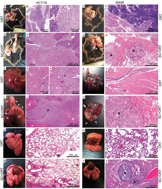
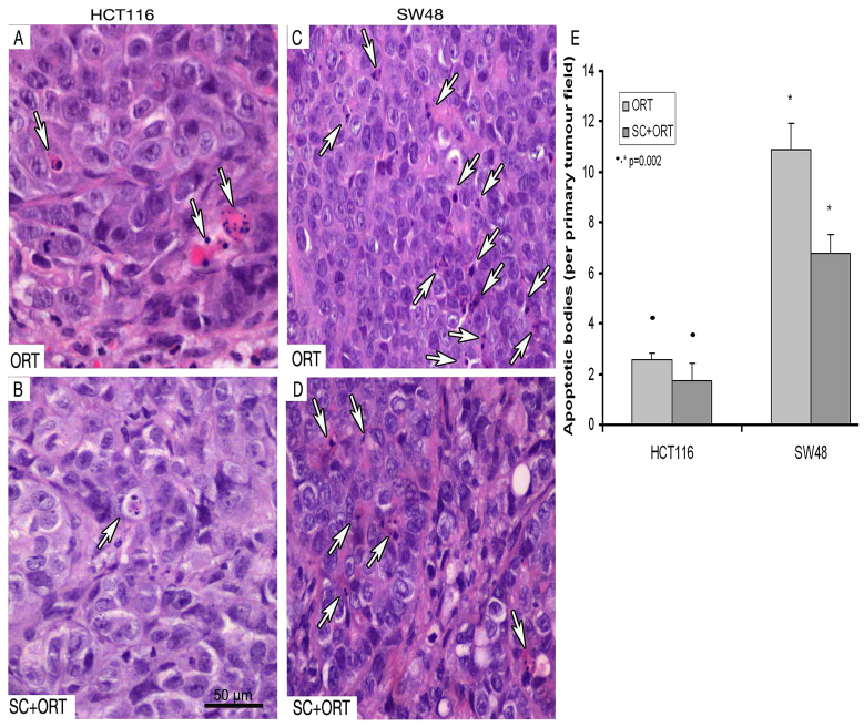
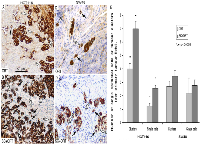
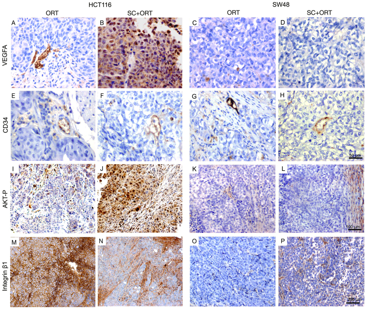
Mouse colorectal cancer (CRC) models generated by orthotopic microinjection of human CRC cell lines reproduce the pattern of lymphatic, haematological and transcoelomic spread but generate low metastatic efficiency. Our aim was to develop a new strategy that could increase the metastatic efficiency of these models. We used subcutaneous implantation of the human CRC cell lines HCT116 or SW48 prior to their orthotopic microinjection in the cecum of nude mice (SC+ORT). This subcutaneous preconditioning significantly enhanced metastatic dissemination. In the HCT116 model it increased the number and size of metastatic foci in lymph nodes, lung, liver and peritoneum, whereas, in the SW48 model, it induced a shift from non-metastatic to metastatic. In both models the number of apoptotic bodies in the primary tumour in the SC+ORT group was significantly reduced compared with that in the direct orthotopic injection (ORT) group. Moreover, in HCT116 tumours the number of keratin-positive tumour buddings and single epithelial cells increased at the invasion front in SC+ORT mice. In the SW48 tumour model, we observed a trend towards a higher number of tumour buds and single cells in the SC+ORT group but this did not reach statistical significance. At a molecular level, the enhanced metastatic efficiency observed in the HCT116 SC+ORT model was associated with an increase in AKT activation, VEGF-A overexpression and downregulation of β1 integrin in primary tumour tissue, whereas, in SW48 SC+ORT mice, the level of expression of these proteins remained unchanged. In summary, subcutaneous preconditioning increased the metastatic dissemination of both orthotopic CRC models by increasing tumour cell survival and invasion at the tumour invasion front. This approach could be useful to simultaneously study the mechanisms of metastases and to evaluate anti-metastatic drugs against CRC.

Keywords: Collective invasion; Colorectal cancer model; Metastasis; Orthotopic injection; Single tumour cell; Subcutaneous preconditioning.


Figures

Fig. 1.
Fig. 1.
Enhanced metastatic efficiency in SC+ORT mice. (A–E, O–R) Lymph node metastases: the HCT116-ORT group showed micro- and macrometastasis (B, C), and visible lymph node metastases (A) that were smaller than those in the SC+ORT group. We recorded a low number of visible metastases (white arrow), involving mainly mesenteric lymph nodes (A), and only one visible peripancreatic lymph node. In contrast, in the HCT116-SC+ORT group (D,E), we observed a high number of visible metastases, affecting the peripancreatic parenchyma and the mesenteric lymph nodes (white arrows, D). In addition to the isolated visible metastases in the SC−ORT group, four animals developed a massive conglomerate of peripancreatic lymph node metastases (white arrows) involving the whole pancreas (D); these metastases were physically connected under the microscope (white asterisk, E). SW48 ORT mice lacked lymphatic dissemination (L,O). In contrast, SW48 SC+ORT mice showed lymphatic micro, macro and visible dissemination (white arrow in Q shows visible metastases and asterisks in R show micrometastases; Q,R). PT, primary tumour. (F–J) Liver metastases: in the liver of mice in the HCT116-ORT group (F–H), all metastases were microscopic (G,H, black asterisks). In contrast, in the HCT116-SC+ORT group we observed a larger number of microscopic metastases than in the ORT group, in addition to macroscopic and single visible metastases. Moreover, in two of the animals in the SC+ORT group (I,J), massive visible liver metastases developed (white arrows), forming a conglomerate (I) that invaded the hepatic parenchyma (white asterisks) and involved 95% of its area. Black asterisk in J shows macrometastasis. No liver metastases were observed in any group of SW48 mice (not shown). (S–V) Peritoneal metastases: no metastases were detected in the SW48-ORT group (S,T). In contrast, SW48 SC+ORT mice developed micro- and visible metastases (U,V, white arrows and black asterisk). (K–N,W–Z) Lung metastases: mice in the HCT116-SC+ORT group (M,N) developed a significantly higher number of microfoci in their lung (N, black asterisks) than mice in the ORT (K,L) group. In the SW48-ORT group, no pulmonary metastases were recorded (W,X). In contrast, SW48 SC+ORT mice developed micro- and macrometastases in the lung (Y,Z). Type of metastasis as a function of its diameter: microfoci <1 mm; macrofoci 1–3 mm; visible >3 mm; H&E staining. Scale bars for magnification.
Fig. 2.
Fig. 2.
Increased number of apoptotic bodies in the primary tumour in the ORT groups. The primary tumours in the HCT116-ORT group (A) showed a significantly (P=0.002) higher number of apoptotic cells (white arrows) than primary tumours in the HCT116-SC+ORT group (B,E). Primary tumours in the SW48-ORT group (C) also displayed an increased number of apoptotic bodies (white arrows) as compared with the SW48-ORT group (D,E). Quantification of apoptotic bodies was performed in a 400× magnified primary tumour field. Scale bar for magnification (A–D all have the same magnification).
Fig. 3.
Fig. 3.
Increased number of tumour cell clusters and single cells at the primary tumour invasive front in SC+ORT tumours. The primary tumours in the HCT116-SC+ORT group (B) showed a significantly (P<0.001) larger number of pan-keratin-positive small clusters of tumour epithelial cells, surrounded by stroma (white arrows), than primary tumours in the HCT116-ORT group (A,E). Moreover, the number of pan-keratin-positive single tumour epithelial cells, completely surrounded by stroma, at the invasive front in HCT116-SC+ORT tumours (B, black arrows) was also significantly (P<0.001) higher than in ORT tumours (A,E). The tumour front in SW48-SC+ORT mice (D) displayed an increased number of clusters (white arrows) and single cells (black arrows) compared with the tumour front in SW48-ORT mice (C,E). (E) Quantification of tumour cell clusters of five or fewer cells (tumour clusters or budding) or single epithelial cells per 200× magnified primary tumour field observed in ORT and SC+ORT tumours. Scale bar for magnification (A–D all have the same magnification).
Fig. 4.
Fig. 4.
Expression of proteins that regulate survival and invasion in primary tumours in ORT and SC+ORT groups. No differences in the pattern and number of vessels, assessed by CD34 immunostaining, were observed between ORT and SC+ORT groups in HCT116 or SW48 mice (E–H). Interestingly, primary tumours in the HCT116-ORT (A) group showed significantly lower VEGFA expression than the SC+ORT group (B), whereas no VEGFA expression was observed in any of the SW48 groups (C,D). Primary tumours in the HCT116-ORT group (I) presented a lower level of AKT activation than that observed in HCT116 SC+ORT tumours (J). No differences in AKT activation were observed in primary tumours between ORT and SC+ORT groups in SW48 mice (K,L). HCT116-ORT primary tumours (M) showed a significantly higher level of β1 integrin expression than SC+ORT primary tumours (N), whereas no expression of β1 integrin was detected in primary tumours in any groups of SW48 mice (O,P). Scale bars for magnification (A–H, I–L and M–P have the same magnification).

Similar articles

Cited by

References

    1. Adell G. C., Zhang H., Evertsson S., Sun X. F., Stål O. H., Nordenskjöld B. A. (2001). Apoptosis in rectal carcinoma: prognosis and recurrence after preoperative radiotherapy. Cancer 91, 1870–1875 - PubMed
    1. Ahluwalia A., Jones M. K., Budnik T. M., Tarnawski A. S. (2013). VEGF and colon cancer growth beyond angiogenesis: Does VEGF directly mediate colon cancer growth via a non-angiogenic mechanism? Curr. Pharm. Des. [Epub ahead of print] 10.2174/1381612819999131218175905 - DOI - PubMed
    1. Bacac M., Stamenkovic I. (2008). Metastatic cancer cell. Annu. Rev. Pathol. 3, 221–247 - PubMed
    1. Bendardaf R., Ristamäki R., Kujari H., Laine J., Lamlum H., Collan Y., Pyrhönen S. (2003). Apoptotic index and bcl-2 expression as prognostic factors in colorectal carcinoma. Oncology 64, 435–442 - PubMed
    1. Bosch R., Moreno M. J., Dieguez-Gonzalez R., Céspedes M. V., Gallardo A., Nomdedeu J., Pavón M. A., Espinosa I., Mangues M. A., Sierra J., et al. (2012). Subcutaneous passage increases cell aggressiveness in a xenograft model of diffuse large B cell lymphoma. Clin. Exp. Metastasis 29, 339–347 - PubMed

Publication types

Substances