. 2014 Apr;80(7):2176-85.
doi: 10.1128/AEM.03627-13. Epub 2014 Jan 31.

Acidic pH strongly enhances in vitro biofilm formation by a subset of hypervirulent ST-17 Streptococcus agalactiae strains

Affiliations

Acidic pH strongly enhances in vitro biofilm formation by a subset of hypervirulent ST-17 Streptococcus agalactiae strains

Nunzia D'Urzo et al. Appl Environ Microbiol. 2014 Apr.

Abstract

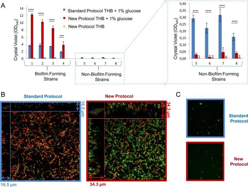
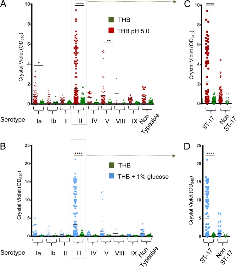
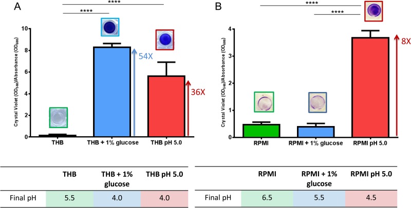
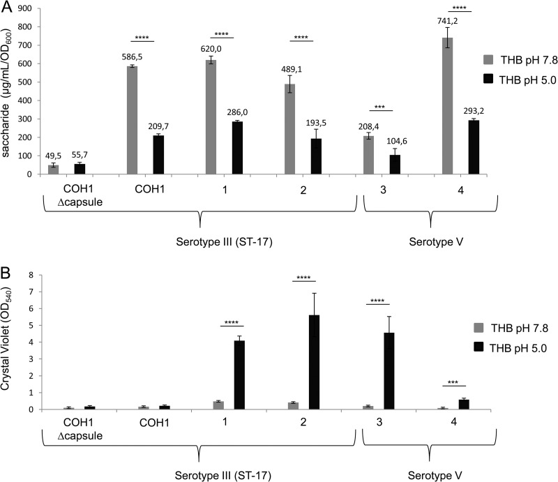
Streptococcus agalactiae, also known as group B Streptococcus (GBS), is a primary colonizer of the anogenital mucosa of up to 40% of healthy women and an important cause of invasive neonatal infections worldwide. Among the 10 known capsular serotypes, GBS type III accounts for 30 to 76% of the cases of neonatal meningitis. In recent years, the ability of GBS to form biofilm attracted attention for its possible role in fitness and virulence. Here, a new in vitro biofilm formation protocol was developed to guarantee more stringent conditions, to better discriminate between strong-, low-, and non-biofilm-forming strains, and to facilitate interpretation of data. This protocol was used to screen the biofilm-forming abilities of 366 GBS clinical isolates from pregnant women and from neonatal infections of different serotypes in relation to medium composition and pH. The results identified a subset of isolates of serotypes III and V that formed strong biofilms under acidic conditions. Importantly, the best biofilm formers belonged to serotype III hypervirulent clone ST-17. Moreover, the abilities of proteinase K to strongly inhibit biofilm formation and to disaggregate mature biofilms suggested that proteins play an essential role in promoting GBS biofilm initiation and contribute to biofilm structural stability.


Figures

FIG 1
FIG 1
Comparison of biofilm formation protocols. (A) Biofilm formation abilities of four strong biofilm-forming strains and four weak biofilm formers or non-biofilm formers produced by the standard protocol and the new protocol. The GBS strains were grown in THB or in THB supplemented with 1% glucose. Surface-attached cells were CV stained and quantified by measuring the OD540. The mean values and standard deviations of three independent experiments are shown. Asterisks denote statistically significant difference determined by Student's t test (***, P < 0.001; ****, P < 0.0001). (B) CLSM of biofilms formed by a strong biofilm-forming strain with the standard protocol and the new protocol in THB in the presence of 1% glucose. (C) CLSM of the biofilm produced by a non-biofilm-forming strain with the standard protocol and the new protocol in THB in the presence of 1% glucose. Biofilms were stained with a LIVE/DEAD viability kit. Live and dead cells are green and red, respectively.
FIG 2
FIG 2
Effects of an acidic pH and glucose on GBS biofilm formation. Shown are the biofilm-forming abilities of 366 GBS clinical isolates of eight different serotypes and nontypeable strains grown in THB (green dots) or pH 5.0 THB (red dots) (A) or in THB (green dots) or THB supplemented with 1% glucose (red dots) (B). Panels C and D focus on serotype III strains clustered into ST-17 and non-ST-17 groups. Biofilm formation was evaluated by CV assay. Each dot represents the mean value of three independent experiments performed with each isolate. Asterisks denote statistically significant differences determined by ANOVA (*, P < 0.05; **, P < 0.01; ****, P < 0.0001).
FIG 3
FIG 3
An acidic pH and not glucose induces GBS biofilm formation. Comparison of the biofilm formation abilities of a strong biofilm-forming strain grown in THB, THB supplemented with 1% glucose, or pH 5.0 THB (A) or in RPMI, RPMI supplemented with 1% glucose, or pH 5.0 RPMI (B). Biofilm formation was evaluated by CV assay. Bacterial growth in planktonic form was evaluated by measuring OD600. pH values were measured with pH test strips (pH increment, 0.2). To normalize the biofilm-forming ability with cell growth, the y axis shows the ratio of CV assay values to OD600 values. The mean values and standard deviations of three independent experiments are shown. Asterisks denote statistically significant difference, determined by Student's t test (****, P < 0.0001).
FIG 4
FIG 4
Correlation between capsule expression and pH. Shown is an evaluation of the capsule amounts (A) and biofilm formation (B) of four serotype III and two serotype V biofilm-forming and non-biofilm-forming strains at pH 7.8 (gray) and pH 5.0 (black). Capsular polysaccharides were isolated and quantified with the resorcinol-hydrochloric acid assay by Svennerholm's method. Surface-attached cells were CV stained, and the OD540 was measured. Unencapsulated COH1 was used as a control. The mean values and standard deviations of three independent experiments are shown. Asterisks denote statistically significant differences determined by Student's t test (***, P < 0.001; ****, P < 0.0001).
FIG 5
FIG 5
Enzymatic inhibition or eradication of GBS biofilm. Shown are the inhibition (A) and eradication (B) of biofilms formed by three serotype III and three serotype V biofilm-forming strains by proteinase K or DNase. Surface-attached cells were quantified by CV assay. The mean values and standard deviations of three independent experiments are shown.

Similar articles

Cited by

References

    1. Wilkinson HW. 1978. Group B streptococcal infection in humans. Annu. Rev. Microbiol. 32:41–57. 10.1146/annurev.mi.32.100178.000353 - DOI - PubMed
    1. Fry RM. 1938. Prevention and control of puerperal sepsis: bacteriological aspects. BMJ 2:340–342. 10.1136/bmj.2.4049.340 - DOI - PMC - PubMed
    1. Schuchat A. 1999. Group B streptococcus. Lancet 353:51–56. 10.1016/S0140-6736(98)07128-1 - DOI - PubMed
    1. Schuchat A. 1998. Epidemiology of group B streptococcal disease in the United States: shifting paradigms. Clin. Microbiol. Rev. 11:497–513 - PMC - PubMed
    1. Whidbey C, Harrell MI, Burnside K, Ngo L, Becraft AK, Iyer LM, Aravind L, Hitti J, Waldorf KM, Rajagopal L. 2013. A hemolytic pigment of group B Streptococcus allows bacterial penetration of human placenta. J. Exp. Med. 210:1265–1281. 10.1084/jem.20122753 - DOI - PMC - PubMed

Publication types

MeSH terms

LinkOut - more resources