

Diallyl trisulfide inhibits estrogen receptor-α activity in human breast cancer cells
- PMID: 24487688
- PMCID: PMC3941172
- DOI: 10.1007/s10549-014-2841-x
Diallyl trisulfide inhibits estrogen receptor-α activity in human breast cancer cells
Abstract
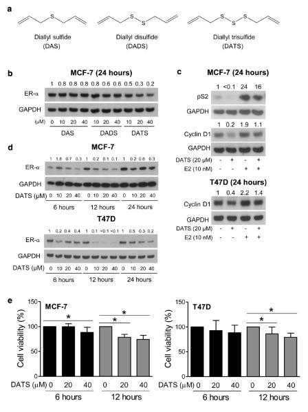
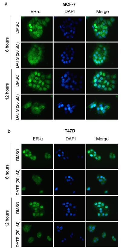
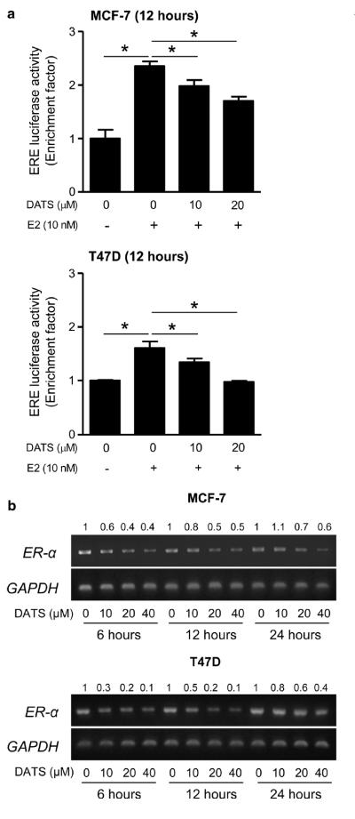
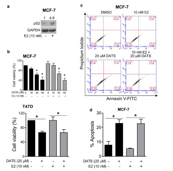
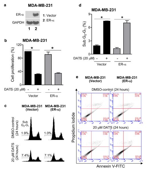
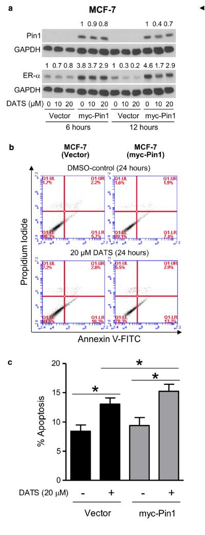
Organosulfur compounds from garlic effectively inhibit growth of transplanted as well as spontaneous cancers in preclinical animal models without any adverse side effects. However, the mechanisms underlying anticancer effect of this class of compounds are not fully understood. This study reports, for the first time, that garlic organosulfide diallyl trisulfide (DATS) inhibits estrogen receptor-α (ER-α) activity in human breast cancer cells. Exposure of MCF-7 and T47D cells to DATS resulted in downregulation of ER-α protein, which peaked between 12- and 24-h post-treatment. DATS was relatively more effective in suppressing ER-α protein expression compared with its mono and disulfide analogs. The 17β-estradiol (E2)-induced expression of pS2 and cyclin D1, ER-α target gene products, was also decreased in the presence of DATS. Downregulation of ER-α protein expression resulting from DATS treatment was accompanied by a decrease in nuclear levels of ER-α protein, ER-α mRNA suppression, and inhibition of ERE2e1b-luciferase reporter activity. DATS-mediated inhibition of cell viability and apoptosis induction were not affected in the presence of E2. In agreement with these results, ectopic expression of ER-α in MDA-MB-231 cell line failed to confer any protection against cell proliferation inhibition or apoptosis induction resulting from DATS exposure. DATS treatment caused a decrease in protein levels of peptidyl-prolyl cis-trans isomerase (Pin1), and overexpression of Pin1 partially attenuated ER-α downregulation by DATS. DATS-induced apoptosis was modestly but significantly augmented by overexpression of Pin1. In conclusion, this study identifies ER-α as a novel target of DATS in mammary cancer cells.
Figures
Similar articles
-
Critical role for reactive oxygen species in apoptosis induction and cell migration inhibition by diallyl trisulfide, a cancer chemopreventive component of garlic.Breast Cancer Res Treat. 2013 Feb;138(1):69-79. doi: 10.1007/s10549-013-2440-2. Epub 2013 Feb 15. Breast Cancer Res Treat. 2013. PMID: 23412769 Free PMC article.
-
Garlic (Allium sativum L.) Organosulfur Compounds Inhibit Breast Cancer Cell Proliferation by Decreasing Steroid Sulfatase Levels.Anticancer Res. 2025 Jan;45(1):145-152. doi: 10.21873/anticanres.17401. Anticancer Res. 2025. PMID: 39740834
-
Garlic constituent diallyl trisulfide induced apoptosis in MCF7 human breast cancer cells.Cancer Biol Ther. 2009 Nov;8(22):2175-85. doi: 10.4161/cbt.8.22.9882. Epub 2009 Nov 22. Cancer Biol Ther. 2009. PMID: 19823037
-
Molecular mechanisms for the anti-cancer effects of diallyl disulfide.Food Chem Toxicol. 2013 Jul;57:362-70. doi: 10.1016/j.fct.2013.04.001. Epub 2013 Apr 9. Food Chem Toxicol. 2013. PMID: 23583486 Review.
-
Garlic-derived compounds: Epigenetic modulators and their antitumor effects.Phytother Res. 2024 Mar;38(3):1329-1344. doi: 10.1002/ptr.8108. Epub 2024 Jan 9. Phytother Res. 2024. PMID: 38194996 Review.
Cited by
-
Spices for Prevention and Treatment of Cancers.Nutrients. 2016 Aug 12;8(8):495. doi: 10.3390/nu8080495. Nutrients. 2016. PMID: 27529277 Free PMC article. Review.
-
Health Benefits of Plant-Derived Sulfur Compounds, Glucosinolates, and Organosulfur Compounds.Molecules. 2020 Aug 21;25(17):3804. doi: 10.3390/molecules25173804. Molecules. 2020. PMID: 32825600 Free PMC article. Review.
-
Diet, Sports, and Psychological Stress as Modulators of Breast Cancer Risk: Focus on OPRM1 Methylation.Front Nutr. 2021 Dec 8;8:747964. doi: 10.3389/fnut.2021.747964. eCollection 2021. Front Nutr. 2021. PMID: 35024367 Free PMC article.
-
The potential role of hydrogen sulfide in cancer cell apoptosis.Cell Death Discov. 2024 Mar 6;10(1):114. doi: 10.1038/s41420-024-01868-w. Cell Death Discov. 2024. PMID: 38448410 Free PMC article. Review.
-
Mechanistic Targets of Diallyl Trisulfide in Human Breast Cancer Cells Identified by RNA-seq Analysis.J Cancer Prev. 2021 Jun 30;26(2):128-136. doi: 10.15430/JCP.2021.26.2.128. J Cancer Prev. 2021. PMID: 34258251 Free PMC article.
References
-
- Rivlin RS. Historical perspective on the use of garlic. J Nutr. 2001;131(3):951S–954S. - PubMed
-
- Rahman K. Historical perspective on garlic and cardio-vascular disease. J Nutr. 2001;131(3):977S–979S. - PubMed
-
- Dausch JG, Nixon DW. Garlic: a review of its relationship to malignant disease. Prev Med. 1990;19(3):346–361. - PubMed
-
- Agarwal KC. Therapeutic actions of garlic constituents. Med Res Rev. 1996;16(1):111–124. - PubMed
-
- El-Bayoumy K, Sinha R, Pinto JT, Rivlin RS. Cancer chemoprevention by garlic and garlic-containing sulfur and selenium compounds. J Nutr. 2006;136(3):864S–869S. - PubMed
Publication types
MeSH terms
Substances
Grants and funding
LinkOut - more resources
Full Text Sources
Other Literature Sources
Medical
Research Materials
Miscellaneous
