. 2014:5:3206.
doi: 10.1038/ncomms4206.

Iridium oxide nanotube electrodes for sensitive and prolonged intracellular measurement of action potentials

Affiliations

Iridium oxide nanotube electrodes for sensitive and prolonged intracellular measurement of action potentials

Ziliang Carter Lin et al. Nat Commun. 2014.

Abstract

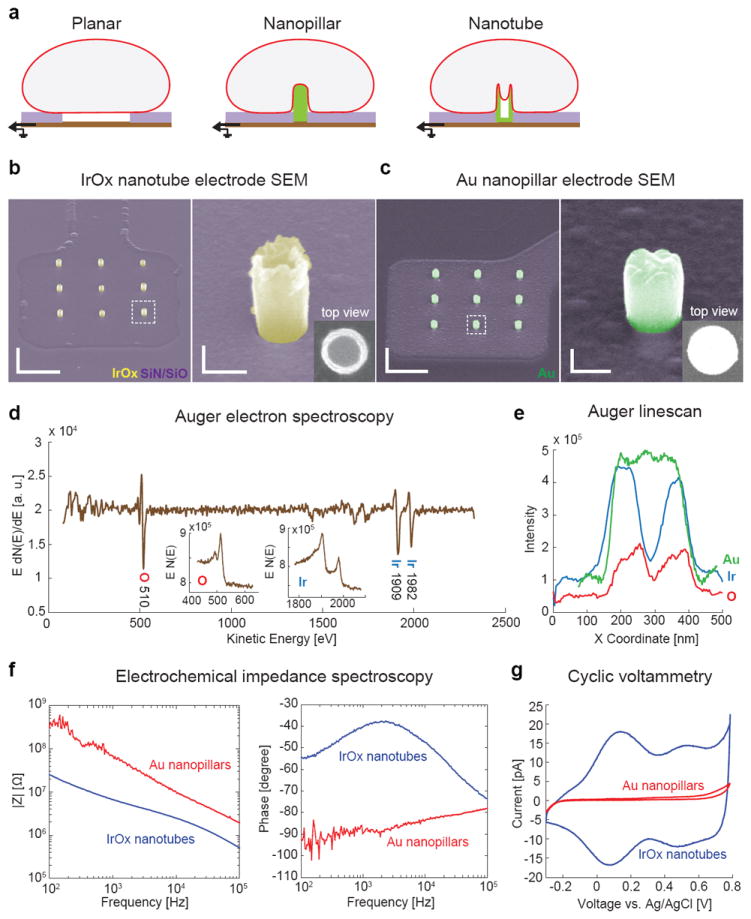
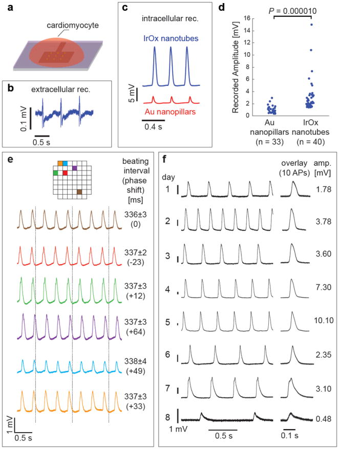
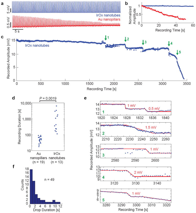
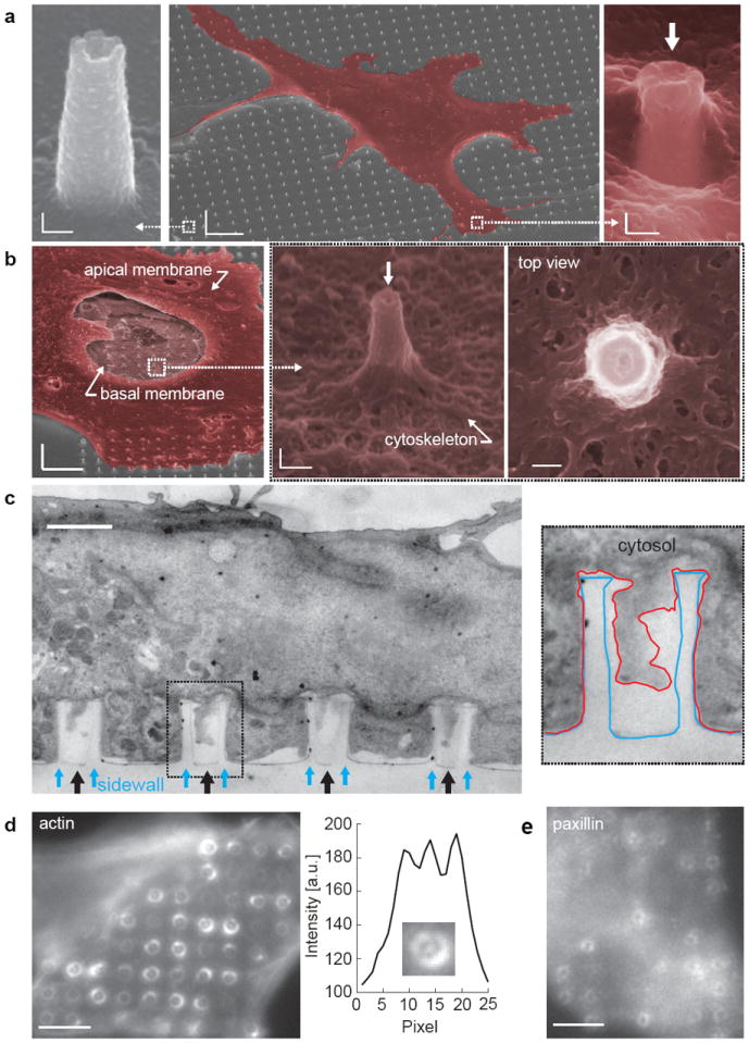
Intracellular recording of action potentials is important to understand electrically-excitable cells. Recently, vertical nanoelectrodes have been developed to achieve highly sensitive, minimally invasive and large-scale intracellular recording. It has been demonstrated that the vertical geometry is crucial for the enhanced signal detection. Here we develop nanoelectrodes of a new geometry, namely nanotubes of iridium oxide. When cardiomyocytes are cultured upon those nanotubes, the cell membrane not only wraps around the vertical tubes but also protrudes deep into the hollow centre. We show that this nanotube geometry enhances cell-electrode coupling and results in larger signals than solid nanoelectrodes. The nanotube electrodes also afford much longer intracellular access and are minimally invasive, making it possible to achieve stable recording up to an hour in a single session and more than 8 days of consecutive daily recording. This study suggests that the nanoelectrode performance can be significantly improved by optimizing the electrode geometry.


Conflict of interest statement

Conflict of Interests The authors declare competing financial interests. A patent provision (13-019) has been filed by Stanford University.

Figures

Figure 1
Figure 1. Characterization of vertical IrOx nanotube electrode
a, Schematics of cells interfacing with planar, vertical nanopillar and vertical nanotube electrodes. The cell membrane not only wraps around the outside of the nanotube but also protrudes inside the hollow center. b, SEM images of a three-by-three array of IrOx nanotubes on a square Pt pad with the rest of the area insulted with Si3N4/SiO2. The hollow center of the nanotube can be clearly seen in expanded 52°-tilted view and the top view. Scale bars: left 2 μm, right 200 nm.c, SEM images of Au nanopillar electrodes show solid pillars. The diameter of the Au nanopillar is designed to be slightly larger than the IrOx nanotube so that they have similar surface area. Scale bars: left 2 μm, right 200 nm.d, Auger electron spectrum of the nanotube electrodes confirms the presence of iridium and oxygen (insets: raw spectra). e, Elemental line scans along the diameter of an IrOx nanotube and a Au nanopillar. Ir and O plots (blue and red) show two peaks at the side wall and a drop at the center, while Au (green) shows a flat top. f, Electrochemical impedance spectroscopy of IrOx nanotube and Au nanopillar electrodes in phosphate buffered saline. g, Cyclic voltammetry of IrOx nanotube and Au nanopillar electrodes in PBS at a scan rate of 30 mV/s.
Figure 2
Figure 2. Intracellular recording of action potentials by the IrOx nanotube electrodes
a, Schematic illustration of electrophysiology recording using vertical IrOx nanotube electrodes. b, Before local electroporation, nanotube electrodes measured extracellular action potentials in HL-1 cardiomyocytes. c, Immediately after local electroporation, both IrOx nanotube and Au nanopillar achieved intracellular recording of action potentials. d, Statistical measurements show that nanotube electrodes recorded much larger signal than nanopillars of similar surface area. e, Simultaneous recording by six different nanotube electrode arrays in the same culture. Adjacent array separation is 100 μm and the array positions on the chip are color labeled. The vertical dashed lines are guides to see the phase shift of action potentials among different electrodes. f, Intracellular recording of a single cell over eight consecutive days (the lifespan of the culture).
Figure 3
Figure 3. Prolonged intracellular access duration by IrOx nanotube electrodes
a, Intracellular recording of HL-1 action potentials by IrOx nanotube and Au nanopillar electrodes for the first 60 s after electroporation. b, Normalized intracellular recorded amplitude for the first 60 s after electroporation. c, Time plot of recorded action potentials by IrOx nanotubes show intracellular access for almost 1 hour in a single recording session. Green arrows indicate sudden amplitude step drops. d, Expanded views of the five amplitude drops. Red lines are drawn to guide eyes. e, Statistical comparison of intracellular access duration between Au nanopillar and IrOx nanotube electrodes. IrOx nanotubes afford more than an order of magnitude longer intracellular access than Au nanopillars. f, Distributions of drop durations.
Figure 4
Figure 4. Cardiomyocyte interfacing with the vertical nanotube arrays
a, SEM images of a cardiomyocyte growing on top of a large IrOx nanotube array show that the cell engulfs the nanotubes. The top membrane dips into the nanotubes (white arrow) at the cell edge. Scale bars: left 100 nm, center 5 μm, right 100 nm. b, SEM image of an unroofed cell with part of the top plasma membrane, the nucleus and the cytosol removed. Expanded images show the bottom membrane and cytoskeleton wrap around the IrOx nanotube and extends into the nanotube. Scale bars: left 5 μm, center 200 nm, right 100 nm.c, TEM vertical section image of a cardiomyocyte growing on top of quartz nanotube arrays (black arrows) show the bottom plasma membrane protrudes into the nanotubes. Inset shows the outline of a nanotube (blue line) and the plasma membrane (red line). Scale bar: 1 μm. d, Immunofluorescent staining of F-actin reveals actin rings surrounding the outside of the quartz nanotubes. The expanded view and a line-plot across the center of the nanotube clearly show the accumulation of the actin inside the nanotube. Scale bar: 4 μm. e, Immunofluorescent staining of paxillin shows that some of the focal adhesions colocalize with the quartz nanotubes. Scale bar: 4 μm.
Figure 5
Figure 5. Simultaneous recording by IrOx nanotube electrodes and patch clamp
a, A bright field image of a cardiomyocyte (yellow dashed-line circle) being simultaneously recorded by the IrOx nanotubes (red arrow) and a patch pipette electrode (blue arrow). Scale bar: 20 μm. b, Before electroporation, patch clamp records intracellular action potentials with -80 mV resting membrane potential, while IrOx nanotube records extracellular potentials that match the intracellular action potentials in the time domain but not in shape. c, After local electroporation, IrOx nanotube records intracellular action potential. The time-dependent decay of the IrOx signal matches well with the decrease of the resting membrane potential recorded by patch clamp. d, Intracellular recording by IrOx nanotubes matches well with intracellular recording by patch clamp after scaling. e, Sudden drops in resting membrane potential detected by patch clamp is faithfully detected by IrOx nanotubes.

References

    1. Sakmann B, Neher E. Single-channel recording. 2. Springer; New York, NY: 2009.
    1. Thomas CA, Jr, Springer PA, Loeb GE, Berwald-Netter Y, Okun LM. A miniature microelectrode array to monitor the bioelectric activity of cultured cells. Exp Cell Res. 1972;74:61–66. - PubMed
    1. Pine J. Recording action potentials from cultured neurons with extracellular microcircuit electrodes. J Neurosci Methods. 1980;2:19–31. - PubMed
    1. Connolly P, Clark P, Curtis AS, Dow JA, Wilkinson CD. An extracellular microelectrode array for monitoring electrogenic cells in culture. Biosens Bioelectron. 1990;5:223–234. - PubMed
    1. Amin AS, Tan HL, Wilde AA. Cardiac ion channels in health and disease. Heart Rhythm. 2010;7:117–126. - PubMed

Publication types