. 2014 Mar 21;289(12):8240-51.
doi: 10.1074/jbc.M113.544965. Epub 2014 Jan 31.

Structure of the chicken CD3εδ/γ heterodimer and its assembly with the αβT cell receptor

Affiliations

Structure of the chicken CD3εδ/γ heterodimer and its assembly with the αβT cell receptor

Richard Berry et al. J Biol Chem. .

Abstract

In mammals, the αβT cell receptor (TCR) signaling complex is composed of a TCRαβ heterodimer that is noncovalently coupled to three dimeric signaling molecules, CD3εδ, CD3εγ, and CD3ζζ. The nature of the TCR signaling complex and subunit arrangement in different species remains unclear however. Here we present a structural and biochemical analysis of the more primitive ancestral form of the TCR signaling complex found in chickens. In contrast to mammals, chickens do not express separate CD3δ and CD3γ chains but instead encode a single hybrid chain, termed CD3δ/γ, that is capable of pairing with CD3ε. The NMR structure of the chicken CD3εδ/γ heterodimer revealed a unique dimer interface that results in a heterodimer with considerable deviation from the distinct side-by-side architecture found in human and murine CD3εδ and CD3εγ. The chicken CD3εδ/γ heterodimer also contains a unique molecular surface, with the vast majority of surface-exposed, nonconserved residues being clustered to a single face of the heterodimer. Using an in vitro biochemical assay, we demonstrate that CD3εδ/γ can assemble with both chicken TCRα and TCRβ via conserved polar transmembrane sites. Moreover, analogous to the human TCR signaling complex, the presence of two copies of CD3εδ/γ is required for ζζ assembly. These data provide insight into the evolution of this critical receptor signaling apparatus.

Keywords: Cell Signaling; Immunology; Protein Assembly; Protein Evolution; Protein Structure.


Figures

FIGURE 1.
FIGURE 1.
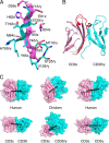
Overview of the ch-CD3 NMR structure. A, ensemble of 10 final NMR structures of ch-CD3. Regions with secondary structure (as defined by STRIDE (54)) are colored red. B, ribbon diagram of ch-CD3 with the secondary structure of CD3ϵ shown in pink, and CD3δ/γ is shown in cyan. The cysteine residues that form intra-domain disulfide bonds are represented as red sticks. The single predicted N-linked glycosylation site in CD3δ/γ (Asn71) is shown as an orange sphere. The structures shown span from Glu-6ϵ to Val70ϵ and from Leu9δ/γ to Met79δ/γ and exclude the 26-amino acid linker. C, secondary structure-matched superimposition of human and chicken CD3ϵ chains (I). In II and III, the structure of ch-CD3δ/γ was aligned to that of human CD3δ and CD3γ, respectively. Human CD3 structures are colored gray, and ch-CD3 structures are in pink (CD3ϵ) or cyan (CD3δ/γ). Only residues that were assigned in the ch-CD3 structure and the corresponding residues in human CD3 are shown. Dashed lines highlight regions of significant structural deviation. D, amino acid sequence alignment of the immunoglobulin domains of human, mouse, and chicken CD3ϵ, δ, and γ chains. The secondary structure for human and chicken variants is displayed above and below the sequences, respectively. Potential N-linked glycosylation sites are highlighted in bold. * indicates absolutely conserved residues.
FIGURE 2.
FIGURE 2.
The ch-CD3 heterodimer. A, interactions at the ch-CD3ϵ-δ/γ interface. The interlocking ladder running along the parallel G-strands comprises residues Asp59ϵ–Tyr65ϵ (pink) and Asn71δγ–His75δ/γ (cyan). The interface is supported by contacts between the F-strand of CD3ϵ (residues Thr52ϵ and Ser54ϵ) and the A-strand of CD3δ/γ (Met11δ/γ–Val13δ/γ). B, the ch-CD3 heterodimer is shown in ribbon representation with the FG loop and G-strand of human CD3ϵ overlaid in red. The bulkier human CD3ϵ loop would clash extensively with the ch-CD3δ/γ subunit. C, surface representation showing side view (top) and top view (bottom) of ch-CD3. For comparison the structures of human CD3ϵδ (1XIW) and CD3ϵγ (1SY6) are shown. All structures were aligned via the CD3ϵ chain (pink). The black line connects the center of mass of the CD3 subunits. Center of mass calculations included only those residues for which NMR spectra were assigned (for ch-CD3) and the corresponding residues in the human CD3 variants. Subunits are colored as in Fig. 1.
FIGURE 3.
FIGURE 3.
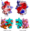
The ch-CD3 molecular surface. A, positive and negative electrostatic surface potentials (blue and red, respectively) of chicken (left) and human (right) CD3ϵ. The view shown is focused on the B, D, and E strands. Although human CD3ϵ possesses a highly electronegative surface, ch-CD3ϵ is relatively neutral (white) in this region. B, surface-exposed conserved residues in human and chicken CD3. The ch-CD3 heterodimer is displayed as a surface representation in two orientations (left, ϵ-ABE, δ/γ-C′CFG; right, ϵ-CFG, δ/γ-ABE) with the CD3 chains colored as in Fig. 1. Residues conserved in human CD3 are colored as follows: CD3ϵ (orange), CD3δ and CD3γ (orange), CD3δ (red), and CD3γ (yellow). Amino acid residues shown are as in Fig. 1.
FIGURE 4.
FIGURE 4.
Alignments of human and chicken TCR and CD3 stalk, transmembrane, and intracellular juxtamembrane sequences. Residues that are identical between human (hu) and chicken sequences are marked with asterisks. In the comparison of chicken CD3δ/γ with human CD3δ and CD3γ, residues identical to human CD3δ are marked above the alignment, and those identical to human CD3γ are marked below the alignment. The predicted membrane-spanning regions based on analysis of TMHMM 2.0 scores (55) are shaded with gray rectangles. The polar TM residues that are known to be essential for assembly of the human TCR and CD3 subunits and the corresponding positions in the chicken sequences are marked in blue (basic), red (acidic), and orange (hydroxyl-bearing).
FIGURE 5.
FIGURE 5.
Both TCRα and TCRβ can bind CD3ϵδ/γ and the interaction is dependent on the central TM lysines. HA-tagged TCR chains were translated with CD3ϵ and CD3δ/γ in in vitro translation and immunoprecipitated with anti-HA-agarose. Shown is a representative gel of the species bound to the beads in each immunoprecipitation and a gel showing 5% of the input before immunoprecipitation. The data presented on the graph represent three repeats of the same experiment. Two-tailed t tests indicated that TCRα could pull down more CD3ϵδ/γ than TCRβ, but that the amount pulled down by TCRβ was still more than the mixing control where TCRβ and the CD3 mRNAs were translated in separate in vitro translation reactions and mixed prior to IP. ** indicates 0.001 < p ≤ 0.01, * indicates 0.01 < p ≤ 0.05, ns indicates p > 0.05.
FIGURE 6.
FIGURE 6.
The central TM lysines of both TCRα and TCRβ are important for CD3ϵδ/γ recruitment in full assembly of the TCR signaling complex. SBP-tagged TCRα and HA-tagged TCRβ were translated with CD3ϵ, CD3δ/γ, and ζ, and a sequential immunoprecipitation for TCRα followed by TCRβ was used to select TCRαβ heterodimers. In the mixing control, TCRα and TCRβ were translated in a separate reaction from the CD3 and ζ components and mixed after stopping the assembly reaction. A 5% sample before immunoprecipitation was taken to ensure that input was similar. Densitometry of TCRαβ heterodimers and CD3 chains was performed, and the results were calculated as a ratio of CD3 heterodimer/TCR heterodimer. The contribution of individual TCRα and TCRβ chains to the CD3 signal was included in the ratio calculations. ** indicates 0.001 < p < 0.01, * indicates 0.01 < p < 0.05, and ns indicates p > 0.05.
FIGURE 7.
FIGURE 7.
ζ incorporation requires the upper TM lysine of TCRα and all CD3 chains. TCRα and TCRβ were translated with CD3ϵ-SBP, CD3δ/γ and ζ-PC with ζ at 10-fold lower concentration than CD3 chains and 5-fold lower than TCR chains. ζ was targeted for immunoprecipitation and the amount of co-precipitated TCR heterodimer was measured by densitometry. In mixing controls, TCRα and TCRβ mRNA were translated separately from CD3 and ζ chains and processed as described in the legend for Fig. 6. A 5% sample of the digitonin extraction was loaded on a gel as an input control. The ratio of TCR heterodimer to ζζ homodimer is graphed. * indicates 0.01 < p < 0.05.
FIGURE 8.
FIGURE 8.
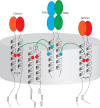
Schematic of the chicken TCR signaling complex. The gray disc represents the plasma membrane. Residues that are important in guiding TCR assembly are color-coded: light blue, lysine; red, aspartic acid. The extracellular domains are color-coded to identify each polypeptide: light blue, TCRα; green, TCRβ; orange, CD3ϵ; red, CD3δ/γ. ITAMs in the cytoplasmic tails of each species are represented as gray rectangles. Although this schematic implies that both CD3ϵ TM domains would fall closest to ζζ in the assembled complex, this information is not known, and this uncertainty is indicated with question marks.

Similar articles

Cited by

References

    1. Call M. E., Pyrdol J., Wucherpfennig K. W. (2004) Stoichiometry of the T-cell receptor-CD3 complex and key intermediates assembled in the endoplasmic reticulum. EMBO J. 23, 2348–2357 - PMC - PubMed
    1. Schamel W. W., Alarcón B. (2013) Organization of the resting TCR in nanoscale oligomers. Immunol. Rev. 251, 13–20 - PubMed
    1. Schamel W. W., Arechaga I., Risueño R. M., van Santen H. M., Cabezas P., Risco C., Valpuesta J. M., Alarcón B. (2005) Coexistence of multivalent and monovalent TCRs explains high sensitivity and wide range of response. J. Exp. Med. 202, 493–503 - PMC - PubMed
    1. Birkinshaw R. W., Kjer-Nielsen L., Eckle S. B. G., McCluskey J., Rossjohn J. (2014) MAITs, MR1 and vitamin B metabolites. Curr. Opin. Immunol. 26, 7–13 - PubMed
    1. Eckle S. B., Turner S. J., Rossjohn J., McCluskey J. (2013) Predisposed αβT cell antigen receptor recognition of MHC and MHC-I like molecules? Curr. Opin. Immunol. 25, 653–659 - PubMed

Publication types

Associated data

LinkOut - more resources