. 2014 Nov;63(11):1711-20.
doi: 10.1136/gutjnl-2013-305964. Epub 2014 Jan 31.

Cell lineage distribution atlas of the human stomach reveals heterogeneous gland populations in the gastric antrum

Affiliations

Cell lineage distribution atlas of the human stomach reveals heterogeneous gland populations in the gastric antrum

Eunyoung Choi et al. Gut. 2014 Nov.

Abstract

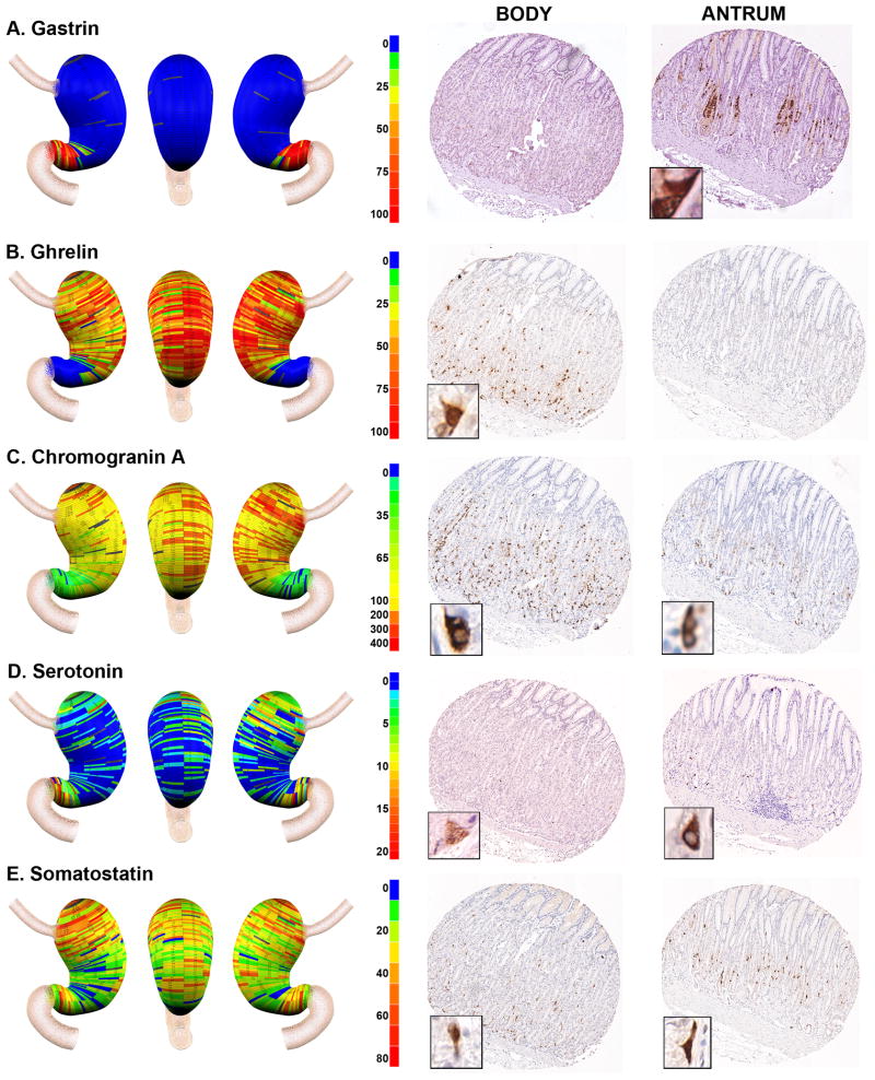
Objective: The glands of the stomach body and antral mucosa contain a complex compendium of cell lineages. In lower mammals, the distribution of oxyntic glands and antral glands define the anatomical regions within the stomach. We examined in detail the distribution of the full range of cell lineages within the human stomach.

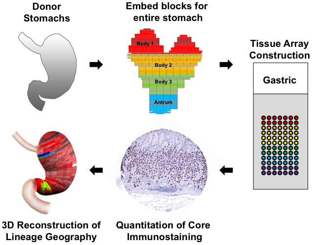
Design: We determined the distribution of gastric gland cell lineages with specific immunocytochemical markers in entire stomach specimens from three non-obese organ donors.

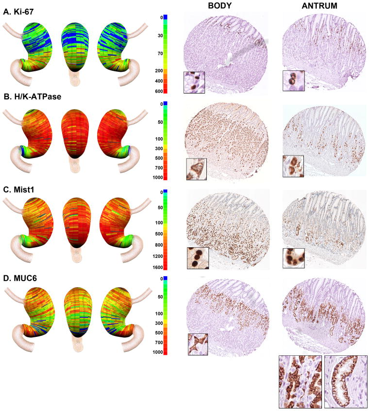
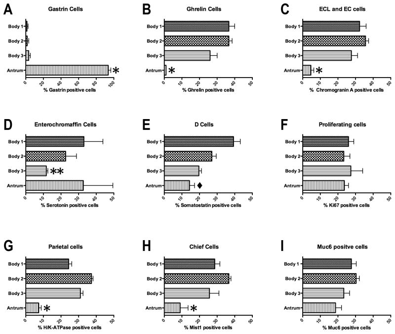
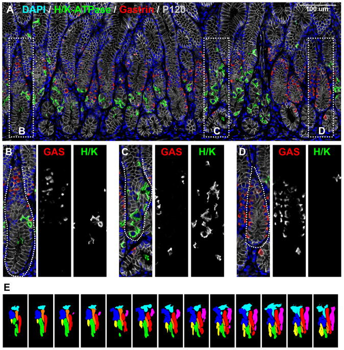
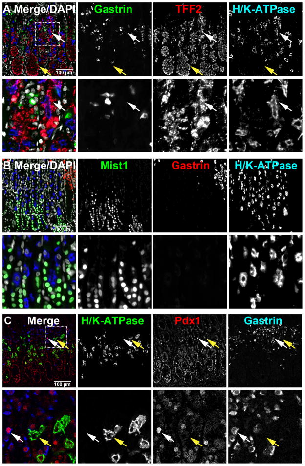
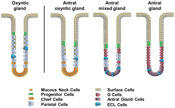
Results: The anatomical body and antrum of the human stomach were defined by the presence of ghrelin and gastrin cells, respectively. Concentrations of somatostatin cells were observed in the proximal stomach. Parietal cells were seen in all glands of the body of the stomach as well as in over 50% of antral glands. MIST1 expressing chief cells were predominantly observed in the body although individual glands of the antrum also showed MIST1 expressing chief cells. While classically described antral glands were observed with gastrin cells and deep antral mucous cells without any parietal cells, we also observed a substantial population of mixed type glands containing both parietal cells and G cells throughout the antrum.

Conclusions: Enteroendocrine cells show distinct patterns of localisation in the human stomach. The existence of antral glands with mixed cell lineages indicates that human antral glands may be functionally chimeric with glands assembled from multiple distinct stem cell populations.

Keywords: Gastric Epithelial Cell FUnction; Gastric Function; Gastric Parietal Cell; Gastric Physiology; Gastrin.


Conflict of interest statement

None of the authors have any conflicts of interest.

Figures

Figure 1
Figure 1. Procedure for geographic mapping of the cell lineages within the human stomach
The three entire donor stomachs were divided into regions of 0.5 cm height X 2 cm wide to embed in paraffin blocks and 1 mm cores from the paraffin blocks were excised and assembled into tissue arrays. These arrays were stained with cell lineage-specific antibodies, and then the cell numbers per core were determined using a digital quantitation system (Ariol SL-50). Finally, the distribution of cell lineages was displayed in three-dimensional projections of the stomach.
Figure 2
Figure 2. Geographic mapping of endocrine cell lineages
Tissue array sections from Donor 2 were stained for A. Gastrin (G cells), B. Ghrelin (X cells), C. Chromogranin A (enteroendocrine cells), D. Serotonin (EC cells) and E. Somatostatin (D cells). Quantitated cell lineage numbers per core were mapped onto three dimensional stomach maps to demonstrate the distribution of cells in the stomach (left panels). The scale bars represent the range of positive cells in a core. Representative cores from the body and antrum are shown at the right of each panel with high magnification insets showing the individual cell staining pattern.
Figure 3
Figure 3. Geographic mapping of progenitor and oxyntic gland cell lineages
Tissue array sections were stained for A. Ki-67 (progenitor cells), B. H/K-ATPase (parietal cells), C. Mist1 (chief cells) and D. Muc6 (mucous neck cells and deep antral gland cells). Quantitated cell lineage numbers per core were mapped onto three dimensional stomach maps to demonstrate the distribution of cells in the stomach (left panels). The scale bars represent the quantitated range of positive cells in a core. Representative cores from the body and antrum are shown at the right of each panel with high magnification insets showing the individual cell staining pattern. In the antral core for MUC6 staining in D, insets show the staining for cells with mucous neck cell morphology at left and cells with deep antral gland cell morphology at right.
Figure 4
Figure 4. Quantitation of the distribution of cell lineages in the human stomach
To quantitate the distribution of cell lineages within the human stomachs the specimens were divided into three body regions (proximal to distal): Body 1 (B1), Body 2 (B2) and Body 3 (B3) and the antrum. The numbers of cells staining for each lineage in each zone were determined as a percentage of the total labeled cells in each stomach specimen. The percentages of labeled cells in each region were compared with ANOVA and Bonferroni’s test for significant means. *p<0.05 between the antrum and all three Body regions; **p<0.05 comparing antrum to B1 and B2; ◆p<0.05 comparing B3 with both B1 and antrum. All bars represent the mean ± SEM.
Figure 5
Figure 5. Immunofluorescence staining for parietal cells in human antrum
Paraffin sections of human gastric antrum were immunostained for H/K-ATPase (parietal cells, green) and gastrin (G cells, red). P120 (grey scale) immunostaining was used for lateral membrane staining and DAPI (blue) was used for nuclear staining. Dotted boxes in panel A indicate regions enlarged in B-D. Three populations of glands were observed in the human antrum: B) oxyntic glands with parietal cells but not gastrin cells, C) mixed glands with both parietal cells and gastrin cells and D) antral-type glands with gastrin cells. Scale bars are as indicated. E. Tracings of glands in 13 serial sections. Glands morphologies were defined as “gastrin” or “mixed” based on triple labeling with antibodies against gastrin (red), H/K-ATPase (green) and p120 (blue). The color-coding for traced gland units was as follows: Red, orange, and green –antral-type glands lacking parietal cells, pink and yellow – mixed-type glands with both parietal cells and gastrin cells, light blue and blue – incompletely mapped glands.
Figure 6
Figure 6. Characterization of gastric glands in human antrum
Paraffin sections of human gastric antrum were immunostained for TFF2 (A), Mist1 (B) or Pdx1 (C) with both gastrin and H/K-ATPase. DAPI was used for nuclear staining. In panel A, the mucous neck cells (white arrow) and the deep antral mucous cells (yellow arrow) were immunostained for TFF2. In panel B, gastrin-expressing G cells were co-localized with Pdx1 (white arrow), however, H/K-ATPase-expressing parietal cells were not co-labeled with Pdx1 (yellow arrow). Dotted boxes depict regions enlarged. Scale bars are as indicated.
Figure 7
Figure 7. A new model of the heterogeneous types of human antral glands
Analysis of the composition of gastric cells in human antrum indicates the existence of three types of glands: 1) the Antral oxyntic gland, 2) the Antral mixed gland and 3) the gastrin cell-containing Antral gland. The oxyntic gland represents the typical gland type present in the body of human stomach (shown at left) and contains more parietal cells than the antral oxyntic gland and a less prominent foveolar region.

References

    1. Karam SM, Leblond CP. Dynamics of epithelial cells in the corpus of the mouse stomach. I. Identification of proliferative cell types and pinpointing of the stem cells. Anat Rec. 1993;236:259–79. - PubMed
    1. Karam SM, Leblond CP. Dynamics of epithelial cells in the corpus of the mouse stomach. II. Outward migration of pit cells. Anat Rec. 1993;236:280–96. - PubMed
    1. Karam SM, Leblond CP. Dynamics of epithelial cells in the corpus of the mouse stomach. III. Inward migration of neck cells followed by progressive transformation into zymogenic cells. Anat Rec. 1993;236:297–313. - PubMed
    1. Karam SM. Dynamics of epithelial cells in the corpus of the mouse stomach. IV. Bidirectional migration of parietal cells ending in their gradual degeneration and loss. Anat Rec. 1993;236:314–32. - PubMed
    1. Karam SM, Leblond CP. Dynamisc of epithelial cells in the corpus of the mouse stomach. V. Behavior of entero-endocrine and caveolated cells:general conclusions of cell kinetics in the oxyntic epithelium. Anat Rec. 1993;236:333–40. - PubMed

Publication types