. 2014 Mar;164(3):1271-82.
doi: 10.1104/pp.113.234187. Epub 2014 Jan 31.

Multiple RNA binding protein complexes interact with the rice prolamine RNA cis-localization zipcode sequences

Affiliations

Multiple RNA binding protein complexes interact with the rice prolamine RNA cis-localization zipcode sequences

Yongil Yang et al. Plant Physiol. 2014 Mar.

Abstract

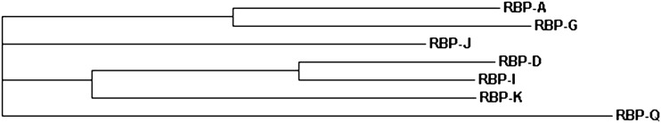
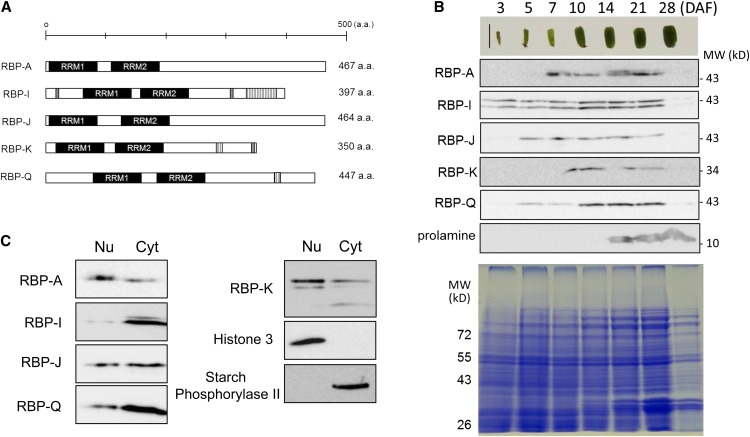
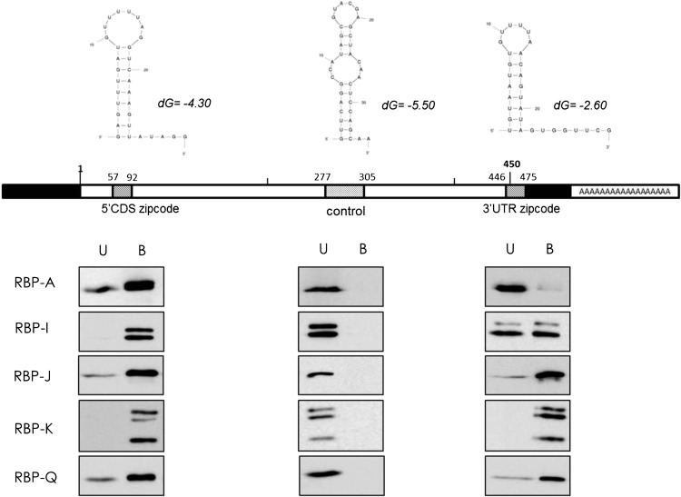
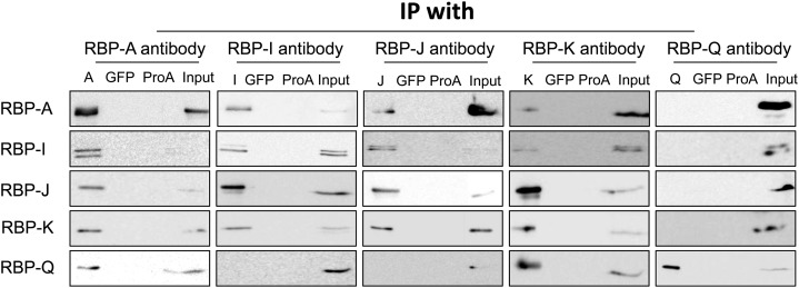
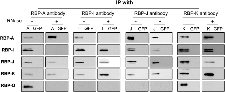
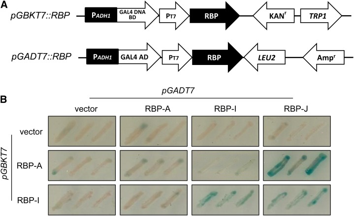
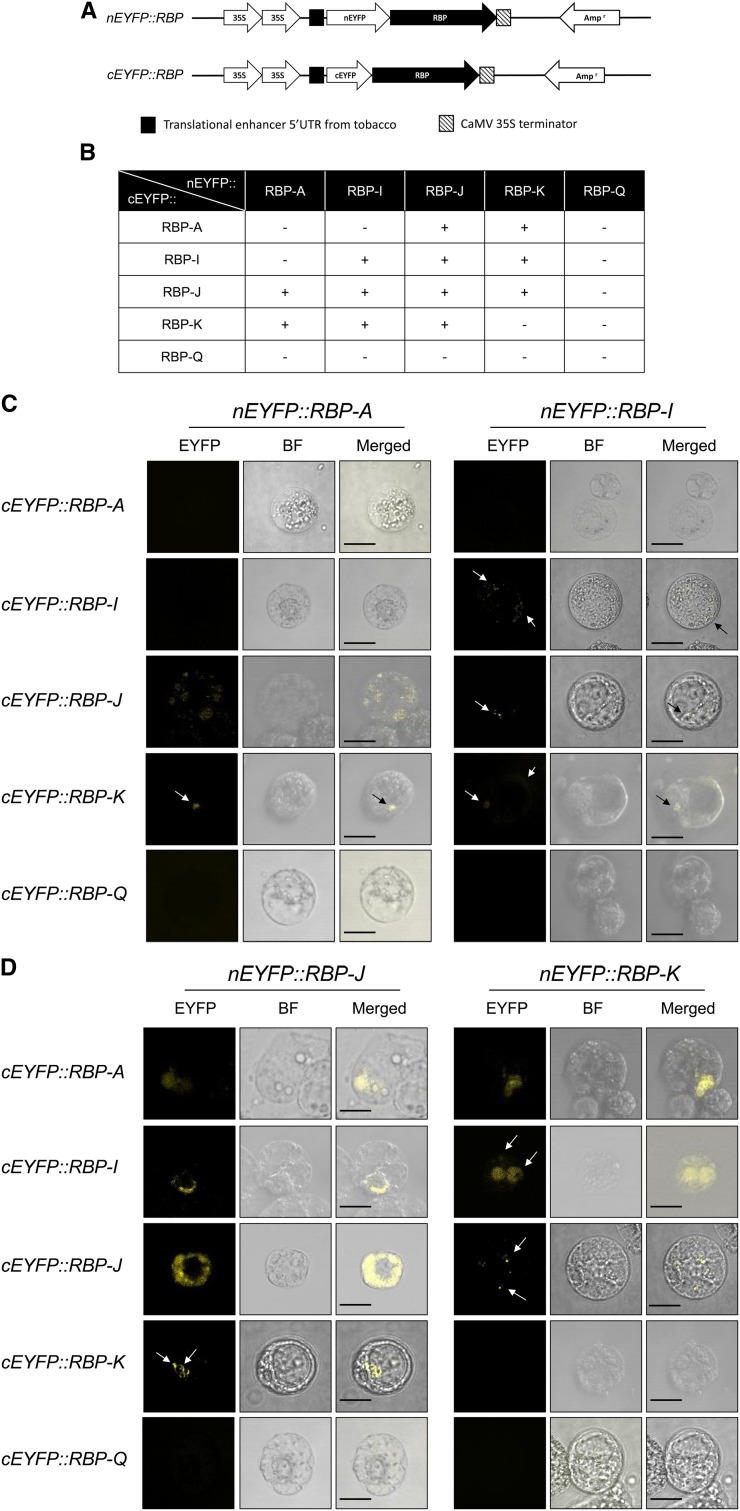
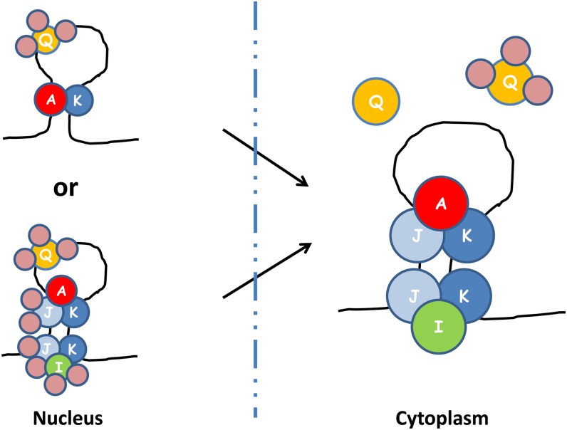
RNAs for the storage proteins, glutelins and prolamines, contain zipcode sequences, which target them to specific subdomains of the cortical endoplasmic reticulum in developing rice (Oryza sativa) seeds. Fifteen RNA binding proteins (RBPs) specifically bind to the prolamine zipcode sequences and are likely to play an important role in the transport and localization of this storage protein RNA. To understand the underlying basis for the binding of multiple protein species to the prolamine zipcode sequences, the relationship of five of these RBPs, RBP-A, RBP-I, RBP-J, RBP-K, and RBP-Q, were studied. These five RBPs, which belong to the heterogeneous nuclear ribonucleoprotein class, bind specifically to the 5' coding regions as well as to the 3' untranslated region zipcode RNAs but not to a control RNA sequence. Coimmunoprecipitation-immunoblot analyses in the presence or absence of ribonuclease showed that these five RBPs are assembled into three multiprotein complexes to form at least two zipcode RNA-protein assemblies. One cytoplasmic-localized zipcode assembly contained two multiprotein complexes sharing a common core consisting of RBP-J and RBP-K and either RBP-A (A-J-K) or RBP-I (I-J-K). A second zipcode assembly of possibly nuclear origin consists of a multiprotein complex containing RBP-Q and modified forms of the other protein complexes. These results suggest that prolamine RNA transport is initiated in the nucleus to form a zipcode-protein assembly, which is remodeled in the cytoplasm to target the RNA to its proper location on the cortical endoplasmic reticulum.


Figures

Figure 1.
Figure 1.
Phylogram of hnRNP homologs in identified prolamine zipcode binding proteins. The phylogenetic tree was generated by ClusterW2 with amino acid sequences of RBP (http://www.ebi.ac.uk/Tools).
Figure 2.
Figure 2.
Five selected hnRNPs that interact with the prolamine zipcode. A, Schematic structural representations of the hnRNPs RBP-A, RBP-I, RBP-J, RBP-K, and RBP-Q, which all contain a pair of RRMs (black rectangle). RBP-I and RBP-K contain RGG (Arg-Gly-Gly) motifs (gray rectangles) and GAY (Gly-Ala-Tyr) motifs (vertical line rectangles), while RBP-Q contains only the GAY motif. B, The temporal expression pattern of RBP-A, RBP-I, RBP-J, RBP-K, and RBP-Q as viewed by immunoblotting. Note that the RBPs are all readily detected when prolamine proteins begin to accumulate at the midstage of seed development. Only the 36-kD polypeptide band for RBP-K is shown, as the presence of the higher molecular-sized polypeptide bands at 45 and 41 kD in C was variable. C, Distribution of the RBPs in nuclear/chromatin and cytosolic fractions prepared from 10- to 13-DAF seed extracts. Histone3 and starch phosphorylase2 were used as markers of the nuclear/chromatin and cytosolic fractions, respectively. Nu, Nucleus; Cyt, cytosol. [See online article for color version of this figure.]
Figure 3.
Figure 3.
Recognition of 5′ CDS and 3′ UTR zipcodes by RBP-A, RBP-I, RBP-J, RBP-K, and RBP-Q. Rice seed extracts were incubated with biotinylated 5′ CDS and 3′ UTR zipcode or control sequences bound to streptavidin-conjugated magnetic beads. The bound (B) and unbound (U) fractions were then subjected to immunoblot analysis using affinity-purified antibodies raised against RBP-A, RBP-I, RBP-J, RBP-K, and RBP-Q. The top portion of the figure depicts the secondary structures of 5′ CDS and 3′ UTR zipcodes and control sequences as predicted by mfold web-based analysis tools (http://mfold.rna.albany.edu). Schematic structure of prolamine7 complementary DNA depicts the location of the 5′ CDS and 3′ UTR zipcodes and control sequence. The bottom portion of the figure depicts immunoblot results, which show that all five RBPs bind to 5′ CDS and 3′ UTR zipcodes but not to the control sequence.
Figure 4.
Figure 4.
Interaction of the five RBPs as viewed by Co-IP analysis. Immunoprecipitates were generated by incubating rice seed extracts with protein A conjugated with various RBP antibodies. The immunoprecipitates were then subjected to immunoblot analysis for the presence of other RBPs. Mock IPs with anti-GFP and antibody-free protein A agarose beads (ProA) were performed as negative controls. Note that RBP-A, RBP-I, RBP-J, and RBP-K are found in reciprocal IPs, while RBP-Q is only found in IPs formed by anti-RBP-A and anti-RBP-K. Note also that RBP-A and RBP-K are not found in IPs generated with anti-RBP-Q, indicating that RBP-Q is present in multiple forms, with one form being masked from interacting with its antibody when associated with RBP-A and RBP-K. Only the 36-kD polypeptide band for RBP-K is shown, as the presence of the higher molecular weight forms seen in Figures 2 and 3 was highly variable and not reproducibly detected.
Figure 5.
Figure 5.
The association of the five RBPs in Co-IPs treated with RNase. Immunoprecipitates formed using antibodies raised against RBP-A, RBP-I, RBP-J, RBP-K, or RBP-Q were treated with or without RNase and then subjected to immunoblot analysis. Protein A beads conjugated with anti-GFP were used as a negative control (GFP). Note that both RBP-A and RBP-I are found in RNase-resistant complexes with RBP-J and RBP-K but not with each other. The association of RBP-Q with RBP-A and RBP-K is RNase sensitive, indicating that the former does not directly interact with the two latter RBPs. Overall, these results support the existence of three RBP-containing multiprotein complexes.
Figure 6.
Figure 6.
The interaction of RBP-A and RBP-I to other hnRNPs as viewed by yeast two-hybrid analysis. A, Schematic representation of gene constructs used for yeast two-hybrid analysis. Y187 yeast cells were transformed with pGBKT7 plasmids harboring RBP-A or RBP-I sequences or as an empty vector. The prey vector, pGADT7, was constructed with RBP-A, RBP-I, or RBP-J sequences and transformed into AH109 yeast strain. B, Yeast two-hybrid screening on Trp and Leu synthetic dextrose media containing X-α-gal. RBP-K sequences and the pGBKT7 containing RBP-J were toxic to yeast cells and, hence, were not further evaluated. Note that RBP-A and RBP-I interact with RBP-J, while RBP-I interacts with itself.
Figure 7.
Figure 7.
Interactions of RBPs in tomato protoplasts as assessed by BiFC. A, Schematic diagrams of RBP gene fusions to the N-terminal and C-terminal EYFP fragments. B, Summary of various protein-protein interactions among the five RBPs. C, BiFC confocal microscopic images of tomato protoplasts expressing nEYFP::RBP-A or nEYFP::RBP-I and cEYFP versions of RBP-A, RBP-I, RBP-J, RBP-K, and RBP-Q. D, BiFC confocal microscopic images of tomato protoplasts expressing nEYFP::RBP-J or nEYFP::RBP-K and cEYFP versions of RBP-A, RBP-I, RBP-J, RBP-K, and RBP-Q. The yellow fluorescence formed by direct interaction of RBPs was observed by confocal microscope. Individual panels for each BiFC pair of plasmids denote presence or absence of fluorescence (EYFP), bright field image (BF), and superimposed BF and EYFP images (Merged). Bar = 20 μm.
Figure 8.
Figure 8.
A proposed model of prolamine zipcode mRNA-RBP protein assembly complexes based on results obtained by IP, yeast two-hybrid analysis, and BiFC. Note that at least three different RBP complexes bind to the prolamine zipcodes to form two ribonucleoprotein assembly complexes. RBP-A, RBP-J, and RBP-K and RBP-I, RBP-J, and RBP-K are assembled in the cytoplasm as two separate multiprotein complexes on the prolamine zipcode. When associated with RBP-A and RBP-K, RBP-Q is not accessible to its antibody, suggesting that it is bound by other proteins that comprise a third multiprotein family. As BiFC results indicate that heterodimer formation occurs in the nucleus (Figure 7), it is likely that multiprotein complexes A-J-K and I-J-K are formed in the nucleus, where they interact with the prolamine zipcode. If so, RBP-I and RBP-J are bound by other proteins so that they are not accessible to their respective antibodies. A simpler ribonucleoprotein assembly complex in the nucleus would consist of two multiprotein complexes, one containing RBP-Q and a second containing RBP-A and RBP-K. Note that the various multiprotein complexes were arbitrarily drawn on the zipcode stem-loop structure. The actual binding specificity of the various multiprotein complexes to the stem-loop structure remains to be resolved.

Similar articles

Cited by

References

    1. Arn EA, Cha BJ, Theurkauf WE, Macdonald PM. (2003) Recognition of a bicoid mRNA localization signal by a protein complex containing Swallow, Nod, and RNA binding proteins. Dev Cell 4: 41–51 - PubMed
    1. Birney E, Kumar S, Krainer AR. (1993) Analysis of the RNA-recognition motif and RS and RGG domains: conservation in metazoan pre-mRNA splicing factors. Nucleic Acids Res 21: 5803–5816 - PMC - PubMed
    1. Böhl F, Kruse C, Frank A, Ferring D, Jansen RP. (2000) She2p, a novel RNA-binding protein tethers ASH1 mRNA to the Myo4p myosin motor via She3p. EMBO J 19: 5514–5524 - PMC - PubMed
    1. Choi SB, Wang C, Muench DG, Ozawa K, Franceschi VR, Wu Y, Okita TW. (2000) Messenger RNA targeting of rice seed storage proteins to specific ER subdomains. Nature 407: 765–767 - PubMed
    1. Crofts AJ, Crofts N, Whitelegge JP, Okita TW. (2010) Isolation and identification of cytoskeleton-associated prolamine mRNA binding proteins from developing rice seeds. Planta 231: 1261–1276 - PubMed

Publication types

MeSH terms

LinkOut - more resources