. 2014 Mar 1;192(5):2156-66.
doi: 10.4049/jimmunol.1300675. Epub 2014 Jan 31.

Type I IFN induces binding of STAT1 to Bcl6: divergent roles of STAT family transcription factors in the T follicular helper cell genetic program

Affiliations

Type I IFN induces binding of STAT1 to Bcl6: divergent roles of STAT family transcription factors in the T follicular helper cell genetic program

Shingo Nakayamada et al. J Immunol. .

Abstract

CD4(+) T follicular helper cells (TFH) are critical for the formation and function of B cell responses to infection or immunization, but also play an important role in autoimmunity. The factors that contribute to the differentiation of this helper cell subset are incompletely understood, although several cytokines including IL-6, IL-21, and IL-12 can promote TFH cell formation. Yet, none of these factors, nor their downstream cognate STATs, have emerged as nonredundant, essential drivers of TFH cells. This suggests a model in which multiple factors can contribute to the phenotypic characteristics of TFH cells. Because type I IFNs are often generated in immune responses, we set out to investigate whether these factors are relevant to TFH cell differentiation. Type I IFNs promote Th1 responses, thus one possibility was these factors antagonized TFH-expressed genes. However, we show that type I IFNs (IFN-α/β) induced B cell lymphoma 6 (Bcl6) expression, the master regulator transcription factor for TFH cells, and CXCR5 and programmed cell death-1 (encoded by Pdcd1), key surface molecules expressed by TFH cells. In contrast, type I IFNs failed to induce IL-21, the signature cytokine for TFH cells. The induction of Bcl6 was regulated directly by STAT1, which bound to the Bcl6, Cxcr5, and Pdcd1 loci. These data suggest that type I IFNs (IFN-α/β) and STAT1 can contribute to some features of TFH cells but are inadequate in inducing complete programming of this subset.


Figures
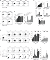
Figure 1
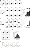
Figure 1. Type I IFNs promote Bcl6 expression
(A, C, D) Bcl6 and T-bet staining in CD4+ T cells cultured in the presence of the indicated cytokines for 5 days. (B) Relative mRNA expression of Bcl6, T-bet, Maf, Irf-4, and Batf was evaluated by qPCR. The data (mean ± SD) are representative of three independent experiments. (E, F) Bcl6 and T-bet staining in CD4+ T cells cultured with increasing doses of IFN-α or IFN-β for 5 days (E) or in neutral conditions or IFN-β for the indicated number of days (F). Data (mean ± SD) are from duplicate cultures of three independent experiments (n=6). The plots are representative of three independent experiments. *, p<0.05; **, p<0.01.
Figure 2
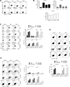
Figure 2. Type I IFNs induce CXCR5 and PD-1
(A-E) CXCR5 and PD-1 staining in CD4+ T cells cultured in the presence of the indicated cytokines for 5 days (A&D), with increasing doses of IFN-α or IFN-β for 5 days (B) or in neutral conditions or IFN-β for the indicated number of days (C). Location of the gates was chosen at each time point based on a negative control sample. Data (mean ± SD) are from duplicate cultures of three independent experiments (n=6). The plots are representative of three independent experiments. *, p<0.05; **, p<0.01. (F) ICOS staining in CD4+ T cells cultured in the presence of the indicated cytokines for 5 days.
Figure 3
Figure 3. Type I IFNs do not induce secretion of IL-21
(A-E) IL-21 and IFN-γ staining in CD4+ T cells cultured in the presence of the indicated cytokines for 5 days (A&B), with increasing doses of IFN-α or IFN-β for 5 days (D) or in neutral conditions or IFN-β for the indicated number of days (E). Data (mean ± SD) are from duplicate cultures of three independent experiments (n=6). The plots are representative of three independent experiments. *, p<0.05; **, p<0.01. (C-E). (F,G) Intracellular cytokine staining of IL-21 and IFN-γ in T cells co-cultured with naïve B cells for 4 days either with (stimulated) or without (unstimulated) anti-CD3 and anti-CD28 stimulation.
Figure 4
Figure 4. STAT1, but not STAT3 or STAT4 is required for type I IFN mediated induction of key TFH cell molecules
(A, B) p-STAT3, p-STAT4, and p-STAT1 staining in CD4+ T cells cultured in the presence of the indicated cytokines without TCR stimulation for 30 minutes. Graphs (means ± SD) are from duplicate cultures and are representative of three independent experiments. (C-E) Bcl6 and T-bet (C), IL-21 and IFN-γ (D), or CXCR5 and PD-1 (E) staining in CD4+ T cells cultured in the presence of the indicated cytokines for 5 days from WT, Stat1−/−, Stat3−/−, or Stat4−/− mice. Data (mean ± SD) are from duplicate cultures of three independent experiments (n=6). The plots are representative of three independent experiments. *, p<0.05; **, p<0.01.
Figure 5
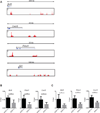
Figure 5. STAT1 binds to TFH cell signature genes
(A) STAT1 binding was mapped by ChIP-seq in IFN-β treated CD4+T cells. Naïve CD4+ T cells were cultured for 3 days with plate bound anti-CD3 and anti-CD28 (10 ug/ml of each) and IFN-β (5000 U/ml). The genome browser views for Bcl6 (chr16:23,961,956-24,227,286), Cxcr5 (chr9:44,319,494-44,365,190 ), Pdcd1 (chr1:95,927,980-95,963,539), and Il21 (chr3:37,092,096-37,201,024) loci are shown. Y axis depicts the normalized tag number, defined as the tag count per one million. (B,C) p300 (B) and STAT1 (C) binding to selected regions of the Bcl6, Pdcd1, and Cxcr5 genes was determined by ChIP-qPCR. The amount of precipitated DNA was calculated as % input. The results are representative of two independent experiments, qPCR was done in triplicate (mean ± SEM). Unpaired t test was perfermed to calculate p values; *<0.1, **<0.01, ***<0.001.

Similar articles

Cited by

References

    1. Abbas AK, Murphy KM, Sher A. Functional diversity of helper T lymphocytes. Nature. 1996;383:787–793. - PubMed
    1. Miller JF, De Burgh PM, Grant GA. Thymus and the production of antibody-plaque-forming cells. Nature. 1965;208:1332–1334. - PubMed
    1. Friedman H. Absence of Antibody Plaque Forming Cells in Spleens of Thymectomized Mice Immunized with Sheep Erythrocytes. Proc Soc Exp Biol Med. 1965;118:1176–1180. - PubMed
    1. Crotty S. Follicular helper CD4 T cells (TFH) Annual review of immunology. 2011;29:621–663. - PubMed
    1. Breitfeld D, Ohl L, Kremmer E, Ellwart J, Sallusto F, Lipp M, Forster R. Follicular B helper T cells express CXC chemokine receptor 5, localize to B cell follicles, support immunoglobulin production. The Journal of experimental medicine. 2000;192:1545–1552. - PMC - PubMed

Publication types

MeSH terms