. 2014 Mar 1;192(5):2252-60.
doi: 10.4049/jimmunol.1301249. Epub 2014 Jan 31.

Glycoengineering of therapeutic antibodies enhances monocyte/macrophage-mediated phagocytosis and cytotoxicity

Affiliations

Glycoengineering of therapeutic antibodies enhances monocyte/macrophage-mediated phagocytosis and cytotoxicity

Sylvia Herter et al. J Immunol. .

Abstract

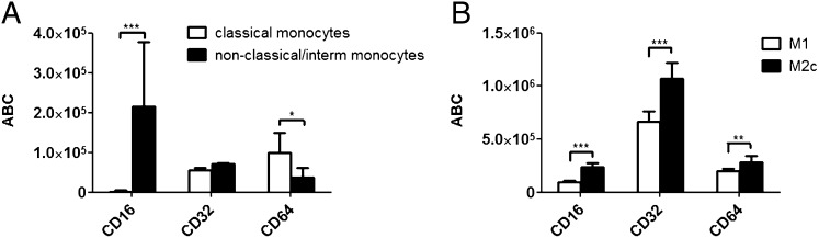
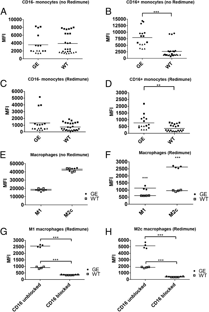
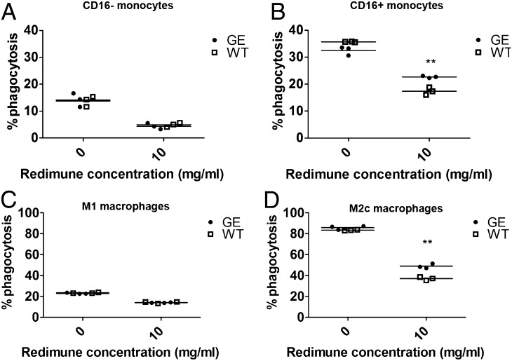
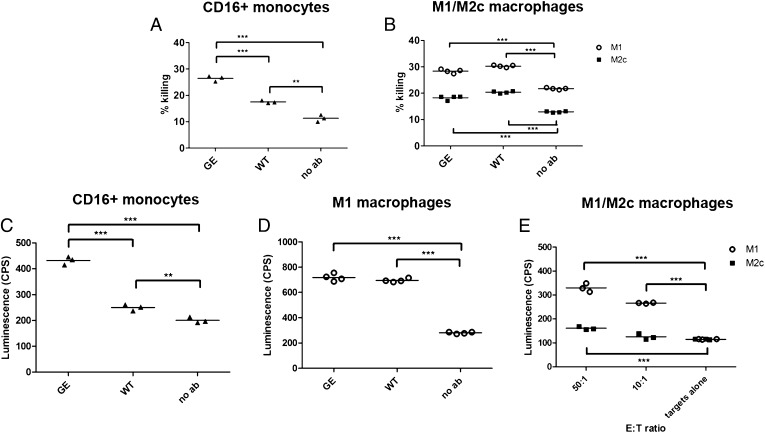
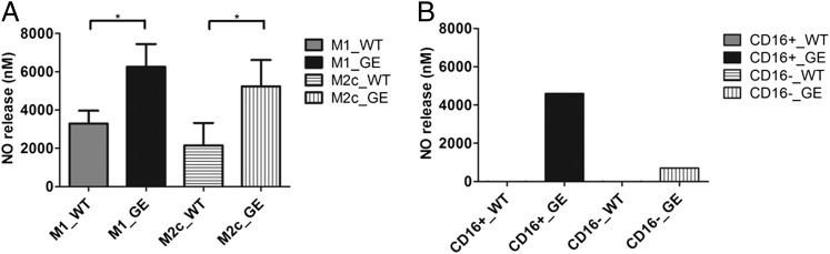
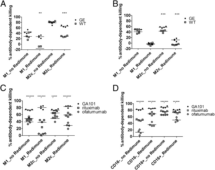
Therapeutic Abs possess several clinically relevant mechanisms of action including perturbation of tumor cell signaling, activation of complement-dependent cytotoxicity, Ab-dependent cellular cytotoxicity (ADCC), Ab-dependent cellular phagocytosis (ADCP), and induction of adaptive immunity. In view of the important role of phagocytic lineage cells in the mechanism of action of therapeutic Abs, we analyzed FcγR receptor-dependent effector functions of monocytes and macrophages triggered by glycoengineered (GE) Abs (having enhanced FcγRIIIa [CD16a] binding affinity) versus their wild-type (WT) counterparts under different experimental conditions. We first defined the precise FcγR repertoire on classical and nonclassical intermediate monocytes--M1 and M2c macrophage populations. We further show that WT and GE Abs display comparable binding and induce similar effector functions (ADCC and ADCP) in the absence of nonspecific, endogenous IgGs. However, in the presence of these IgGs (i.e., in a situation that more closely mimics physiologic conditions), GE Abs display significantly superior binding and promote stronger monocyte and macrophage activity. These data show that in addition to enhancing CD16a-dependent NK cell cytotoxicity, glycoengineering also enhances monocyte and macrophage phagocytic and cytotoxic activities through enhanced binding to CD16a under conditions that more closely resemble the physiologic setting.


Figures

FIGURE 1.
FIGURE 1.
Quantitative assessment of FcγRs surface expression on human monocytes and M1 and M2c macrophages. (A) Freshly isolated monocytes or (B) M1- and M2c-polarized macrophages were analyzed with flow cytometry to quantify the surface expression of CD64, CD32, and CD16. Ordinate denotes the ABC obtained for each receptor. Data represent the mean ABC fluorescence ±SD derived from analysis of four different donors. Statistical analysis, unpaired t test: *p < 0.05, **p < 0.01, ***p < 0.001.
FIGURE 2.
FIGURE 2.
Binding of therapeutic Abs to human monocytes and M1 and M2c macrophages. The binding of AlexaFluor647 directly labeled WT and GE Abs was assessed using classical (CD16) monocytes (A, C), nonclassical intermediate (CD16+) monocytes (B, D) and M1 and M2c macrophages (E, F). The binding of each Ab was assessed in absence (A, B, E) or presence (C, D, F) of competing endogenous human IgGs (10 mg/ml Redimune). Each symbol in (A)–(D) corresponds to median fluorescence intensity of single data points derived from three independent experiments (donors). Donors are identified by the following symbols: donor 1 = black circles; donor 2 = black squares; donor 3 = black triangles. Two GE Abs were used (GA101 and GA201) as duplicates or triplicates, therefore resulting in four to six symbols per donor per condition. Three WT Abs were used as duplicates or triplicates (GA101 WT, GA201 WT, and ofatumumab), resulting in six to nine symbols per donor per condition. (G and H) Binding of directly labeled WT and GE Abs in the presence of saturating concentrations of blocking CD16 Ab and 10 mg/ml Redimune to M1 (G) and M2c (H) macrophages. Data in (E)–(H) correspond to median fluorescence intensity of duplicates derived from one representative experiment out of a total of three independent ones using GA101 GE, GA201 GE, GA101 WT, and ofatumumab. Two of these experiments included the blocking anti-CD16 Ab. Statistical analysis, unpaired t test: *p < 0.05, **p < 0.01, ***p < 0.001.
FIGURE 3.
FIGURE 3.
Ab-dependent phagocytosis. CFSE-labeled Raji cells were cocultured for 4 h with isolated human classical monocytes (A) or nonclassical, intermediate monocytes (B) (E:T ratio, 3:1) in presence of GA101 GE or WT Abs and in the absence or presence of 10 mg/ml Redimune. KPL-4 cells were cocultured for 4 h with M1 (C) or M2c macrophages (D) (E:T ratio, 3:1) in the presence of GE or WT trastuzumab and 10 mg/ml Redimune. The percentage of phagocytized target cells was determined with flow cytometry as described in Materials and Methods. Triplicates of representative experiments (performed using different targets and GE/WT Ab pairs (GA101, trastuzumab) are shown. Statistical analysis, unpaired t test: **p < 0.01.
FIGURE 4.
FIGURE 4.
Macrophage-mediated cytotoxicity. (A) A549 cells were incubated with isolated human intermediate, nonclassical (CD16+) monocytes (E:T ratio, 4:1) for 24 h in the presence of 1 μg/ml GA201 GE or WT Abs. (B) MKN45 cells were incubated with M1 or M2c macrophages (E:T ratio, 3:1) for 24 h in the presence of 1 μg/ml GA201 GE or cetuximab Abs. The percent killing was determined by setting the LDH released from target cells to 0% and LDH released by target cells lysed with 2% Triton X-100 to 100%. Triplicates and quadruplicates referring to one representative experiment out of three are shown. (C) 1833-PPOP233 cells were incubated with isolated human intermediate, nonclassical (CD16+) monocytes (E:T ratio, 30:1) for 8 h in the presence of 1 μg/ml GA201 GE or WT Abs. (D) 1833-PPOP233 cells were incubated with polarized human M1 macrophages (E:T ratio, 30:1) for 24 h in the presence of 1 μg/ml GA201 GE or WT Abs. (E) 1833-PPOP233 cells were incubated with polarized human M1 or M2c macrophages (E:T ratio, 10:1 or 50:1) for 24 h in the presence of 1 μg/ml GA201 GE. Triplicates and quadruplicates referring to one representative experiment out of three are shown. Statistical analysis, unpaired t test: *p < 0.05, **p < 0.01, ***p < 0.001.
FIGURE 5.
FIGURE 5.
NO release. (A) Raji or WIL2S cells incubated with human M1 or M2c macrophages (E:T ratio, 1:1) for 24 or 48 h in the presence of 1 μg/ml GA101 GE or WT Abs and ofatumumab. NO release in culture supernatants was assessed with Griess reagent and corresponds to the average of pooled triplicates derived from three independent experiments (two using Raji and one using WIL2S as targets). (B) Raji cells incubated with human classical or CD16+ monocytes (E:T ratio, 10:1) for 22 h in the presence of 1 μg/ml GA101 GE or WT Abs. NO release in culture supernatants was assessed by Griess reagent using pooled quadruplicates derived from one experiment. Statistical analysis, unpaired t test: *p < 0.05, **p < 0.01, ***p < 0.001.
FIGURE 6.
FIGURE 6.
Assessment of the overall macrophage activity. (A) The overall macrophage activity was assessed in a 48-h coculture assay of MKN45 tumor cells with human M1 or M2c macrophages (E:T ratio, 3:1) in the presence of GA201 or cetuximab and Redimune (10 mg/ml). (B) The overall macrophage activity assessed in a 48-h coculture assay of KPL-4 tumor cells with human M1 or M2c macrophages (E:T ratio, 4:1) in the presence of trastuzumab GE and WT Abs and Redimune (10 mg/ml). Quadruplicates of two representative experiments out of a total of four with different Abs and tumor target cells are shown. Statistical analysis, unpaired t test: *p < 0.05, **p < 0.01, ***p < 0.001. The overall macrophage (C) or monocyte (D) activity in combination with direct Ab effects was assessed in a 24-h coculture assay of Raji tumor cells with human isolated CD16+ or CD16 monocytes or monocyte-derived M1 or M2c macrophages (E:T ratio, 3:1) in the presence of GA101, rituximab and ofatumumab, and Redimune (10 mg/ml). Quadruplicates of two representative experiments out of a total of six (three with monocytes and three with macrophages as effectors) from different donors are shown. Statistical analysis, unpaired t test: *p < 0.05, **p < 0.01, ***p < 0.001, comparison of GA101 to rituximab/comparison GA101 to ofatumumab indicated on top of the graphs.

Similar articles

Cited by

References

    1. Shuptrine C. W., Surana R., Weiner L. M. 2012. Monoclonal antibodies for the treatment of cancer. Semin. Cancer Biol. 22: 3–13. - PMC - PubMed
    1. Weiner L. M., Murray J. C., Shuptrine C. W. 2012. Antibody-based immunotherapy of cancer. Cell 148: 1081–1084. - PMC - PubMed
    1. Weiner L. M., Surana R., Wang S. 2010. Monoclonal antibodies: versatile platforms for cancer immunotherapy. Nat. Rev. Immunol. 10: 317–327. - PMC - PubMed
    1. Reichert J. M. 2011. Antibody-based therapeutics to watch in 2011. MAbs 3: 76–99. - PMC - PubMed
    1. Herter S., Herting F., Mundigl O., Waldhauer I., Weinzierl T., Fauti T., Muth G., Ziegler-Landesberger D., Van Puijenbroek E., Lang S., et al. 2013. Preclinical activity of the type II CD20 antibody GA101 (obinutuzumab) compared with rituximab and ofatumumab in vitro and in xenograft models. Mol. Cancer Ther. 12: 2031–2042. - PubMed