. 2014 Apr;42(7):4741-54.
doi: 10.1093/nar/gku085. Epub 2014 Jan 30.

Structure of human RNA N⁶-methyladenine demethylase ALKBH5 provides insights into its mechanisms of nucleic acid recognition and demethylation

Affiliations

Structure of human RNA N⁶-methyladenine demethylase ALKBH5 provides insights into its mechanisms of nucleic acid recognition and demethylation

WeiShen Aik et al. Nucleic Acids Res. 2014 Apr.

Abstract

ALKBH5 is a 2-oxoglutarate (2OG) and ferrous iron-dependent nucleic acid oxygenase (NAOX) that catalyzes the demethylation of N(6)-methyladenine in RNA. ALKBH5 is upregulated under hypoxia and plays a role in spermatogenesis. We describe a crystal structure of human ALKBH5 (residues 66-292) to 2.0 Å resolution. ALKBH5₆₆₋₂₉₂ has a double-stranded β-helix core fold as observed in other 2OG and iron-dependent oxygenase family members. The active site metal is octahedrally coordinated by an HXD…H motif (comprising residues His204, Asp206 and His266) and three water molecules. ALKBH5 shares a nucleotide recognition lid and conserved active site residues with other NAOXs. A large loop (βIV-V) in ALKBH5 occupies a similar region as the L1 loop of the fat mass and obesity-associated protein that is proposed to confer single-stranded RNA selectivity. Unexpectedly, a small molecule inhibitor, IOX3, was observed covalently attached to the side chain of Cys200 located outside of the active site. Modelling substrate into the active site based on other NAOX-nucleic acid complexes reveals conserved residues important for recognition and demethylation mechanisms. The structural insights will aid in the development of inhibitors selective for NAOXs, for use as functional probes and for therapeutic benefit.


Figures

Scheme 1.
Scheme 1.
ALKBH5 and FTO catalyze m6A demethylation.
Figure 1.
Figure 1.
(a) Ribbons representation of the ALKBH566–292 structure showing the active site metal Mn(II) (purple sphere) substituting for Fe(II); residues His204, Asp206 and His266 (white sticks); NRL1 and NRL2 (pink fonts); disordered residues (dashed line); the DSBH β-strands I–VIII (yellow); other β-strands (salmon); and α- and 310-helices (blue). (b) Topology of the structure of ALKBH566–292. β-strands are shown as triangles, α-helices as large circles and 310-helices as small circles. The βIV-V loop is highlighted in purple, the DSBH in yellow and the NRLs in salmon. (c) Active site residues of ALKBH566–292 with representative electron density (3.0σ mFo-DFc OMIT; green mesh) for side chains of His204, Asp 206, His266 and water molecules (red spheres) all coordinated (black dashed lines) to the active site Mn(II) ion (purple sphere) (d) A ClustalW2 (67) sequence alignment of ALKBH5 homologues from various organisms [from top: Homo sapiens: human (sequence identity 100%, PDB ID 4NJ4, Uniprot ID Q6P6C2); Mus musculus: mouse (sequence identity 97%, Uniprot ID Q3TSG4); Gallus gallus: chicken (sequence identity 86%, F1NIA5); Xenopus laevis: frog (sequence identity 78%, Uniprot ID Q6GPB5); Danio rerio: zebrafish (sequence identity 72%, Uniprot ID Q08BA6); Strigamia maritima: centipede (sequence identity 56%, Uniprot ID T1JJ71); and Strongylocentrotus purpuratus: purple sea urchin (sequence identity 52%, Uniprot ID H3I4D7)]; combined with structure-based sequence alignment (68) with ALKBH3 (PDB ID 2IUW); ALKBH2 (PDB ID 3BUC); ALKBH8 (PDB ID 3THT); FTO (PDB ID 3LFM); and AlkB (PDB ID 3I3Q). A few selected manual adjustments were made to the alignment to correct for likely automated errors. Note: Variant residues from the reported ALKBH2 (PDB ID 3BUC) structure sequence were changed to reflect the wild-type ALKBH2 sequence (Uniprot Q6NS38). Residues highlighted as conserved (dark blue); semi-conserved (light blue); weakly conserved (grey); conserved 2OG oxygenase catalytic triad HXD…H, red; conserved 2OG binding arginine (green). Boxed residues indicate those forming NRL1 (red); NRL2 (blue); the βIV-V loop of ALKBH5 (purple); the L1 loop of FTO (black). Secondary structural elements of H. sapiens ALKBH5 are represented as light blue sinusoidal waves (α-helices), red arrows (β-strands excluding the DSBH core), yellow arrows (β-strands of the DSBH core) and single light blue arcs (310-helix). (e) Schematic domain representations of human NAOXs for which structures have been reported. DSBH, double-stranded β-helix core domain; RRM, RNA recognition motif; MT, methyltransferase domain; and CTD, C-terminal domain.
Figure 1.
Figure 1.
(a) Ribbons representation of the ALKBH566–292 structure showing the active site metal Mn(II) (purple sphere) substituting for Fe(II); residues His204, Asp206 and His266 (white sticks); NRL1 and NRL2 (pink fonts); disordered residues (dashed line); the DSBH β-strands I–VIII (yellow); other β-strands (salmon); and α- and 310-helices (blue). (b) Topology of the structure of ALKBH566–292. β-strands are shown as triangles, α-helices as large circles and 310-helices as small circles. The βIV-V loop is highlighted in purple, the DSBH in yellow and the NRLs in salmon. (c) Active site residues of ALKBH566–292 with representative electron density (3.0σ mFo-DFc OMIT; green mesh) for side chains of His204, Asp 206, His266 and water molecules (red spheres) all coordinated (black dashed lines) to the active site Mn(II) ion (purple sphere) (d) A ClustalW2 (67) sequence alignment of ALKBH5 homologues from various organisms [from top: Homo sapiens: human (sequence identity 100%, PDB ID 4NJ4, Uniprot ID Q6P6C2); Mus musculus: mouse (sequence identity 97%, Uniprot ID Q3TSG4); Gallus gallus: chicken (sequence identity 86%, F1NIA5); Xenopus laevis: frog (sequence identity 78%, Uniprot ID Q6GPB5); Danio rerio: zebrafish (sequence identity 72%, Uniprot ID Q08BA6); Strigamia maritima: centipede (sequence identity 56%, Uniprot ID T1JJ71); and Strongylocentrotus purpuratus: purple sea urchin (sequence identity 52%, Uniprot ID H3I4D7)]; combined with structure-based sequence alignment (68) with ALKBH3 (PDB ID 2IUW); ALKBH2 (PDB ID 3BUC); ALKBH8 (PDB ID 3THT); FTO (PDB ID 3LFM); and AlkB (PDB ID 3I3Q). A few selected manual adjustments were made to the alignment to correct for likely automated errors. Note: Variant residues from the reported ALKBH2 (PDB ID 3BUC) structure sequence were changed to reflect the wild-type ALKBH2 sequence (Uniprot Q6NS38). Residues highlighted as conserved (dark blue); semi-conserved (light blue); weakly conserved (grey); conserved 2OG oxygenase catalytic triad HXD…H, red; conserved 2OG binding arginine (green). Boxed residues indicate those forming NRL1 (red); NRL2 (blue); the βIV-V loop of ALKBH5 (purple); the L1 loop of FTO (black). Secondary structural elements of H. sapiens ALKBH5 are represented as light blue sinusoidal waves (α-helices), red arrows (β-strands excluding the DSBH core), yellow arrows (β-strands of the DSBH core) and single light blue arcs (310-helix). (e) Schematic domain representations of human NAOXs for which structures have been reported. DSBH, double-stranded β-helix core domain; RRM, RNA recognition motif; MT, methyltransferase domain; and CTD, C-terminal domain.
Figure 2.
Figure 2.
Binding of 1-chloro-4-hydroxyisoquinoline-3-carbonyl)glycine (IOX3) to ALKBH5 involves covalent attachment (Figure S8). (a) The small molecule IOX3 (cyan sticks) reacts and forms a covalent bond with the side chain of Cys200 (white sticks); the electron density map (3.0σ mFo–DFc OMIT; green mesh) is shown. (b) Two protein molecules in an asymmetric unit of an ALKBH566–292 crystal. Covalently attached IOX3 molecules from each protein molecule stack against each other via π–π interactions.
Figure 3.
Figure 3.
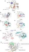
Comparison of NAOX structures reveals differences in their nucleotide recognition lids. NRL1 (red) and NRL2 (blue); (a) ALKBH5 (PDB ID 4NJ4), (b) FTO (PDB ID 3LFM), (c) ALKBH2 in complex with dsDNA (PDB ID 3BUC), (d) ALKBH3 (PDB ID 2IUW) and (e) AlkB in complex with dsDNA (PDB ID 3BIE). (f) The NRL sequences of ALKBH8 (PDB ID 3THT) are mostly disordered. (g) TYW5 (PDB ID 3AL5) from the JmjC oxygenase subfamily; note the different structural elements for tRNA substrate recognition; potential substrate contact regions are coloured brown. (h) Superimposition of ALKBH5 (light orange ribbon), FTO (grey ribbon) and ALKBH2 (not shown) in complex with double-stranded DNA (green). The βIV-V loop (purple) of ALKBH5 and L1 loop (black) of FTO overlap with the ‘unrepaired’ DNA strand (light green), potentially conferring single-strand selectivity for ALKBH5 and FTO.
Figure 4.
Figure 4.
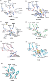
Comparison of the active site of ALKBH5 with those of other nucleic acid oxygenases. Active site residues of (a) ALKBH5 (white sticks), (b) ALKBH2 (light blue sticks), (c) FTO (grey sticks), (d) ALKBH3 (green sticks), (e) ALKBH8 (pink sticks), (f) AlkB (cyan sticks) and (g) TYW5 (teal sticks) (PDB ID 3AL6). Oxygen (red), nitrogen (blue), phosphorous (orange), sulphur (yellow), m1A base carbon (yellow), Mn(II) (purple sphere), Fe(II) (orange sphere), Ni(II) (green sphere) and water molecule (red sphere) and electrostatic interaction (black dashed line) are indicated.
Figure 5.
Figure 5.
(a and c) ALKBH5 electrostatic surface representation (basic in blue; acidic in red) with 90° rotation along the X-axis and (b and d) corresponding ribbon representation. The substrate-binding groove around the active site is largely basic for the binding of the negatively charged ssRNA phosphate backbone. The basic region between the βIV-V loop and α1-helix forms a potential substrate binding groove.

References

    1. Carell T, Brandmayr C, Hienzsch A, Muller M, Pearson D, Reiter V, Thoma I, Thumbs P, Wagner M. Structure and function of noncanonical nucleobases. Angew. Chem. Int. Ed. Engl. 2012;51:7110–7131. - PubMed
    1. Cantara WA, Crain PF, Rozenski J, McCloskey JA, Harris KA, Zhang X, Vendeix FA, Fabris D, Agris PF. The RNA Modification Database, RNAMDB: 2011 update. Nucleic Acids Res. 2011;39:D195–D201. - PMC - PubMed
    1. Machnicka MA, Milanowska K, Osman Oglou O, Purta E, Kurkowska M, Olchowik A, Januszewski W, Kalinowski S, Dunin-Horkawicz S, Rother KM, et al. MODOMICS: a database of RNA modification pathways–2013 update. Nucleic Acids Res. 2013;41:D262–D267. - PMC - PubMed
    1. Sibbritt T, Patel HR, Preiss T. Mapping and significance of the mRNA methylome. Wiley Interdiscip. Rev. RNA. 2013;4:397–422. - PubMed
    1. Furuichi Y, Shatkin AJ. Viral and cellular mRNA capping: past and prospects. Adv. Virus Res. 2000;55:135–184. - PMC - PubMed

Publication types

Associated data