

doi: 10.1136/bjophthalmol-2013-304822.
Epub 2014 Jan 31.
A novel RPGR mutation masquerading as Stargardt disease
Affiliations
- PMID: 24489377
- PMCID: PMC4170590
- DOI: 10.1136/bjophthalmol-2013-304822
Item in Clipboard
A novel RPGR mutation masquerading as Stargardt disease
Br J Ophthalmol.
2014 May.
No abstract available
Keywords: Genetics; Retina; Treatment other.
Figures
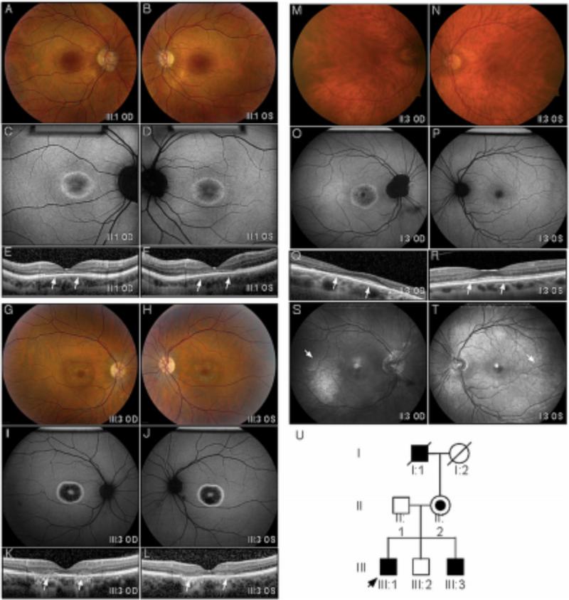
A-F. Proband (III:1). Color fundus images showed perifoveal pigment atrophy in a bull's eye pattern that was pronounced in autofluorescent imaging. SDOCT revealed foveal thinning (arrows), loss of outer plexiform layer, ellipsoid and interdigitation line in both eyes with preservation of this structure outside macular. G-L. Affected Brother (III:3). A similar bull's eye pattern with marked RPE atrophy was also observed in color fundus, autofluorescent, and SD-OCT images. M- T. Manifest carrier (II:2). Fundus images showed minimal RPE alteration in her right fovea and a normal appearance in her left. AF revealed a Bull's eye pattern with peripapillary atrophy in her right eye. The left eye appeared normal. A minimal hyperautofluorescent patch was observed in the temporal macula of both eyes. This corresponded to a prominent tapetal reflex (arrow) on red free imaging with a radial pattern of hyperreflectance in the posterior and middle periphery. U. The pedigree was consistent with an x-linked inheritance pattern. The proband is indicated with an arrow. Black symbols represent clinically affected subjects with a Bull's eye pattern from cone dystrophy. Open symbols represent unaffected subjects. The carrier subject is shown with a smaller black symbol inside an open symbol. Deceased individuals are marked by a slash. (RPE – retinal pigment atrophy; SD-OCT spectral domain OCT.)
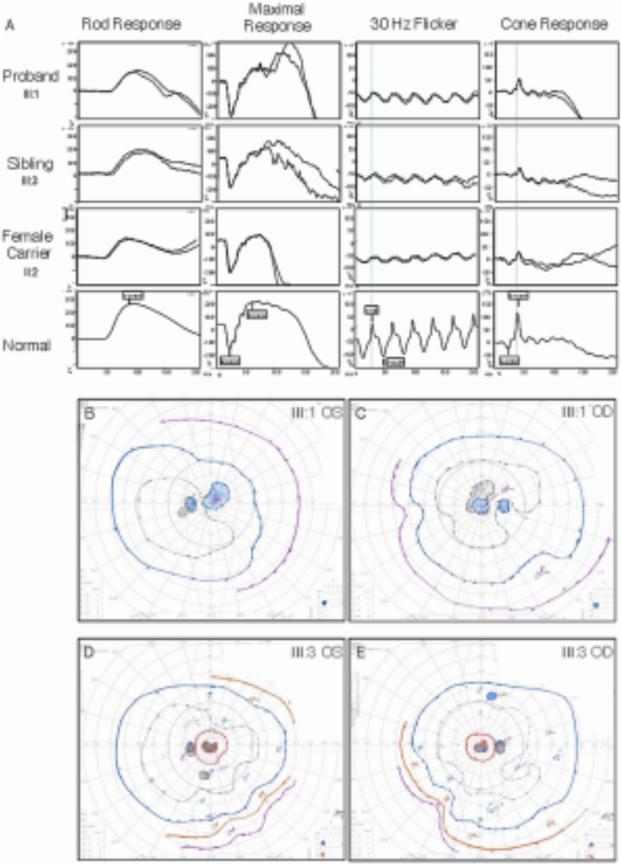
A. Electroretinography. Replicated dual ERG tracing of the proband, affected brother, and mother (manifest carrier) were compared to a normal subject. The traces showed implicit time delays and amplitude reduction in the cone system with a relatively normal rod system. The carrier female ERG showed asymmetry where the right eye was more affected and corresponded with the fundus findings. B – E. Goldmann visual fields. Visual field testing showed that both the proband (B, C) and the affected brother (D, E) had central scotomas with a small island of preservation inside central scotoma of both eyes. Minimal visual field constrictions were also noted in both patients.
References
-
- Laboratory for the Molecular Diagnosis of Inherited Eye Diseases, The University of Texas - Houston Health Science Center. Daiger SP. RetNet: Summaries of Genes and Loci Causing Retinal Diseases. [August 2013];Secondary RetNet: Summaries of Genes and Loci Causing Retinal Diseases. https://sph.uth.edu/retnet/sum-dis.htm.
-
- Audo I, Bujakowska KM, Leveillard T, et al. Development and application of a next-generation-sequencing (NGS) approach to detect known and novel gene defects underlying retinal diseases. [December 2013];Orphanet J Rare Dis. 2012 7:8. http://www.ncbi.nlm.nih.gov/pmc/articles/PMC3352121/. doi: 10.1186/1750-1172-7-8. - PMC - PubMed
Publication types
MeSH terms
Substances
Grants and funding
LinkOut - more resources
Full Text Sources
Other Literature Sources