. 2013 Sep;8(5):583-597.
doi: 10.2217/fnl.13.37.

Diffusion tensor imaging and related techniques in tuberous sclerosis complex: review and future directions

Affiliations

Diffusion tensor imaging and related techniques in tuberous sclerosis complex: review and future directions

Jurriaan M Peters et al. Future Neurol. 2013 Sep.

Abstract

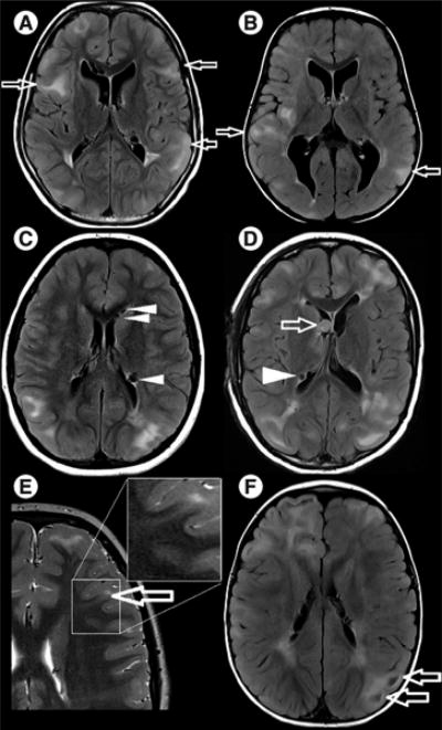
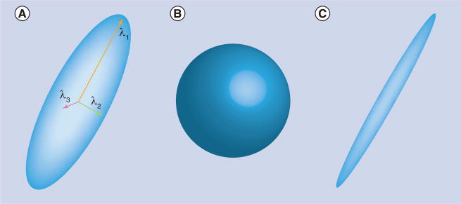
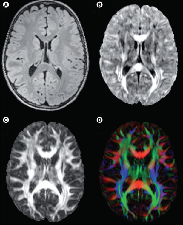
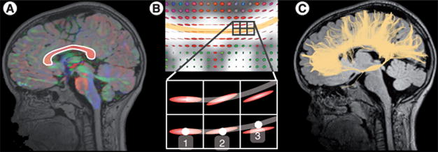
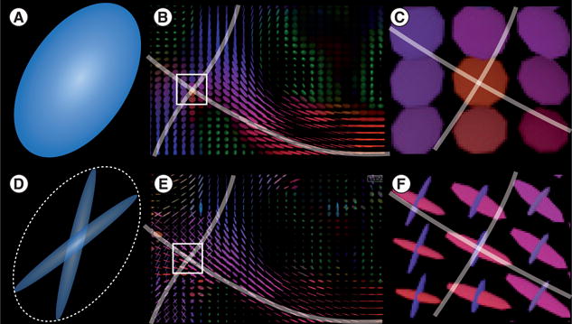
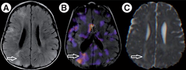
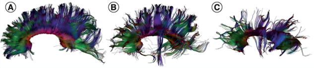
In this article, the authors aim to introduce the nonradiologist to diffusion tensor imaging (DTI) and its applications to both clinical and research aspects of tuberous sclerosis complex. Tuberous sclerosis complex is a genetic neurocutaneous syndrome with variable and unpredictable neurological comorbidity that includes refractory epilepsy, intellectual disability, behavioral abnormalities and autism spectrum disorder. DTI is a method for modeling water diffusion in tissue and can noninvasively characterize microstructural properties of the brain. In tuberous sclerosis complex, DTI measures reflect well-known pathological changes. Clinically, DTI can assist with detecting the epileptogenic tuber. For research, DTI has a putative role in identifying potential disease biomarkers, as DTI abnormalities of the white matter are associated with neurocognitive morbidity including autism. If indeed DTI changes parallel phenotypical changes related to the investigational treatment of epilepsy, cognition and behavior with mTOR inhibitors, it will facilitate future clinical trials.

Keywords: MRI; autism spectrum disorders; behavior; cognition; diffusion tensor imaging; epilepsy; mTOR serine–threonine kinases; tuberous sclerosis complex.


Figures

Figure 1
Figure 1. Conventional MRI findings in tuberous sclerosis complex
(A & B) Axial fluid attenuation inversion recovery images. Both patients have subcortical tubers (arrows) of comparable size and distribution (not all tubers shown in current plane), but patient (A) has severe autism, no active seizure disorder and is nonverbal, while patient (B) has mild motor and language delays, no autism and refractory seizures despite multiple antiepileptic drugs. (C & D) Axial fluid attenuation inversion recovery images. Hypointense partially calcified subependymal nodules are seen lining the ependyma (arrowheads) and a subependymal giant cell astrocytoma is seen in (D), at the level of the foramen of Monro (arrow). (E) Axial T2-weighted image shows a radial migration line tracking from the tuber into the deep white matter (arrow, and zoom frame). (F) Axial fluid attenuation inversion recovery image. Cyst-like appearance of a tuber (arrows).
Figure 2
Figure 2. Diffusion tensor imaging is the most common model of the diffusion
(A) Diffusion tensor imaging can be represented as an ellipsoid that consists of three axes of diffusion and the corresponding diffusivities (here λ1, λ2 and λ3). The shape of the ellipsoids provides information about the type of diffusion present in the voxel. (B) An isotropic diffusion leads to a spherical tensor. (C) Diffusion that is highly restricted in two directions and favored in one direction will present as an elongated tensor with very small second and third diffusivities.
Figure 3
Figure 3. Diffusion tensor imaging and diffusion tensor imaging-based measures
(A) Fluid attenuation inversion recovery MRI structural image, axial plane. (B) Mean diffusivity image. Mean diffusivity is especially large in the corpus callosum and in corticospinal tracts. (C) Fractional anisotropy (FA) image shows where in the brain diffusion is more (white) or less (black) anisotropic. Owing to the presence of highly structured white matter fascicles with aligned axons and myelin sheath, FA in the white matter is high. By contrast, gray matter present in axons with various directions, results in a lower FA. (D) FA can be colored based on the directions of the fascicle in each voxel: red means the fascicle is oriented left to right, green represents fascicles that are oriented along the anterior–posterior axis and blue represents the superior–inferior axis. For color images please see www.futuremedicine.com/doi/pdf/10.2217/fnl.13.37.
Figure 4
Figure 4. Tractography allows detection of white matter pathways in the brain
(A) A seeding region is first defined (here the corpus callosum) from where tracts will start growing. (B) From each voxel of the seeding region, tracts grow in the direction of the tensor. (C) Step by step, from voxel to voxel, tracts keep growing until they reach the gray matter where they stop. This yields 3D maps of the fascicles in the brain, all connected to the seeding region. In order to avoid spurious fibers when performing tractography, using two seed points instead of one can be more accurate [122]. A correction for the density of tracts can also be applied, in which spurious tracts get largely ignored in calculating an average of a diffusion metric [23].
Figure 5
Figure 5. Single tensor and multifascicle models
(A–C) Unlike assumptions of the diffusion tensor models, fascicles in the voxels may have more than one preferential direction. Diffusion tensor imaging model assumes that a single fascicle is present at each voxel. This assumption is violated in regions where fascicles cross, such as (B) the corona radiata. In those regions, tensors are abnormally inflated to capture the signal arising from (C) each fascicle, resulting in a lower fractional anisotropy that may be misleadingly interpreted. (D–F) By contrast, multifascicle models represent each fascicle independently and are, therefore, able to characterize regions with crossing fascicles.
Figure 6
Figure 6. A 2-year-old patient with tuberous sclerosis complex with an epileptogenic right parietal tuber
Visible on (A) axial fluid attenuation inversion recovery image, arrow indicates large right parietal tuber. (B) Single-photon emission CT scan with an injection shortly after onset of a seizure, demonstrating focal increased tracer uptake in the epileptogenic tuber (note the difference in angulation of the image). (C) Mean diffusivity image, revealing elevated mean diffusivity (0.0015 mm2/s) at the tuber compared with elsewhere in white matter (0.0008 mm2/s). The tuber was resected and the patient has been seizure-free for over 6 months.
Figure 7
Figure 7. Diffusion tensor imaging of the corpus callosum in tuberous sclerosis complex
(A–C) Tractography renderings of the corpus callosum of three subjects: (A) healthy control, mean fractional anisotropy (FA) is 0.46; (B) patient with tuberous sclerosis complex, no autism spectrum disorder, mean FA is 0.50; and (C) patient with tuberous sclerosis complex and autism spectrum disorder, mean FA is 0.34. The corpus callosum of (B) and (C) are more ragged owing to tubers interfering with streamlines, but only in the patient with autism spectrum disorder is the mean FA is lower. (C) Reproduced with permission from [23].

References

    1. Crino PB, Nathanson KL, Henske EP. The tuberous sclerosis complex. N Engl J Med. 2006;355(13):1345–1356. - PubMed
    1. Osborne JP, Fryer A, Webb D. Epidemiology of tuberous sclerosis. Ann NY Acad Sci. 1991;615:125–127. - PubMed
    1. Ess KC. The neurobiology of tuberous sclerosis complex. Semin Pediatr Neurol. 2006;13(1):37–42. - PubMed
    1. Jentarra G, Snyder SL, Narayanan V. Genetic aspects of neurocutaneous disorders. Semin Pediatr Neurol. 2006;13(1):43–47. - PubMed
    1. Curatolo P, Bombardieri R, Jozwiak S. Tuberous sclerosis. Lancet. 2008;372(9639):657–668. - PubMed

LinkOut - more resources