

Isolation, characterization, and molecular modeling of a rheumatoid factor from a Hepatitis C virus infected patient with Sjögren's syndrome
- PMID: 24489505
- PMCID: PMC3892945
- DOI: 10.1155/2013/516516
Isolation, characterization, and molecular modeling of a rheumatoid factor from a Hepatitis C virus infected patient with Sjögren's syndrome
Abstract
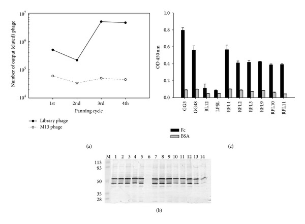
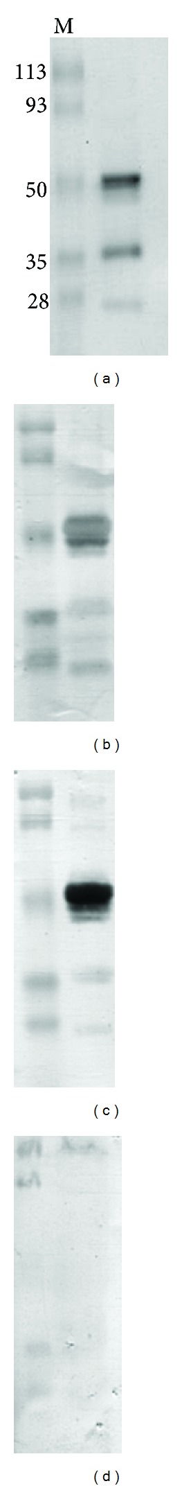
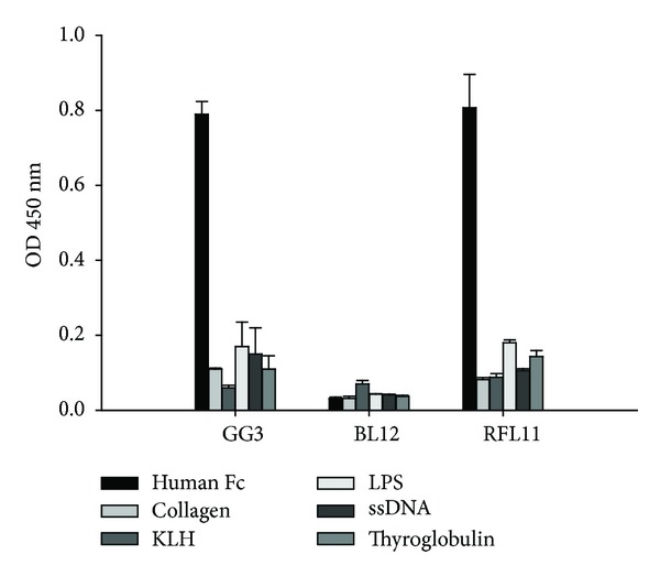
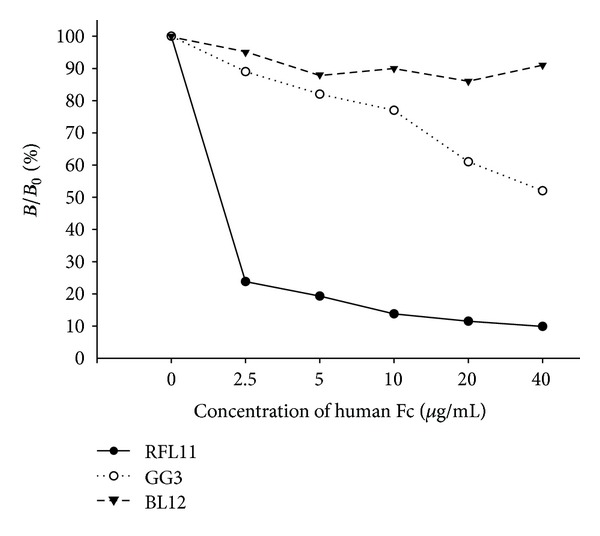
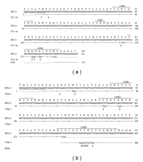
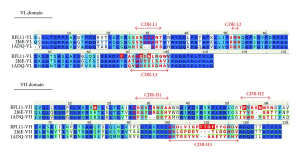
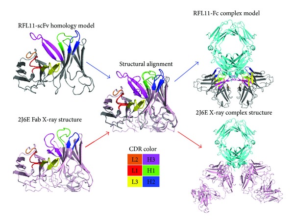
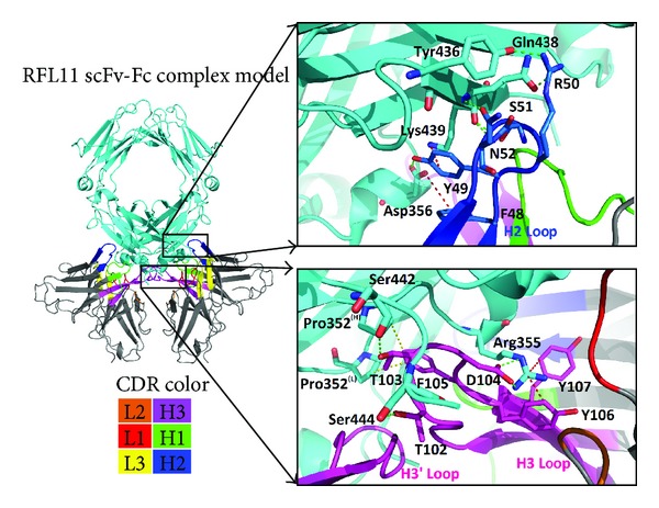
We have previously isolated several IgG rheumatoid factors (RFs) from patients with both rheumatoid arthritis and idiopathic thrombocytopenia purpura using phage display system. To study IgG RFs in patients with other autoimmune diseases, phage display antibody libraries from a hepatitis C virus infected patient with Sjögren's syndrome were constructed. After panning, a specific clone RFL11 was isolated for characterization in advance. The binding activity and specificity of RFL11 to IgG Fc fragment were comparable to those of RFs previously isolated. The analysis with existed RF-Fc complex structures indicated the homology model of RFL11 is similar to IgM RF61 complex with high binding affinity of about 6 × 10⁻⁸ M. This effect resulted from longer complementarity-determining region (CDR) combining key somatic mutations. In the RFL11-Fc interfaces, the CDR-H3 loop forms a finger-like structure extending into the bottom of Fc pocket and resulting in strong ion and cation-pi interactions. Moreover, a process of antigen-driven maturation was proven by somatically mutated VH residues on H2 and H3 CDR loops in the interfaces. Taken together, these results suggested that high affinity IgG RFs can be generated in patients with Sjögren's syndrome and may play an important role in the pathogenesis of this autoimmune disease.
Figures
Similar articles
-
Crystal structure of a human autoimmune complex between IgM rheumatoid factor RF61 and IgG1 Fc reveals a novel epitope and evidence for affinity maturation.J Mol Biol. 2007 May 18;368(5):1321-31. doi: 10.1016/j.jmb.2007.02.085. Epub 2007 Mar 6. J Mol Biol. 2007. PMID: 17395205 Free PMC article.
-
Salivary gland B cell lymphoproliferative disorders in Sjögren's syndrome present a restricted use of antigen receptor gene segments similar to those used by hepatitis C virus-associated non-Hodgkins's lymphomas.Eur J Immunol. 2002 Mar;32(3):903-10. doi: 10.1002/1521-4141(200203)32:3<903::AID-IMMU903>3.0.CO;2-D. Eur J Immunol. 2002. PMID: 11870635
-
Salivary Gland Mucosa-Associated Lymphoid Tissue-Type Lymphoma From Sjögren's Syndrome Patients in the Majority Express Rheumatoid Factors Affinity-Selected for IgG.Arthritis Rheumatol. 2020 Aug;72(8):1330-1340. doi: 10.1002/art.41263. Epub 2020 Jul 8. Arthritis Rheumatol. 2020. PMID: 32182401 Free PMC article.
-
The structure of a human rheumatoid factor bound to IgG Fc.Adv Exp Med Biol. 1998;435:41-50. doi: 10.1007/978-1-4615-5383-0_4. Adv Exp Med Biol. 1998. PMID: 9498063 Review.
-
Hypergammaglobulinemic purpura in patients with Sjögren's syndrome: a report of nine cases and a review of the Japanese literature.Jpn J Med. 1989 Mar-Apr;28(2):148-55. doi: 10.2169/internalmedicine1962.28.148. Jpn J Med. 1989. PMID: 2659854 Review.
References
-
- Daniels TE, Fox PC. Salivary and oral components of Sjogren’s syndrome. Rheumatic Disease Clinics of North America. 1992;18(3):571–589. - PubMed
-
- Ramos-Casals M, la Civita LLA, de Vita S, et al. Characterization of B cell lymphoma in patients with Sjögren’s syndrome and hepatitis C virus infection. Arthritis & Rheumatism. 2007;57(1):161–170. - PubMed
-
- Brito-Zerón P, Ramos-Casals M, Nardi N, et al. Circulating monoclonal immunoglobulins in Sjögren syndrome: prevalence and clinical significance in 237 patients. Medicine. 2005;84(2):90–97. - PubMed
-
- Ramos-Casals M, de Vita S, Tzioufas AG. Hepatitis C virus, Sjögren’s syndrome and B-cell lymphoma: linking infection, autoimmunity and cancer. Autoimmunity Reviews. 2005;4(1):8–15. - PubMed
-
- Ramos-Casals M, Loustaud-Ratti V, de Vita S, et al. Sjögren syndrome associated with hepatitis C virus: a multicenter analysis of 137 cases. Medicine. 2005;84(2):81–89. - PubMed
Publication types
MeSH terms
Substances
LinkOut - more resources
Full Text Sources
Other Literature Sources
Medical
Research Materials
Miscellaneous
