Review
. 2013:2013:606703.
doi: 10.1155/2013/606703. Epub 2013 Dec 31.

Missing links in antibody assembly control

Affiliations
Review

Missing links in antibody assembly control

Tiziana Anelli et al. Int J Cell Biol. 2013.

Abstract

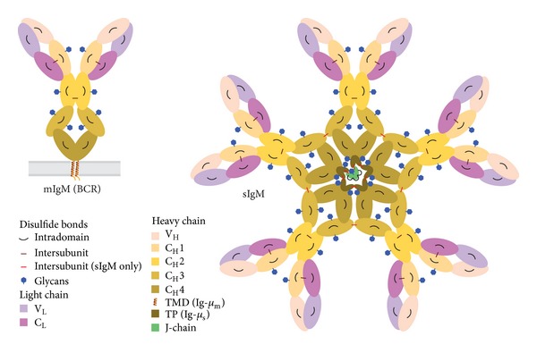
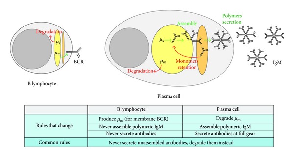
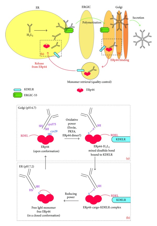
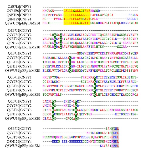
Fidelity of the humoral immune response requires that quiescent B lymphocytes display membrane bound immunoglobulin M (IgM) on B lymphocytes surface as part of the B cell receptor, whose function is to recognize an antigen. At the same time B lymphocytes should not secrete IgM until recognition of the antigen has occurred. The heavy chains of the secretory IgM have a C-terminal tail with a cysteine instead of a membrane anchor, which serves to covalently link the IgM subunits by disulfide bonds to form "pentamers" or "hexamers." By virtue of the same cysteine, unassembled secretory IgM subunits are recognized and retained (via mixed disulfide bonds) by members of the protein disulfide isomerase family, in particular ERp44. This so-called "thiol-mediated retention" bars assembly intermediates from prematurely leaving the cell and thereby exerts quality control on the humoral immune response. In this essay we discuss recent findings on how ERp44 governs such assembly control in a pH-dependent manner, shuttling between the cisGolgi and endoplasmic reticulum, and finally on how pERp1/MZB1, possibly as a co-chaperone of GRP94, may help to overrule the thiol-mediated retention in the activated B cell to give way to antibody secretion.


Figures

Figure 1
Figure 1
Schematic representation of membrane bound IgM and secretory IgM. B lymphocytes display membrane bound IgM (mIgM)—shown left—on their surface as the core part of the BCR. Once committed to the plasma cell stage they secrete secretory IgM (sIgM) in either “pentameric”—shown right—or “hexameric”—not shown—form. HCs and LCs consist of various Ig-fold domains VH, CH1–4; VL, CL that are color-coded as indicated. Glycans and intra- and intersubunit disulfide bonds between HCs, LCs, and J-chain are depicted. Note that the Ig-μ m HC (in mIgM) differs from the Ig-μ s HC (in sIgM) in their C-termini, having a transmembrane domain (TMD) or, respectively, a cysteine containing tail piece (TP) as indicated.
Figure 2
Figure 2
Rules for IgM processing in B lymphocytes and plasma cells.B lymphocytes do not produce secretory antibodies. In B lymphocytes Ig-μ m HC (μ m) is synthesized in the ER; it is assembled in the early secretory pathway (in yellow) and then proceeds through the Golgi (orange) to be exposed on the plasma membrane as part of the BCR; Ig-μ s HC (μ s), instead, is retrotranslocated from the ER and degraded. Conversely, plasma cells are hardworking antibodies factories; they degrade μ m but efficiently assemble μ s into polymeric IgM and secrete them in bulk. Incompletely assembled IgM subunits are retained in the secretory pathway for another chance of being inserted into a polymer or to be finally degraded.
Figure 3
Figure 3
ERp44 quality control and IgM polymerization. IgM monomers are assembled into polymers in the early secretory pathway with the help of the hexameric lectin ERGIC-53. IgM polymers are released from ERGIC-53 in the Golgi and can then be secreted. In the Golgi, ERp44—in an open conformation thanks to the slightly acidic pH—interacts with unassembled IgM subunits exposing the C-terminal cysteine 575 in the SH conformation (inset (a)): the covalent binding between ERp44's active site cysteine 29 and the IgM subunits requires an oxidative power whose source is still unknown. The interaction with the cargo favors the exposure of ERp44's C-terminal RDEL for binding to the KDEL receptor and the complex KDEL receptor-ERp44-cargo (IgM H2L2 “monomer”) is then transported back to the ER. Here, a reducing power disentangles the disulfide bond between ERp44 and the IgM “monomer.” The neutral pH in the ER, which also inhibits the KDEL receptor activity, now stabilizes ERp44 in a more closed conformation (inset (b)). Keeping ERp44 in a closed conformation in the ER likely is important to avoid that ERp44 interacts with subunits that productively take part in the polymerization process.
Figure 4
Figure 4
Alignment of pERp1/MZB1 and members of the CNPY protein family. The alignment of the five human protein sequences with UniProt identifiers as indicated was generated with the ClustalW algorithm and manually curated. The signal peptide encoding residues are highlighted in yellow, the C-terminal KDEL-like tetrapeptides are highlighted in grey, and the conserved cysteines in green. Standard color coding for the residues is as follows: alkaline in pink; acidic in blue; other hydrophilic in green; and hydrophobic in red.

References

    1. Hendershot LM, Sitia R. Immunoglobulin assembly and secretion. In: Honjo T, Alt FW, Neuberger MS, editors. Molecular Biology of B Cells. Amsterdam, The Netherlands: Elsevier Academic Press; 2005. pp. 261–273.
    1. Sitia R, Neuberger M, Alberini C, et al. Developmental regulation of IgM secretion: the role of the carboxy-terminal cysteine. Cell. 1990;60(5):781–790. - PubMed
    1. Braakman I, Bulleid NJ. Protein folding and modification in the mammalian endoplasmic reticulum. Annual Review of Biochemistry. 2011;80:71–99. - PubMed
    1. Haas IG, Wabl M. Immunoglobulin heavy chain binding protein. Nature. 1983;306(5941):387–389. - PubMed
    1. Melnick J, Aviel S, Argon Y. The endoplasmic reticulum stress protein GRP94, in addition to BiP, associates with unassembled immunoglobulin chains. Journal of Biological Chemistry. 1992;267(30):21303–21306. - PubMed