

Landmarking the brain for geometric morphometric analysis: an error study
- PMID: 24489689
- PMCID: PMC3904856
- DOI: 10.1371/journal.pone.0086005
Landmarking the brain for geometric morphometric analysis: an error study
Abstract
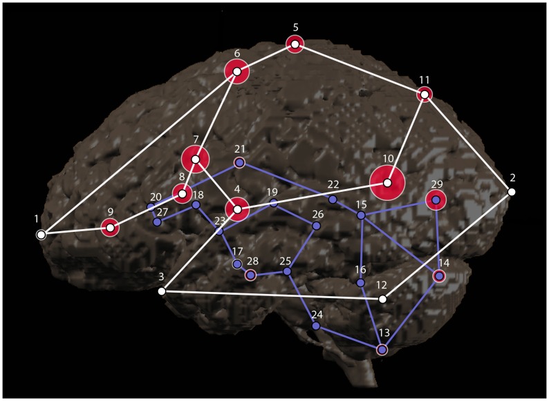
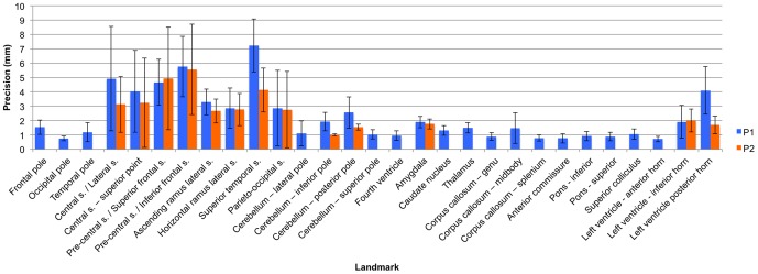
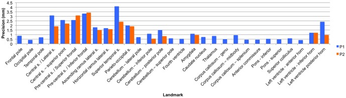
Neuroanatomic phenotypes are often assessed using volumetric analysis. Although powerful and versatile, this approach is limited in that it is unable to quantify changes in shape, to describe how regions are interrelated, or to determine whether changes in size are global or local. Statistical shape analysis using coordinate data from biologically relevant landmarks is the preferred method for testing these aspects of phenotype. To date, approximately fifty landmarks have been used to study brain shape. Of the studies that have used landmark-based statistical shape analysis of the brain, most have not published protocols for landmark identification or the results of reliability studies on these landmarks. The primary aims of this study were two-fold: (1) to collaboratively develop detailed data collection protocols for a set of brain landmarks, and (2) to complete an intra- and inter-observer validation study of the set of landmarks. Detailed protocols were developed for 29 cortical and subcortical landmarks using a sample of 10 boys aged 12 years old. Average intra-observer error for the final set of landmarks was 1.9 mm with a range of 0.72 mm-5.6 mm. Average inter-observer error was 1.1 mm with a range of 0.40 mm-3.4 mm. This study successfully establishes landmark protocols with a minimal level of error that can be used by other researchers in the assessment of neuroanatomic phenotypes.
Conflict of interest statement
Figures
Similar articles
-
The effect of automated landmark identification on morphometric analyses.J Anat. 2019 Jun;234(6):917-935. doi: 10.1111/joa.12973. Epub 2019 Mar 22. J Anat. 2019. PMID: 30901082 Free PMC article.
-
Measurement error in μCT-based three-dimensional geometric morphometrics introduced by surface generation and landmark data acquisition.J Anat. 2019 Aug;235(2):357-378. doi: 10.1111/joa.12999. Epub 2019 May 7. J Anat. 2019. PMID: 31062345 Free PMC article.
-
Surface landmark quantification of embryonic mouse craniofacial morphogenesis.BMC Dev Biol. 2014 Jul 24;14:31. doi: 10.1186/1471-213X-14-31. BMC Dev Biol. 2014. PMID: 25059626 Free PMC article.
-
Robust automated constellation-based landmark detection in human brain imaging.Neuroimage. 2018 Apr 15;170:471-481. doi: 10.1016/j.neuroimage.2017.04.012. Epub 2017 Apr 6. Neuroimage. 2018. PMID: 28392490 Free PMC article. Review.
-
Quantification of Facial Traits.Front Genet. 2019 May 24;10:397. doi: 10.3389/fgene.2019.00397. eCollection 2019. Front Genet. 2019. PMID: 31178890 Free PMC article. Review.
Cited by
-
A Systematist's Guide to Estimating Bayesian Phylogenies From Morphological Data.Insect Syst Divers. 2019 May;3(3):2. doi: 10.1093/isd/ixz006. Epub 2019 Jun 18. Insect Syst Divers. 2019. PMID: 31355348 Free PMC article. Review.
-
Conformal invariants for multiply connected surfaces: Application to landmark curve-based brain morphometry analysis.Med Image Anal. 2017 Jan;35:517-529. doi: 10.1016/j.media.2016.09.001. Epub 2016 Sep 6. Med Image Anal. 2017. PMID: 27639215 Free PMC article.
-
Macroanatomical Landmarks Featuring Junctions of Major Sulci and Fissures and Scalp Landmarks Based on the International 10-10 System for Analyzing Lateral Cortical Development of Infants.Front Neurosci. 2017 Jul 11;11:394. doi: 10.3389/fnins.2017.00394. eCollection 2017. Front Neurosci. 2017. PMID: 28744192 Free PMC article.
-
A Baboon Brain Atlas for Magnetic Resonance Imaging and Positron Emission Tomography Image Analysis.Front Neuroanat. 2022 Jan 14;15:778769. doi: 10.3389/fnana.2021.778769. eCollection 2021. Front Neuroanat. 2022. PMID: 35095430 Free PMC article.
-
Accurate MR Image Registration to Anatomical Reference Space for Diffuse Glioma.Front Neurosci. 2020 Jun 5;14:585. doi: 10.3389/fnins.2020.00585. eCollection 2020. Front Neurosci. 2020. PMID: 32581699 Free PMC article.
References
-
- Casey BJ, Tottenham N, Liston C, Durston S (2005) Imaging the developing brain: what have we learned about cognitive development? Trends Cogn Sci 9: 104–110. - PubMed
-
- Gold SM, Dziobek I, Sweat V, Tirsi A, Rogers K, et al. (2007) Hippocampal damage and memory impairments as possible early brain complications of type 2 diabetes. Diabetologia 50: 711–719. - PubMed
-
- Lorenzetti V, Allen NB, Fornito A, Yucel M (2009) Structural brain abnormalities in major depressive disorder: a selective review of recent MRI studies. J Affect Disord 117: 1–17. - PubMed
Publication types
MeSH terms
Grants and funding
LinkOut - more resources
Full Text Sources
Other Literature Sources
