. 2014 Jan 29;9(1):e86673.
doi: 10.1371/journal.pone.0086673. eCollection 2014.

Spiegelzymes® mirror-image hammerhead ribozymes and mirror-image DNAzymes, an alternative to siRNAs and microRNAs to cleave mRNAs in vivo?

Affiliations

Spiegelzymes® mirror-image hammerhead ribozymes and mirror-image DNAzymes, an alternative to siRNAs and microRNAs to cleave mRNAs in vivo?

Eliza Wyszko et al. PLoS One. .

Erratum in

  • PLoS One. 2014;9(3):e93798

Abstract

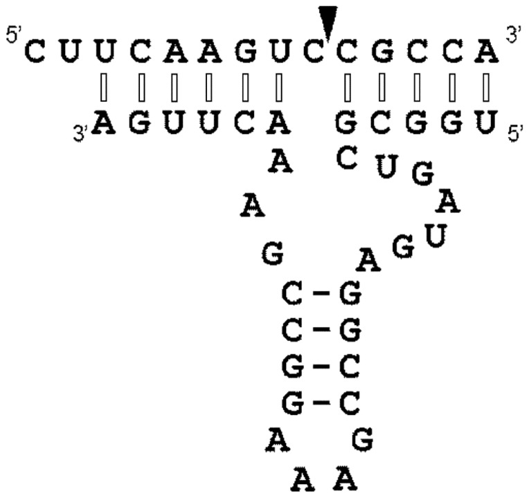
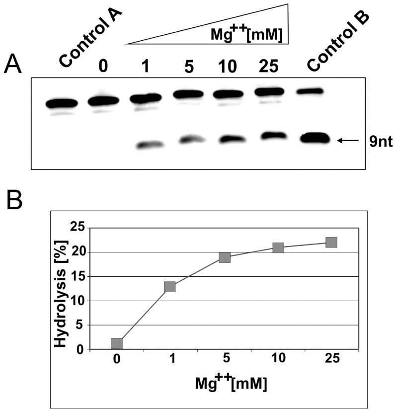
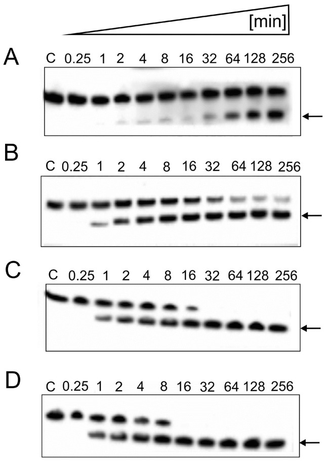
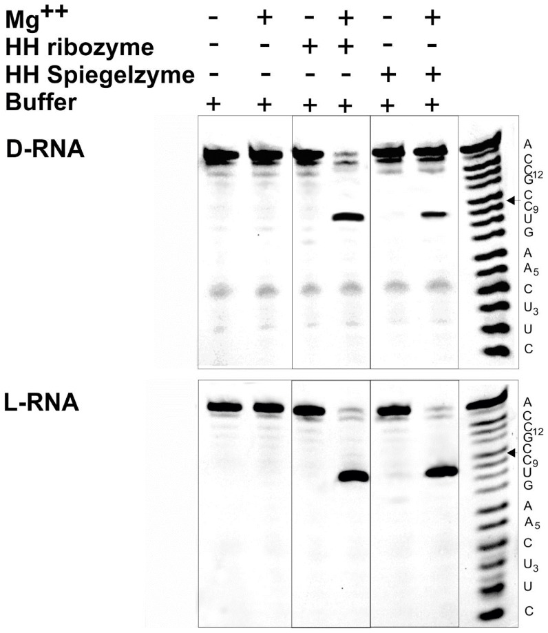
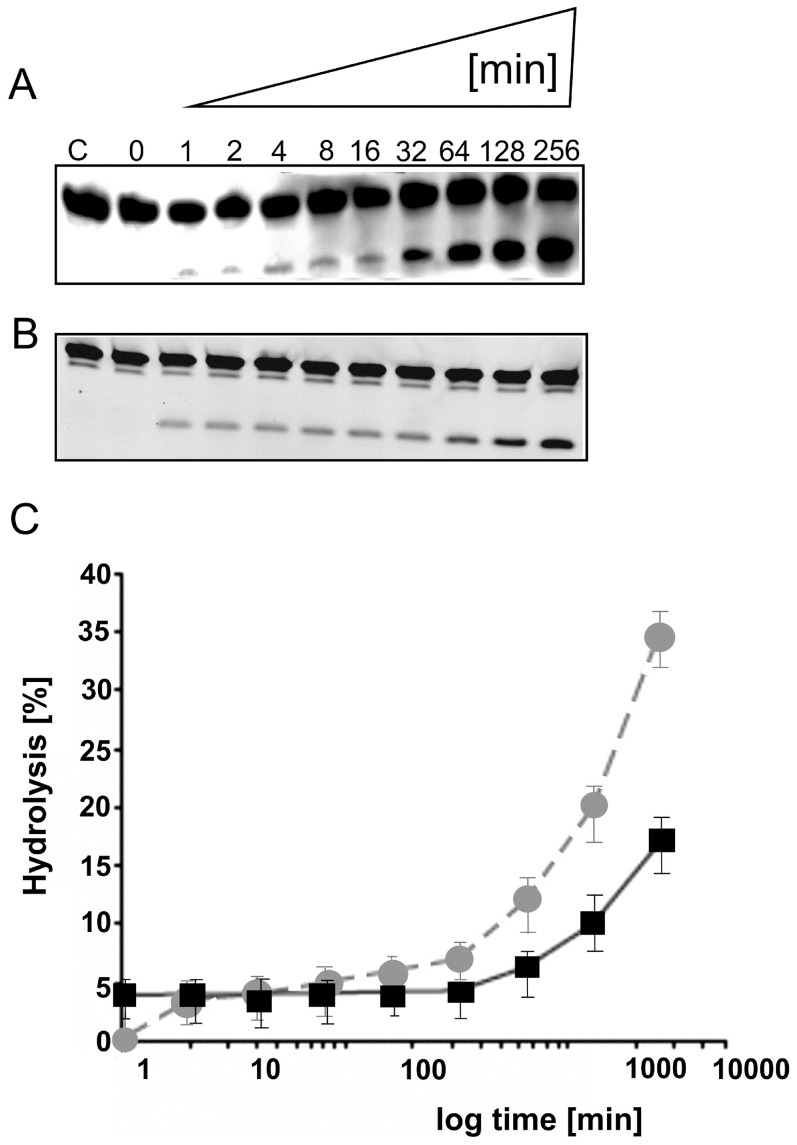
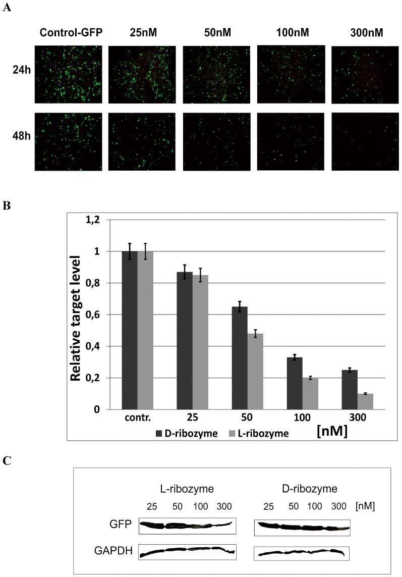
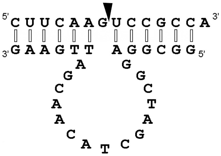
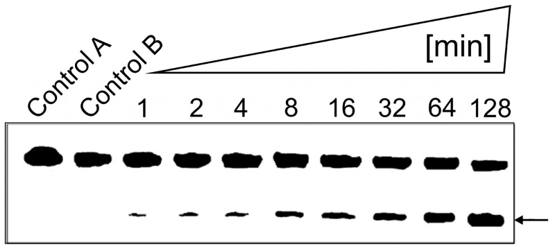
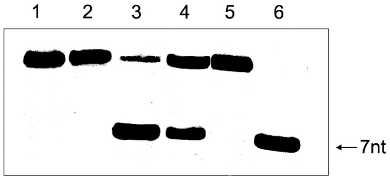
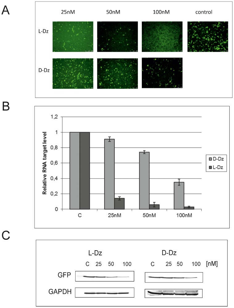
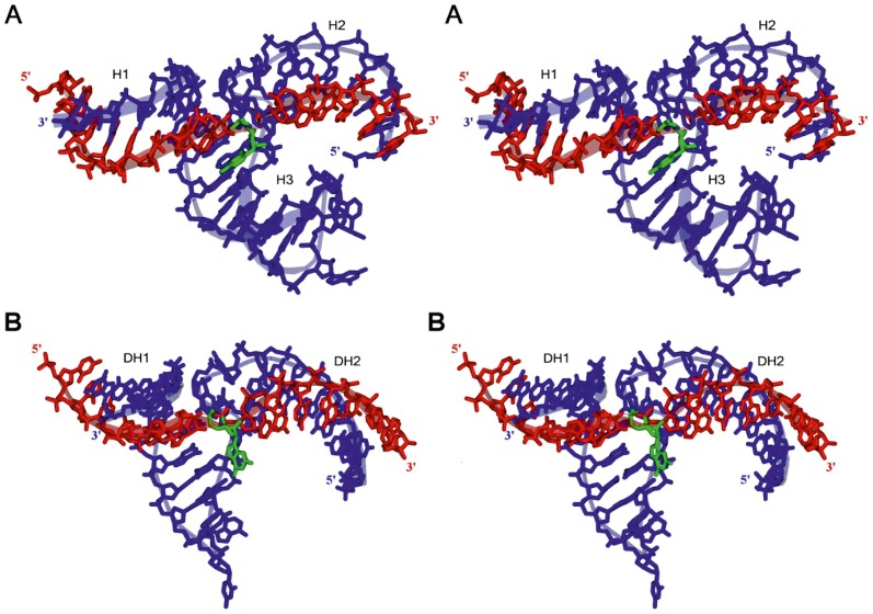
With the discovery of small non-coding RNA (ncRNA) molecules as regulators for cellular processes, it became intriguing to develop technologies by which these regulators can be applied in molecular biology and molecular medicine. The application of ncRNAs has significantly increased our knowledge about the regulation and functions of a number of proteins in the cell. It is surprising that similar successes in applying these small ncRNAs in biotechnology and molecular medicine have so far been very limited. The reasons for these observations may lie in the high complexity in which these RNA regulators function in the cells and problems with their delivery, stability and specificity. Recently, we have described mirror-image hammerhead ribozymes and DNAzymes (Spiegelzymes®) which can sequence-specifically hydrolyse mirror-image nucleic acids, such as our mirror-image aptamers (Spiegelmers) discovered earlier. In this paper, we show for the first time that Spiegelzymes are capable of recognising complementary enantiomeric substrates (D-nucleic acids), and that they efficiently hydrolyse them at submillimolar magnesium concentrations and at physiologically relevant conditions. The Spiegelzymes are very stable in human sera, and do not require any protein factors for their function. They have the additional advantages of being non-toxic and non-immunogenic. The Spiegelzymes can be used for RNA silencing and also as therapeutic and diagnostic tools in medicine. We performed extensive three-dimensional molecular modelling experiments with mirror-image hammerhead ribozymes and DNAzymes interacting with D-RNA targets. We propose a model in which L/D-double helix structures can be formed by natural Watson-Crick base pairs, but where the nucleosides of one of the two strands will occur in an anticlinal conformation. Interestingly enough, the duplexes (L-RNA/D-RNA and L-DNA/D-RNA) in these models can show either right- or left-handedness. This is a very new observation, suggesting that molecular symmetry of enantiomeric nucleic acids is broken down.


Conflict of interest statement

Competing Interests: The authors have the following interests: Dr. Volker A. Erdmann and Dr. Eliza Wyszko are inventors of the following patent: Patent: “RNAzym” (PCT/DE2010/000159). Titel: Pharmazeutische Zusammensetzung zur Behandlung von Nebenwirkungen durch Gabe von Spiegelmeren Inventor: ERDMANN, V.A. [DE]; WYSZKO, Eliza [Pll 1. Registered by: Freie Universität Berlin rDEl. Registration date: 08.02.2010 l. Volker A. Erdmann is inventor of the following patent: Patent: “L-DNA" (PCT/DE2012/000008). Titel: Pharmazeutische Zusammensetzung enthalten1:t L-DNA. Inventor: ERDMANN, V.A. [DE]. Registered by: ERDMANN, V.A. [DE]. Registration date: 02.01 .2012 1. Both patents have been transferred to the “Erdmann Technologies GmbH,” a company founded by Dr. Volker A. Erdmann in July, 2013. The company's goals are to develop anticancer and antiviral pharmaceuticals on the basis of Spiegelzymes. Dr. Florian F. Mueller is the founder and owner of PENTAFOLIUM-SOFT, a company dedicated to develop computer software to predict secondary structures in DNA and RNA. This does not alter the authors' adherence to all the PLOS ONE policies on sharing data and materials.

Figures

Figure 1
Figure 1. The secondary structural model of a hammerhead Spiegelzyme complexed with an D-RNA substrate containing GUC↓N, the required cleavage site for hammerhead ribozymes.
The cleavage site is indicated with an arrow. Interactions between the hammerhead Spiegelzyme (L-RNA) and the D-RNA substrate of opposite chirality show modified Watson-Crick base pairs with nucleotides in anticlinal conformation (see Figs. 12–14) and are marked with open bars. The normal Watson-Crick bases in antiperiplanar conformation are indicated with closed bars.
Figure 2
Figure 2. Magnesium dependence of D-RNA hydrolysis by hammerhead Spiegelzymes.
A. D-RNA (0.2 µM) was incubated with 2 µM of the Spiegelzyme in 50 mM Tris-HCl, pH 7.5, buffer containing 1, 5, 10, and 25 mM MgCl2 for 1 h at 37°C. The reaction products were separated by 20% polyacrylamide gel electrophoresis with 7 M urea. Control reactions were carried out with target D-RNA alone in buffer (lane Control A) or with D-RNA hammerhead ribozyme in buffer with 10 mM Mg2+ (lane Control B). B. Hydrolytic activity in % of hammerhead Spiegelzyme with the D-RNA target at different magnesium concentrations.
Figure 3
Figure 3. Time-dependent cleavage of the D-RNA substrates by the hammerhead Spiegelzyme.
Reactions were carried out at 37°C in a 50 mM Tris-HCl, pH 7.5, buffer, containing 10 mM MgCl2. Reaction products were separated by a 20% polyacrylamide gel electrophoresis containing 7 M urea in 0.09 M Tris-borate buffer at pH 8.3. The fluorescence was analysed by a Fuji Film FLA 5100 phosphoimager. The hydrolysis of 0.2 µM fluorescein-labelled D-RNA with hammerhead Spiegelzyme to substrate ratios of 10∶1 (A) 25∶1 (B), 50∶1 (C), and 100∶1 (D) are shown in this figure. The arrows indicate the expected hydrolysis product of 9-nucleotide length.
Figure 4
Figure 4. Hydrolysis of D-RNA and L-RNA substrates by hammerhead ribozymes and hammerhead Spiegelzymes.
The substrates (0.2 µM) were incubated with 2 µM of hammerhead ribozymes or hammerhead Spiegelzymes in 50 mM Tris-HCl, pH 7.5, buffer, at 37°C for 2 h. 10 mM MgCl2 and hammerhead ribozymes or hammerhead Spiegelzymes were added as indicated. In the top panel, D-RNA substrates and in the bottom panel, L-RNA substrates were used. Hydrolysis products were separated on 20% polyacrylamide gel electrophoreses with 7 M urea. The ladder (right) was generated by alkaline hydrolysis.
Figure 5
Figure 5. Time-dependent cleavage of the D-RNA target with the hammerhead Spiegelzyme (heterochiral complex).
The incubation of 0.2 µM target fluorescein-labelled D-RNA, with 2 µM or 0.02 µM hammerhead Spiegelzyme for single (A) and multiple (B) turnover reactions. The reactions were carried out in 50 mM Tris-HCl, pH 7.5, buffer containing 10 mM MgCl2 at 37°C and different incubation times as indicated. Control reaction was carried out with target D-RNA alone in buffer for 256 min (lane C). Reaction products were separated by 20% polyacrylamide gel electrophoreses containing 7 M urea and analysed by a Fuji Film FLA 5100 phosphoimager. (C) A plot of single (squares, black solid line) and multiple (circle, grey broken line) turnover reactions. The time of reaction is shown in logarithmic scale.
Figure 6
Figure 6. Hammerhead Spiegelzyme stability analysis in COS-7 cells.
A. 5′-fluorescein-labelled Spiegelzyme (1 µg) was transfected into cells with Lipofectamine 2000. The microscope images were taken at 24, 72, 96, and 120 h after transfection. B. The RNA isolated from transfected cells (Trizol, Ambion) was analysed on 20% PAGE with 7 M urea. Lane 1 control hammerhead Spiegelzyme. Spiegelzyme after 120 h of incubation in COS-7 cells (lane 2).
Figure 7
Figure 7. GFP expression in HeLa cells after 24 and 48(L-ribozyme).
(A) Hydrolysis of GFP mRNAs in HeLA cells after 24 and 48(B) Quantification the GFP mRNA hydrolysis by L- and D- hammerhead ribozymes. HeLa cells were transfected with 10, 25, 100, and 300 nM ribozymes and pEGFP. After 48 h of incubation, total RNA was isolated and cDNA was obtained. PCR reactions were performed to assess the GFP expression relative to Hprt and Actb genes. All experiments were repeated at least three times. (C) Western blot analysis of proteins from HeLa cells with antibodies against GFP and GAPDH (control).
Figure 8
Figure 8. The general model of the secondary structure of D- or L-DNAzyme in the homo- or heterochiral complex with target D-or L-RNA.
The arrow shows the anticipated cleavage site. We propose that the interactions between the mirror-image DNAzyme and the D-RNA substrate in the heterochiral complex are Watson-Crick type base pairs in which ribose rings of one strand occur in a rotated orientation (see Figs 12 and also in the Supplementary Figures S1 and S2 and discussion).
Figure 9
Figure 9. Time-dependent D-RNA hydrolysis by L-DNAzyme.
Time-dependent hydrolysis of D-RNA target by L-DNAzyme after 1, 2, 4, 8, 16, 32, 64, and 128 min incubation in Tris-HCl buffer, pH 7.5, 10 mM MgCl2, at 37°C. Incubation was performed with 5 µM L-DNAzyme and 0.2 µM target. Control A incubation of D-RNA in Tris-HCl buffer, pH 7.5, without MgCl2; Control B with 10 mM MgCl2. Both controls were incubated for 128 min. Separation of hydrolysis products was carried out under standard conditions on 20% polyacrylamide gels with 7 M urea. The arrow identifies 7 nt cleavage product.
Figure 10
Figure 10. Incubation of D- and L-RNA targets with D-and L-DNAzymes.
Lanes 1 and 2: D- and L-RNA targets (0.2 µM) incubated in 50 mM Tris-HCl buffer, pH 7.5, and 10 mM MgCl2. Lanes 3 and 4 incubation of D- RNA targets (0.2 µM) with D-and L-DNAzyme (2 µM) in the presence of 10 mM MgCl2, lanes 5 and 6 incubation of L-RNA target (0.2 µM) with D-and L-DNAzyme (2 µM) in the presence of 10 mM MgCl2. All incubations were carried out for 5 h at 37°C. The arrow shows products of hydrolysis as expected when the RNA target is hydrolyzed by the Spiegelzymes as indicated in Figure 8. Separation of reaction products was accomplished by 20% polyacrylamide gel electrophoresis and 7 M urea.
Figure 11
Figure 11. The activity of D-DNAzyme and L-DNAzyme in HeLa cells.
(A) HeLa cells were transfected with 1 ug EGFP-plasmid and 25, 50 and 100 nM of D-DNAzyme or L-DNAzyme. Fluorescence measurements were carried out 48 h after the transfection. (B) Quantification by real-time PCR of the L- and D-DNAzyme hydrolytic activities of GFP mRNAs in HeLa cells. After 48 h, total RNA was isolated and a reverse transcription cDNA was done. PCR reactions were performed to assess GFP expression relative to Hprt and Actb genes in a thermocycler Stratagene Mx3005P instrument. All experiments were repeated at least three times. (C) Western blot analysis of GFP gene expression in HeLa cells after transfection with 25, 50 and 100 nM of L-DNAzyme and D-DNAzyme. After 48 h, incubation cells were sonicated in 10 mM Tris HCl, pH 7.5. Proteins were separated and Western blot analysis was carried out.
Figure 12
Figure 12. Atomic models proposed for the L-hammerhead ribozyme and the L-DNAzyme interacting with their target D-RNA.
A. L-HHRz 5′-U1GGCGCUGAUGAGGCCGAAAGGCCGAAACUUGA33-3′ (shown in blue) with D-RNA target nucleotide sequence 5′-C1UUCAAGUCCGCCA14-3′ (shown in red) with the cleavage site at nucleotide C9 (shown in green). B. L-DNAzyme 5′-G1GCGGAGGCTAGCTACAACGATTGAAG27-3′ (shown in blue) with same D-RNA target nucleotide sequence 5′-C1UUCAAGUCCGCCA14-3′ (shown in red) with the cleavage site at nucleotide G7 (shown in green). See text for detailed discussions of the models.

References

    1. Cava F, Lam H, de Pedro MA, Waldor MK (2011) Emerging knowledge of regulatory roles of D-amino acids in bacteria. Cell Mol Life Sci 68: 817–831. - PMC - PubMed
    1. Wyszko E, Szymanski M, Zeichhardt H, Mueller F, Barciszewski J, et al. (2013) Spigelzymes: Sequence specific hydrolysis with mirror image hammerhead ribozymes and DNAzymes. PLoS One (8)1: e5471. - PMC - PubMed
    1. Klussmann S, Nolte A, Bald R, Erdmann VA, Fürste JP (1996) Mirror-image RNA that binds D-adenosine. Nat Biotechnol 14: 1112–1115. - PubMed
    1. Nolte A, Klussmann S, Bald R, Erdmann VA, Fürste JP (1996) Mirror-design of L-oligonucleotide ligands binding to L-arginine. Nat Biotechnol 14: 1116–1119. - PubMed
    1. van Rooij E, Olson EN (2012) MicroRNA therapeutics for cardiovascular disease: opportunities and obstacles. Nat Rev Drug Discov 11: 860–872. - PMC - PubMed

Publication types

MeSH terms